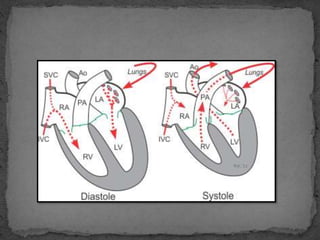
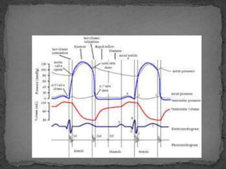
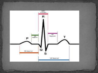
Neeraja A
 Changes that occur in the heart during one beat are
repeated in the same order in the next beat.
 This cyclic repetition is called cardiac cycle.
 Since the heart rate in man is 70 to 80 per minute,
each cycle lasts for 0.8 sec.
 A single cycle of cardiac activity can be divided into
two basic phases - diastole and systole.
 There are two basic events- systole and diastole
 Systole represents the time during which the left and
right ventricles contract and eject blood into the aorta
and pulmonary artery, respectively.
 During systole, the aortic and pulmonic valves open to
permit ejection into the aorta and pulmonary artery.
 The atrioventricular valves are closed during systole,
therefore no blood is entering the ventricles; however,
blood continues to enter the atria though the vena
cavae and pulmonary veins.
 Diastole represents the period of time when the ventricles
are relaxed (not contracting).
 Throughout most of this period, blood is passively flowing
from the left atrium (LA) and right atrium (RA) into the
left ventricle (LV) and right ventricle (RV), respectively (see
figure at right).
 The blood flows through atrioventricular valves (mitral
and tricuspid) that separate the atria from the ventricles.
 The RA receives venous blood from the body through the
superior vena cava (SVC) and inferior vena cava (IVC).
 The LA receives oxygenated blood from lungs through four
pulmonary veins that enter the LA.
 At the end of diastole, both atria contract, which propels an
additional amount of blood into the ventricles.
 The three main events in the cardiac cycle are:
# auricular systole
# ventricular systole
# diastole
 It is also called as atrial systole.
 The cardiac cycle is initiated by auricular systole.
 The contraction of the auricles is called as auricular
systole.
 The two auricles contract simultaneously.
 The duration is 0.1 sec.
 The auricular systole forces the blood into the
ventricles.
 Atrial systole is the contraction of atrial syncytium of
cardiac muscle cells of the left and right atria,
following electrical stimulation.
 Electrical systole is the electrical activity that
stimulates the heart chambers to make them contract.
 This is soon followed by an mechanical systole, which
is the mechanical contraction of the heart.
 As the atria contracts, the blood pressure in each
atrium increases, forcing additional blood flow into
the ventricles.
 This additional blood flow is called as atrial kick.
 80% of the blood flows passively down to the
ventricles, so the atria do not have to contract a great
amount.
 Atrial kick is absent if there is loss of normal electrical
conductionin the geart, such as during atrial
fibrillation, atrial flutter and complete heart block.
DETECTION:
 Electrical systole of the atria begins with the onset of
P wave on ECG. Sino atrial node can be detected by
catheterization.
 The auricular systole is followed by the ventricular
systole.
 Contraction of ventricles is called as ventricular
systole.
 The two ventricles contract simultaneously.
 The duration of ventricular systole is 0.3 sec.
 The ventricular systole forces the blood into the aorta.
 The first sound of the heart is produced by the
ventricular systole.
 Ventricular systole is the contraction of the ventricular
syncytium of the cardiac muscle cells of the right and
left ventricles, following electrical stimulation.
 At the later part of the ejection phase, although the
ventricular pressure falls below the aortic pressure, the
aortic valve remains open because of the inertial
energy of the ejected blood
DETECTION:
 In an ECG electrical systole of the ventricles begins at
the beginning of the QRS complex
 Cardiac diastole is the period of time when the heart
relaxes after contraction in preparing for the next
refilling of circulating blood.
 Together all this they are called as complete cardiac
cycle
Thank you

Cardiac cycle - systole & diastole

  • 1.
  • 2.
     Changes thatoccur in the heart during one beat are repeated in the same order in the next beat.  This cyclic repetition is called cardiac cycle.  Since the heart rate in man is 70 to 80 per minute, each cycle lasts for 0.8 sec.  A single cycle of cardiac activity can be divided into two basic phases - diastole and systole.
  • 3.
     There aretwo basic events- systole and diastole  Systole represents the time during which the left and right ventricles contract and eject blood into the aorta and pulmonary artery, respectively.  During systole, the aortic and pulmonic valves open to permit ejection into the aorta and pulmonary artery.  The atrioventricular valves are closed during systole, therefore no blood is entering the ventricles; however, blood continues to enter the atria though the vena cavae and pulmonary veins.
  • 4.
     Diastole representsthe period of time when the ventricles are relaxed (not contracting).  Throughout most of this period, blood is passively flowing from the left atrium (LA) and right atrium (RA) into the left ventricle (LV) and right ventricle (RV), respectively (see figure at right).  The blood flows through atrioventricular valves (mitral and tricuspid) that separate the atria from the ventricles.  The RA receives venous blood from the body through the superior vena cava (SVC) and inferior vena cava (IVC).  The LA receives oxygenated blood from lungs through four pulmonary veins that enter the LA.  At the end of diastole, both atria contract, which propels an additional amount of blood into the ventricles.
  • 6.
     The threemain events in the cardiac cycle are: # auricular systole # ventricular systole # diastole
  • 7.
     It isalso called as atrial systole.  The cardiac cycle is initiated by auricular systole.  The contraction of the auricles is called as auricular systole.  The two auricles contract simultaneously.  The duration is 0.1 sec.  The auricular systole forces the blood into the ventricles.  Atrial systole is the contraction of atrial syncytium of cardiac muscle cells of the left and right atria, following electrical stimulation.
  • 8.
     Electrical systoleis the electrical activity that stimulates the heart chambers to make them contract.  This is soon followed by an mechanical systole, which is the mechanical contraction of the heart.  As the atria contracts, the blood pressure in each atrium increases, forcing additional blood flow into the ventricles.  This additional blood flow is called as atrial kick.  80% of the blood flows passively down to the ventricles, so the atria do not have to contract a great amount.
  • 9.
     Atrial kickis absent if there is loss of normal electrical conductionin the geart, such as during atrial fibrillation, atrial flutter and complete heart block. DETECTION:  Electrical systole of the atria begins with the onset of P wave on ECG. Sino atrial node can be detected by catheterization.
  • 10.
     The auricularsystole is followed by the ventricular systole.  Contraction of ventricles is called as ventricular systole.  The two ventricles contract simultaneously.  The duration of ventricular systole is 0.3 sec.  The ventricular systole forces the blood into the aorta.  The first sound of the heart is produced by the ventricular systole.
  • 11.
     Ventricular systoleis the contraction of the ventricular syncytium of the cardiac muscle cells of the right and left ventricles, following electrical stimulation.  At the later part of the ejection phase, although the ventricular pressure falls below the aortic pressure, the aortic valve remains open because of the inertial energy of the ejected blood DETECTION:  In an ECG electrical systole of the ventricles begins at the beginning of the QRS complex
  • 14.
     Cardiac diastoleis the period of time when the heart relaxes after contraction in preparing for the next refilling of circulating blood.  Together all this they are called as complete cardiac cycle
  • 15.