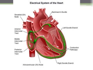
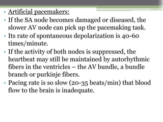
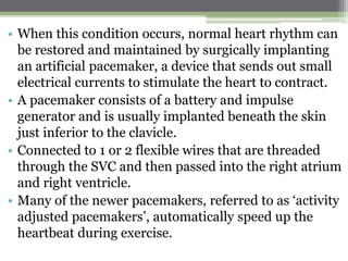
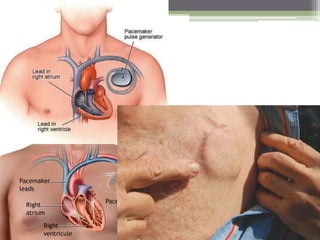
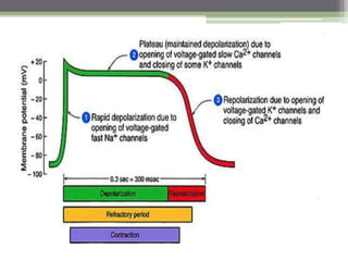
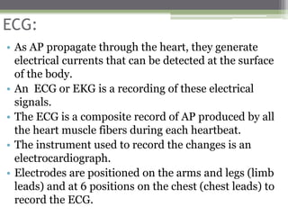
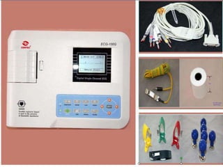
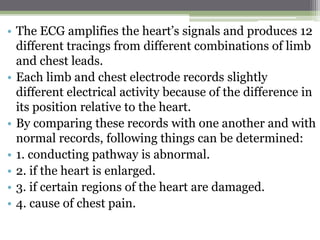
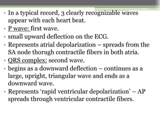
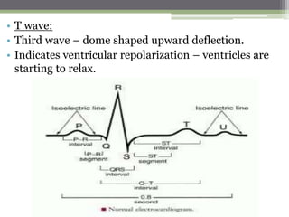
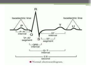
Conduction system and ecg
Conduction system:
• Inherent and rhythmical electrical activity is the
reason for the heart’s life long beat.
• Source: network of specialized cardiac muscle fibers
called ‘autorhythmic fibers’ because they are self-
excitable.
• Repeatedly generate action potentials that trigger
heart contractions.
• Continue to stimulate a heart to beat even after its
removed form the body.
• Autorhythmic fibers – 2 important functions:
• 1. act as a pacemaker, setting the rhythm of electrical
excitation that causes contraction of the heart.
• 2. form the conduction system, a network of
specialized cardiac muscle fibers that provide a
path for each cycle of cardiac excitation to
progress through the heart.
• Conduction system ensures that cardiac
chambers become stimulated to contract in a
coordinated manner, which makes the heart an
effective pump.
• Cardiac action potentials propagate through the
conduction system in the following sequence:
• 1. cardiac excitation normally begins in the SA node,
located in the right atrial wall just inferior to the
opening of the SVC.
• SA node cells do not have a stable resting potential.
• Rather, they repeatedly depolarize to threshold
spontaneously.
• Spontaneous depolarization is a ‘pacemaker potential’.
• When the pacemaker potential reaches threshold, it
triggers an action potential.
• Each AP from the SA node propagates throughout
both atria via gap junctions in the intercalated discs of
atrial muscle fibers.
• Following the AP, the atria contract.
• 2. by conducting along atrial muscle fibers, the AP
reaches the AV node, located in the septum between
the 2 atria, just anterior to the opening of the coronary
sinus.
• 3. from the AV node, the AP enters the AV bundle (or
bundle of HIS).
• This bundle is the only site where action potentials can
conduct from the atria to the ventricles.
• 4. after propagating along the AV bundle, the AP
enters both the right and left bundle branches.
• The bundle branches extend through the
interventricular septum towards the apex of the heart.
• 5. finally, the large-diameter purkinje fibers rapidly
conduct the AP from the apex of the heart upward to
the remainder of the ventricular myocardium.
• Then the ventricular contract, pushing the blood
upward toward the semilunar valves.
• On their own, autorhythmic fibers in the SA node
would initiate an AP about every 0.6 sec, or 100 times
per minute.
• This rate is faster than that of any other autorhythmic
fibers.
• Because AP from the SA node spread through the
conduction system and stimulate other areas before
the other areas are able to generate an AP at their own,
slower rate; the SA node acts as a natural pacemaker
of the heart.
• Nerve impulses from the ANS and blood borne
hormones modify the timing and strength of each
heart beat, but they don’t establish the fundamental
rhythm.
• In a person at rest, Ach released by the
parasympathetic division of the ANS slows SA node
pacing to about 75 APs per minute, or one every 0.8
sec.
• Artificial pacemakers:
• If the SA node becomes damaged or diseased, the
slower AV node can pick up the pacemaking task.
• Its rate of spontaneous depolarization is 40-60
times/minute.
• If the activity of both nodes is suppressed, the
heartbeat may still be maintained by autorhythmic
fibers in the ventricles – the AV bundle, a bundle
branch or purkinje fibers.
• Pacing rate is so slow (20-35 beats/min) that blood
flow to the brain is inadequate.
• When this condition occurs, normal heart rhythm can
be restored and maintained by surgically implanting
an artificial pacemaker, a device that sends out small
electrical currents to stimulate the heart to contract.
• A pacemaker consists of a battery and impulse
generator and is usually implanted beneath the skin
just inferior to the clavicle.
• Connected to 1 or 2 flexible wires that are threaded
through the SVC and then passed into the right atrium
and right ventricle.
• Many of the newer pacemakers, referred to as ‘activity
adjusted pacemakers’, automatically speed up the
heartbeat during exercise.
Action potential and contraction of
contractile fibers:
• AP initiated by the SA node travels along the
conduction system and spreads out to excite the
‘working’ atrial and ventricular muscle fibers, called
contractile fibers.
• An AP occurs in a contractile fiber as follows:
• 1. depolarization:
• Unlike autorhythmic fibers, contractile fibers have a
stable resting membrane potential that is close to -
90mv.
• When a contractile fiber is brought to threshold by an
AP from neighbouring fibers, its ‘voltage gated fast
Na+ channels’ open.
• These Na+ ion channels are referred to as ‘fast’
because they open very rapidly in response to a
threshold-level depolarization.
• Opening of these channels allows Na+ inflow because
the cytosol of contractile fibers is electrically more
negative than interstitial fluid and Na+ conc. Is higher
in interstitial fluid.
• Inflow of Na+ down the electrochemical gradient
produces a ‘rapid depolarization’.
• Within a few millisecs, the fast Na+ channels
automatically inactivate and Na+ inflow decreases.
• 2. plateau:
• Next phase of an AP in a contractile fiber.
• Period of maintained depolarization.
• Due in part to opening of ‘voltage-gated slow Ca2+
channels’ in the sarcolemma.
• When these channels open, calcium move from the
interstitial fluid into the cytosol.
• This inflow of Ca2+ causes even more Ca2+ to pour
out of the SR into the cytosol through additional Ca2+
channels in the SR membrane.
• Increased Ca2+ conc. In the cytosol ultimately triggers
contraction.
• Several different types of voltage gated K+ channels
are also found in the sarcolemma of a contractile fiber.
• Just before the plateau phase begins, some of these K+
channels open, allowing potassium ions to leave the
contractile fiber.
• Therefore, depolarization is sustained during the
plateau because Ca2+ inflow just balances K+ outflow.
• The plateau phase lasts for about 0.25sec, and the
membrane potential of the contractile fiber is close to
0 mV.
• By comparision, depolarization in a neuron or skeletal
muscle is much briefer, about 1 msec, because it lacks
a plateau phase.
• 3. repolarization:
• Recovery of the RMP.
• Resembles other excitable cells.
• After a delay, voltage gated K+ channels open.
• Outflow of K+ restores the negative RMP.
• At the same time, the Ca2+ channels in the
sarcolemma and the SR are closing, which also
contribute to repolarization.
• Mechanism of contraction is similar in cardiac and
skeletal muscle.
• Electrical activity leads to the mechanical response
after a short delay.
• As Ca2+ conc. Rises inside a contractile fiber, Ca2+
binds to the regulatory protein troponin, which allows
the actin and myosin filaments to begin sliding past
one another, and tension starts to develop.
• Subs. That alter the movement of Ca2+ through slow
Ca2+ channels influence the strength of heart
contractions.
• E.g: epinephrine – increases contraction force by
enhancing Ca2+ inflow into the cytosol.
• In muscle, the ‘refractory period’ is the time interval
during which a second contraction cannot be
triggered.
• Refractory period of a cardiac muscle fiber lasts longer
than the contraction itself.
• As a result, another contraction cannot begin until
relaxation is well underway.
• For this reason, tetanus cannot occur in cardiac
muscle as it can in skeletal muscle.
• Imp. For pumping action of heart as alternating
contraction and relaxation are needed to pump blood
flow.
ATP production in cardiac muscle:
• Cardiac muscle relies almost exclusively on aerobic
cellular respiration.
• O2 needed diffuses from blood in the coronary
circulation and is released from myoglobin inside
cardiac muscle fibers.
• In a person at rest, the heart’s ATP comes mainly from
oxidation of fatty acids (60%) and glucose (35%), with
smaller contributions from lactic acid, aminoacids,
and ketone bodies.
• During exercise, the heart’s use of lactic acid,
produced by actively contracting skeletal muscles,
rises.
• Like skeletal muscle, cardiac muscle also produces
some ATP from ‘creatine phosphate’.
• Creatine kinase – enzyme that catalyzes transfer of a
phosphate group from creatine phosphate to ADP to
make ATP.
• Normally, CK and other enzymes are confined within
cells.
• Blood levels of creatine kinase indicates MI.
• Injured or dying cardiac or skeletal muscle fibers
release CK into the blood.
ECG:
• As AP propagate through the heart, they generate
electrical currents that can be detected at the surface
of the body.
• An ECG or EKG is a recording of these electrical
signals.
• The ECG is a composite record of AP produced by all
the heart muscle fibers during each heartbeat.
• The instrument used to record the changes is an
electrocardiograph.
• Electrodes are positioned on the arms and legs (limb
leads) and at 6 positions on the chest (chest leads) to
record the ECG.
• The ECG amplifies the heart’s signals and produces 12
different tracings from different combinations of limb
and chest leads.
• Each limb and chest electrode records slightly
different electrical activity because of the difference in
its position relative to the heart.
• By comparing these records with one another and with
normal records, following things can be determined:
• 1. conducting pathway is abnormal.
• 2. if the heart is enlarged.
• 3. if certain regions of the heart are damaged.
• 4. cause of chest pain.
• In a typical record, 3 clearly recognizable waves
appear with each heart beat.
• P wave: first wave.
• small upward deflection on the ECG.
• Represents atrial depolarization – spreads from the
SA node thorugh contractile fibers in both atria.
• QRS complex: second wave.
• begins as a downward deflection – continues as a
large, upright, triangular wave and ends as a
downward wave.
• Represents ‘rapid ventricular depolarization’ – AP
spreads through ventricular contractile fibers.
• T wave:
• Third wave – dome shaped upward deflection.
• Indicates ventricular repolarization – ventricles are
starting to relax.
• T wave is smaller and wider than the QRS complex
because repolarization occurs more slowly than
depolarization.
• During the plateau period of steady depolarization, the
ECG tracing is flat.
• Size of the waves can provide clues to abnormalities.
• Larger P waves indicate enlargement of an atrium.
• Enlarged Q wave may indicate a MI.
• Enlarged R wave indicates enlarged ventricles.
• T wave is flatter than normal when the heart muscle is
receiving insufficient oxygen.
• E.g: CAD
• T wave may be elevated in hyperkalemia.
• Analysis of an ECG also involves measuring the time
spans between waves – ‘intervals or segments’.
• P-Q interval: is the time from the beginning of the P
wave to the beginning of QRS complex.
• Represents the conduction time from the beginning of
atrial excitation to be beginning of ventricular
excitation.
• another way: time required for the AP to travel
through the atria, AV NODE, and the remaining fibers
of the conduction system.
• In CAD and RF – AP forced to detour around scar
tissue caused by then – PQ interval lengthens.
• ST segment – begins at the end of the S wave and ends
at the beginning of the T wave.
• Represents the time when the ventricular contractile
fibers are depolarized during the plateau phase of the
AP.
• Elevated in acute MI.
• Depressed when the heart muscle receives insufficient
oxygen.
• QT interval:
• Extends form the start of QRS complex to the end of
the T wave.
• Time from the beginning of ventricular depolarization
to the end of ventricular repolarization.
• Lengthened by myocardial damage, myocardial
ischemia or conduction abnormalities.
Conduction system and ecg

Conduction system and ecg

  • 1.
  • 2.
    Conduction system: • Inherentand rhythmical electrical activity is the reason for the heart’s life long beat. • Source: network of specialized cardiac muscle fibers called ‘autorhythmic fibers’ because they are self- excitable. • Repeatedly generate action potentials that trigger heart contractions. • Continue to stimulate a heart to beat even after its removed form the body. • Autorhythmic fibers – 2 important functions: • 1. act as a pacemaker, setting the rhythm of electrical excitation that causes contraction of the heart.
  • 3.
    • 2. formthe conduction system, a network of specialized cardiac muscle fibers that provide a path for each cycle of cardiac excitation to progress through the heart. • Conduction system ensures that cardiac chambers become stimulated to contract in a coordinated manner, which makes the heart an effective pump.
  • 5.
    • Cardiac actionpotentials propagate through the conduction system in the following sequence: • 1. cardiac excitation normally begins in the SA node, located in the right atrial wall just inferior to the opening of the SVC. • SA node cells do not have a stable resting potential. • Rather, they repeatedly depolarize to threshold spontaneously. • Spontaneous depolarization is a ‘pacemaker potential’. • When the pacemaker potential reaches threshold, it triggers an action potential. • Each AP from the SA node propagates throughout both atria via gap junctions in the intercalated discs of atrial muscle fibers.
  • 7.
    • Following theAP, the atria contract. • 2. by conducting along atrial muscle fibers, the AP reaches the AV node, located in the septum between the 2 atria, just anterior to the opening of the coronary sinus. • 3. from the AV node, the AP enters the AV bundle (or bundle of HIS). • This bundle is the only site where action potentials can conduct from the atria to the ventricles. • 4. after propagating along the AV bundle, the AP enters both the right and left bundle branches. • The bundle branches extend through the interventricular septum towards the apex of the heart.
  • 9.
    • 5. finally,the large-diameter purkinje fibers rapidly conduct the AP from the apex of the heart upward to the remainder of the ventricular myocardium. • Then the ventricular contract, pushing the blood upward toward the semilunar valves. • On their own, autorhythmic fibers in the SA node would initiate an AP about every 0.6 sec, or 100 times per minute. • This rate is faster than that of any other autorhythmic fibers.
  • 11.
    • Because APfrom the SA node spread through the conduction system and stimulate other areas before the other areas are able to generate an AP at their own, slower rate; the SA node acts as a natural pacemaker of the heart. • Nerve impulses from the ANS and blood borne hormones modify the timing and strength of each heart beat, but they don’t establish the fundamental rhythm. • In a person at rest, Ach released by the parasympathetic division of the ANS slows SA node pacing to about 75 APs per minute, or one every 0.8 sec.
  • 13.
    • Artificial pacemakers: •If the SA node becomes damaged or diseased, the slower AV node can pick up the pacemaking task. • Its rate of spontaneous depolarization is 40-60 times/minute. • If the activity of both nodes is suppressed, the heartbeat may still be maintained by autorhythmic fibers in the ventricles – the AV bundle, a bundle branch or purkinje fibers. • Pacing rate is so slow (20-35 beats/min) that blood flow to the brain is inadequate.
  • 14.
    • When thiscondition occurs, normal heart rhythm can be restored and maintained by surgically implanting an artificial pacemaker, a device that sends out small electrical currents to stimulate the heart to contract. • A pacemaker consists of a battery and impulse generator and is usually implanted beneath the skin just inferior to the clavicle. • Connected to 1 or 2 flexible wires that are threaded through the SVC and then passed into the right atrium and right ventricle. • Many of the newer pacemakers, referred to as ‘activity adjusted pacemakers’, automatically speed up the heartbeat during exercise.
  • 16.
    Action potential andcontraction of contractile fibers: • AP initiated by the SA node travels along the conduction system and spreads out to excite the ‘working’ atrial and ventricular muscle fibers, called contractile fibers. • An AP occurs in a contractile fiber as follows: • 1. depolarization: • Unlike autorhythmic fibers, contractile fibers have a stable resting membrane potential that is close to - 90mv. • When a contractile fiber is brought to threshold by an AP from neighbouring fibers, its ‘voltage gated fast Na+ channels’ open.
  • 17.
    • These Na+ion channels are referred to as ‘fast’ because they open very rapidly in response to a threshold-level depolarization. • Opening of these channels allows Na+ inflow because the cytosol of contractile fibers is electrically more negative than interstitial fluid and Na+ conc. Is higher in interstitial fluid. • Inflow of Na+ down the electrochemical gradient produces a ‘rapid depolarization’. • Within a few millisecs, the fast Na+ channels automatically inactivate and Na+ inflow decreases.
  • 19.
    • 2. plateau: •Next phase of an AP in a contractile fiber. • Period of maintained depolarization. • Due in part to opening of ‘voltage-gated slow Ca2+ channels’ in the sarcolemma. • When these channels open, calcium move from the interstitial fluid into the cytosol. • This inflow of Ca2+ causes even more Ca2+ to pour out of the SR into the cytosol through additional Ca2+ channels in the SR membrane. • Increased Ca2+ conc. In the cytosol ultimately triggers contraction. • Several different types of voltage gated K+ channels are also found in the sarcolemma of a contractile fiber.
  • 21.
    • Just beforethe plateau phase begins, some of these K+ channels open, allowing potassium ions to leave the contractile fiber. • Therefore, depolarization is sustained during the plateau because Ca2+ inflow just balances K+ outflow. • The plateau phase lasts for about 0.25sec, and the membrane potential of the contractile fiber is close to 0 mV. • By comparision, depolarization in a neuron or skeletal muscle is much briefer, about 1 msec, because it lacks a plateau phase.
  • 23.
    • 3. repolarization: •Recovery of the RMP. • Resembles other excitable cells. • After a delay, voltage gated K+ channels open. • Outflow of K+ restores the negative RMP. • At the same time, the Ca2+ channels in the sarcolemma and the SR are closing, which also contribute to repolarization.
  • 26.
    • Mechanism ofcontraction is similar in cardiac and skeletal muscle. • Electrical activity leads to the mechanical response after a short delay. • As Ca2+ conc. Rises inside a contractile fiber, Ca2+ binds to the regulatory protein troponin, which allows the actin and myosin filaments to begin sliding past one another, and tension starts to develop. • Subs. That alter the movement of Ca2+ through slow Ca2+ channels influence the strength of heart contractions. • E.g: epinephrine – increases contraction force by enhancing Ca2+ inflow into the cytosol.
  • 29.
    • In muscle,the ‘refractory period’ is the time interval during which a second contraction cannot be triggered. • Refractory period of a cardiac muscle fiber lasts longer than the contraction itself. • As a result, another contraction cannot begin until relaxation is well underway. • For this reason, tetanus cannot occur in cardiac muscle as it can in skeletal muscle. • Imp. For pumping action of heart as alternating contraction and relaxation are needed to pump blood flow.
  • 31.
    ATP production incardiac muscle: • Cardiac muscle relies almost exclusively on aerobic cellular respiration. • O2 needed diffuses from blood in the coronary circulation and is released from myoglobin inside cardiac muscle fibers. • In a person at rest, the heart’s ATP comes mainly from oxidation of fatty acids (60%) and glucose (35%), with smaller contributions from lactic acid, aminoacids, and ketone bodies. • During exercise, the heart’s use of lactic acid, produced by actively contracting skeletal muscles, rises.
  • 32.
    • Like skeletalmuscle, cardiac muscle also produces some ATP from ‘creatine phosphate’. • Creatine kinase – enzyme that catalyzes transfer of a phosphate group from creatine phosphate to ADP to make ATP. • Normally, CK and other enzymes are confined within cells. • Blood levels of creatine kinase indicates MI. • Injured or dying cardiac or skeletal muscle fibers release CK into the blood.
  • 33.
    ECG: • As APpropagate through the heart, they generate electrical currents that can be detected at the surface of the body. • An ECG or EKG is a recording of these electrical signals. • The ECG is a composite record of AP produced by all the heart muscle fibers during each heartbeat. • The instrument used to record the changes is an electrocardiograph. • Electrodes are positioned on the arms and legs (limb leads) and at 6 positions on the chest (chest leads) to record the ECG.
  • 36.
    • The ECGamplifies the heart’s signals and produces 12 different tracings from different combinations of limb and chest leads. • Each limb and chest electrode records slightly different electrical activity because of the difference in its position relative to the heart. • By comparing these records with one another and with normal records, following things can be determined: • 1. conducting pathway is abnormal. • 2. if the heart is enlarged. • 3. if certain regions of the heart are damaged. • 4. cause of chest pain.
  • 37.
    • In atypical record, 3 clearly recognizable waves appear with each heart beat. • P wave: first wave. • small upward deflection on the ECG. • Represents atrial depolarization – spreads from the SA node thorugh contractile fibers in both atria. • QRS complex: second wave. • begins as a downward deflection – continues as a large, upright, triangular wave and ends as a downward wave. • Represents ‘rapid ventricular depolarization’ – AP spreads through ventricular contractile fibers.
  • 38.
    • T wave: •Third wave – dome shaped upward deflection. • Indicates ventricular repolarization – ventricles are starting to relax.
  • 39.
    • T waveis smaller and wider than the QRS complex because repolarization occurs more slowly than depolarization. • During the plateau period of steady depolarization, the ECG tracing is flat. • Size of the waves can provide clues to abnormalities. • Larger P waves indicate enlargement of an atrium. • Enlarged Q wave may indicate a MI. • Enlarged R wave indicates enlarged ventricles. • T wave is flatter than normal when the heart muscle is receiving insufficient oxygen. • E.g: CAD • T wave may be elevated in hyperkalemia.
  • 41.
    • Analysis ofan ECG also involves measuring the time spans between waves – ‘intervals or segments’. • P-Q interval: is the time from the beginning of the P wave to the beginning of QRS complex. • Represents the conduction time from the beginning of atrial excitation to be beginning of ventricular excitation. • another way: time required for the AP to travel through the atria, AV NODE, and the remaining fibers of the conduction system. • In CAD and RF – AP forced to detour around scar tissue caused by then – PQ interval lengthens.
  • 43.
    • ST segment– begins at the end of the S wave and ends at the beginning of the T wave. • Represents the time when the ventricular contractile fibers are depolarized during the plateau phase of the AP. • Elevated in acute MI. • Depressed when the heart muscle receives insufficient oxygen.
  • 45.
    • QT interval: •Extends form the start of QRS complex to the end of the T wave. • Time from the beginning of ventricular depolarization to the end of ventricular repolarization. • Lengthened by myocardial damage, myocardial ischemia or conduction abnormalities.