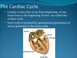
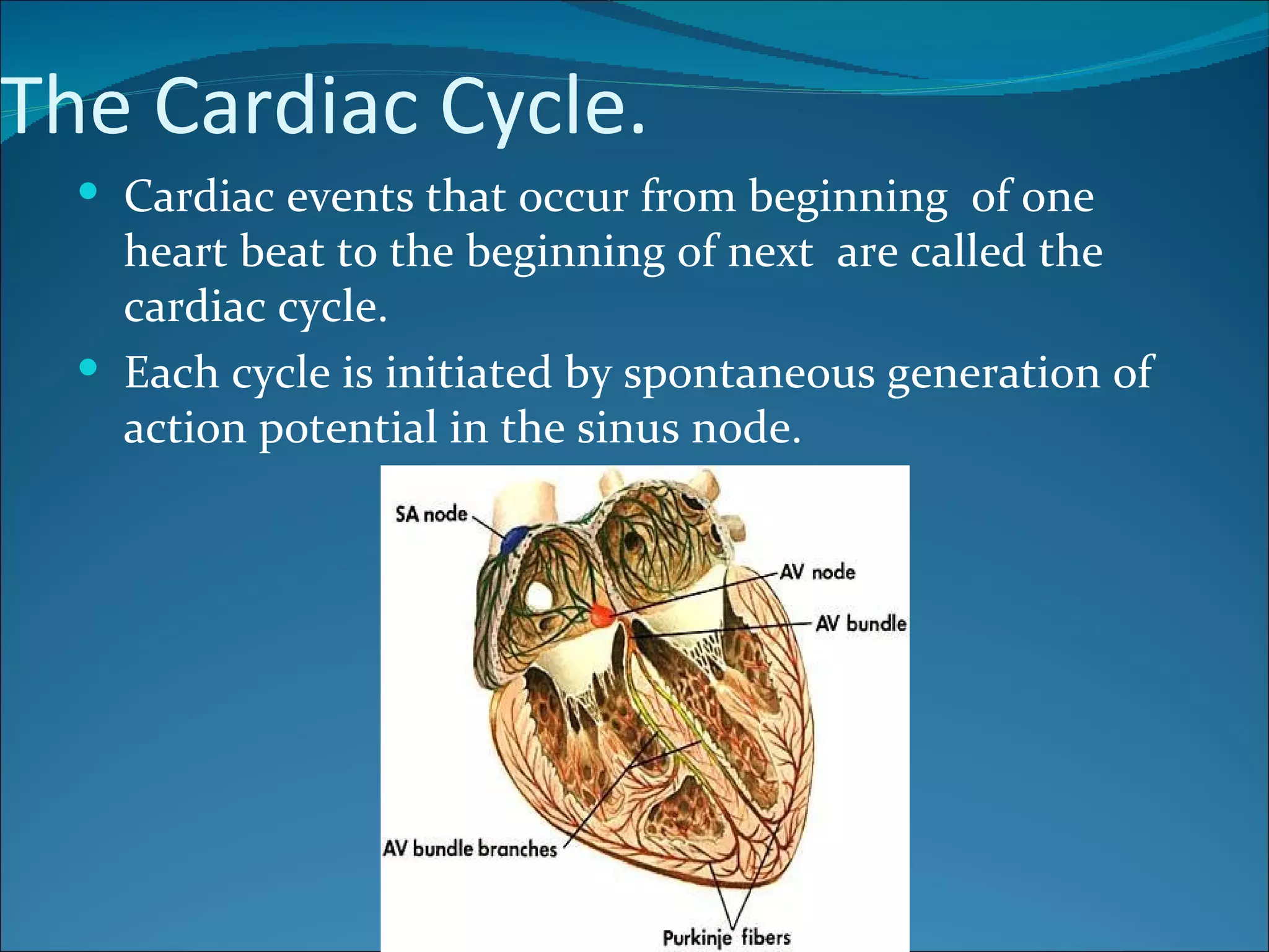
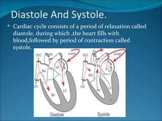
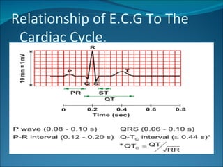
Call Girls Nashik Gayatri 7001305949 Independent Escort Service Nashik
Cardiac cycle.full
1. The Cardiac Cycle.
Cardiac events that occur from beginning of one
heart beat to the beginning of next are called the
cardiac cycle.
Each cycle is initiated by spontaneous generation of
action potential in the sinus node.
2. Diastole And Systole.
Cardiac cycle consists of a period of relaxation called
diastole, during which ,the heart fills with
blood,followed by period of contraction called
systole.
5. P,Q,R,s and T waves are electrical voltages generated
by the heart and recorded by E.C.G from the body
surface.
P wave is caused by spread of depolarization through
atria and this is followed by atrial contraction,which
causes a slight rise in atrial pressure.
About 0.16 sec after onset of p wave,QRS waves
appear as a result of electrical depolarization of
ventricles, which initiate ventricular contraction and
cause ventricular pressure to rise.therefore QRS
complex begins slightly before onset of ventricular
systole.
T wave represent stage of repolarization of ventricles
,therefore T wave occurs slightly before the end of
ventricular contraction.
6.
7. Function Of Atria As A Primer
Pump
About 80%of blood flows directly through atria into
ventricles even before atrial contraction.
Then atrial contraction usually causes an additional
20% ventricular filling.
Therefore atria simply functions as a primer pump.
8. Function Of Ventricles As Pumps.
Filling of Ventricles
During ventricular systole, large amount of
blood accumulate in rt. And lt. atria b/c of
closed A.V valves. Therefore as soon as systole
is over & ventricular pressure
decreases,moderately increased pressure that
have developed in atria during ventricular
systole immediately push A.V valves open and
allow blood to flow rapidly into ventricle.this is
called period of (rapid filling of ventricles).
The period of rapid ventricular filling lasts for about 1/3 of
diastole. During middle third,only a small amount of blood
normally flows into ventricles;this is blood that continues
to empty into atria from veins & passes through atria
directly into ventricles.
During last 3rd, atria contract and give an additional thrust
to inflow of blood into ventricles;this accounts for about
20% of ventricular filling during each heart cycle.
9. Emptying f ventricles during
systole
Period of isovolumic (isometric) contraction
Immediately after ventricular contraction begins,
ventricular pressure rises abruptly,causing A.V valves to
close.then an additional 0.02 to 0.03 sec is required for the
ventricle to build up sufficient pressure to push
semilunar valves open against the pressure in aorta &
pulmonary artery.therefore during this period,contraction
is occuring in ventricles, but there is no emptying.this is
called the period of isovolumic or isometric contraction,
meaning that tension is increasing in muscle but little or
no shortening of muscle fiber is occuring.
10. Period of ejection.
When the left ventricular pressure rises slightly above
80mm Hg(& rt. Ventricular pressure slightly above
8mm Hg), vent. Pressure push semilunar valves
open. Immediately , blood begins to pour out of
ventricles , with about 70% of blood emptying
occuring during the 1st third of period of ejection,&
remaining 30% emptying during next two thirds.
Therefore 1st third is called period of rapid ejection &
last two thirds , period of slow ejection.
11. Period of Isovolumic(Isometric)
Relaxation
At the end of systole,vent. relaxation begins ,allowing
both the rt. & lt. intraventricular pressure to
decrease rapidly. Elevated pressure in distended large
arteries that have just been filled with blood from
contracted ventricles immediately push blood back
towards ventricles ,which close aortic & pulmonary
valves. For another 0.03 to 0.06 sec, ventr. Muscles
continue to relax, even though vent. Volume does
not change, giving rise to period of isovolumic or
isometric relaxation. during this period, intra ventr.
Pressure decrease rapidly back to their low diastolic
levels. Then A,V valves open to begin a new cycle of
ventr. Pumping.
12. End –Diastolic volume
during diastole, normal ventricular filling increases the
volume of each ventricle to about 110 to 120 ml. this
volume is called end- diastolic volume.
Stroke volume output
As ventricles empty during systole, volume decreases
abt 70 ml, which is called stroke volume output.
End- Systolic volume
Remaining volume in each ventricle, abt 40 to 50 ml, is
called end systolic volume.
13. Ejection Fraction
fraction of end diastolic volume that is ejected is called
ejection fraction., usually equal to about 60%
When heart contracts strongly, end systolic vol. can be
decreased to as little as 10 to 20 ml. conversely,
when large amount of blood flows into ventricles
during diastole,ventr. diastolic volumes can become
as great as 150 to 180 ml .
14. Function Of Valves
Atrioventricular valves
The A-V valves (the tricuspid and mitral valves) prevent
backflow of blood from the ventricles to the atria during
systole, and the semilunar valves (the aortic and
pulmonary artery valves) prevent backflow from the aorta
and pulmonary arteries into the ventricles during diastole.
they close when a backward pressure gradient pushes
blood backward, and they open when a forward pressure
gradient forces blood in the forward direction.
15. Aortic And Pulmonary Artery
valves.
High pressures in the arteries at the end of systole
cause the semilunar valves to snap to the closed
position. because of the rapid closure and rapid
ejection, the edges of the aortic and pulmonary
valves are subjected to much greater mechanical
abrasion than are the A-V valves.
16. Relationship Of Heart Sounds To
heart Pumping.
when the valves close, the vanes of the valves and the
surrounding fluids vibrate under the influence of sudden
pressure changes, giving off sound that travels in all
directions through the chest. When the ventricles
contract, a sound produces,caused by closure of the A-
V valves. The vibration is low in pitch and relatively long-
lasting and is known as the first heart sound. When the
aortic and pulmonary valves close at the end of systole,
a rapid snap occurs because these valves close rapidly,
and the surroundings vibrate for a short period. This
sound is called the second heart sound.
17. Work Output Of The Heart
stroke work output of the heart is the amount of
energy that the heart converts to work during each
heartbeat while pumping blood into the arteries.
18. Volume Pressure Diagram Of
Cardiac Cycle
It is divided into four phases.
Phase I: Period of filling. This phase in the volume-
pressure diagram begins at a ventricular volume of about
50 ml and a diastolic pressure of 2 to 3 mm Hg. The
amount of blood that remains in the ventricle after the
previous heartbeat, 50 ml, is called the end-systolic
volume. As venous blood flows into the ventricle from the
left atrium, the ventricular volume normally increases to
about 120 ml, called the end-diastolic volume, an
increase of 70 ml
19. Phase II:
(Period of isovolumic contraction).
During isovolumic contraction, the volume of the
ventricle does not change because all valves are closed.
However, the pressure inside the ventricle increases to
equal the pressure in the aorta, at a pressure value of
about 80 mm Hg.
Phase III:
( Period of ejection).
During ejection, the systolic pressure rises even higher
because of still more contraction of the ventricle. At the
same time, the volume of the ventricle decreases
because the aortic valve has now opened and blood
flows out of the ventricle into the aorta.
20. Phase IV:
( Period of isovolumic relaxation). At the end of
the period of ejection the aortic valve closes, and
the ventricular pressure falls back to the diastolic
pressure level. Thus, the ventricle returns to its
starting point, with about 50 ml of blood left in
the ventricle and at an atrial pressure of 2 to 3
mm Hg.
21. Concepts of Preload and Afterload
(Preload):
The degree of tension on the muscle when it begins to
contract, is called the preload.
(Afterload)
The load against which the muscle exerts its contractile
force, is called the afterload.
.
22. Chemical Energy Required for
Cardiac Contraction: Oxygen
Utilization by the Heart
Heart muscle, like skeletal muscle, uses chemical
energy to provide the work of contraction.
Approximately 70 to 90 percent of this energy is
normally derived from oxidative metabolism of fatty
acids with about 10 to 30 percent coming from other
nutrients, especially lactate and glucose.
23. Regulation Of Heart Pumping
Regulation of Heart Pumping
When a person is at rest, the heart pumps only 4 to 6
liters of blood each minute. During severe exercise,
the heart may be required to pump four to seven
times this amount. The basic means by which the
volume pumped by the heart is regulated are (1)
intrinsic cardiac regulation of pumping in response to
changes in volume of blood flowing into the heart
and (2) control of heart rate and strength of heart
pumping by the autonomic nervous system.
24. Intrinsic Regulation of Heart
Pumping-
The Frank-Starling Mechanism
under most conditions, the amount of blood pumped by
the heart each minute is normally determined almost
entirely by the rate of blood flow into the heart from the
veins, which is called venous return. That is, each
peripheral tissue of the body controls its own local blood
flow, and all the local tissue flows combine and return by
way of the veins to the right atrium. The heart, in turn,
automatically pumps this incoming blood into the arteries
so that it can flow around the circuit again. This intrinsic
ability of the heart to adapt to increasing volumes of
inflowing blood is called the Frank-Starling mechanism of
the heart
25. Basically, the Frank-Starling mechanism means that
the greater the heart muscle is stretched during
filling, the greater is the force of contraction and the
greater the quantity of blood pumped into the aorta.
Or, stated another way: Within physiologic limits, the
heart pumps all the blood that returns to it by the way
of the veins.
26. Control of the Heart by the
Sympathetic and
Parasympathetic Nerves
The pumping effectiveness of the heart also is
controlled by the sympathetic and parasympathetic
(vagus) nerves, which abundantly supply the heart,
the amount of blood pumped each minute (cardiac
output) often can be increased by sympathetic
stimulation. By contrast, the output can be decreased
by (parasympathetic) stimulation.
27. Mechanisms of Excitation of the
Heart by the Sympathetic Nerves
Strong sympathetic stimulation can increase the
heart rate in young adult humans from the normal
rate of 70 beats/min up to 180 to 200 and, rarely, even
250 beats/min. Also, sympathetic stimulation
increases the force of heart contraction to as much as
double normal, thereby increasing the volume of
blood pumped and increasing the ejection pressure.
Thus, sympathetic stimulation often can increase the
maximum cardiac output as much as twofold to
threefold.
28. inhibition of the sympathetic nerves to the heart can
decrease cardiac pumping . when the activity of the
sympathetic nervous system is depressed below
normal, this decreases both heart rate and strength of
ventricular muscle contraction.
29. Parasympathetic (Vagal)
Stimulation of the Heart
Strong stimulation of the parasympathetic nerve
fibers in the vagus nerves to the heart can stop the
heartbeat for a few seconds, but then the heart
usually "escapes" and beats at a rate of 20 to 40 beats/
min as long as the parasympathetic stimulation
continues. In addition, strong vagal stimulation can
decrease the strength of heart muscle contraction by
20 to 30 percent.
30. The vagal fibers are distributed mainly to the atria
and not much to the ventricles, where the power
contraction of the heart occurs. This explains the
effect of vagal stimulation mainly to decrease heart
rate rather than to decrease greatly the strength of
heart contraction.
31. Effect of Potassium Ions On Heart
Function.
Excess potassium in the extracellular fluids causes
the heart to become dilated and flaccid and also
slows the heart rate. Large quantities also can block
conduction of the cardiac impulse from the atria to
the ventricles through the A-V bundle. Elevation of
potassium concentration to only 8 to 12 mEq/L-two
to three times the normal value-can cause such
weakness of the heart and abnormal rhythm that
death occurs.
32. These effects result partially from the fact that a high
potassium concentration in the extracellular fluids
decreases the resting membrane potential in the
cardiac muscle fibers. high extracellular fluid
potassium concentration partially depolarizes the cell
membrane, causing the membrane potential to be
less negative. As the membrane potential decreases,
the intensity of the action potential also decreases,
which makes contraction of the heart progressively
weaker.
33. Effect of Calcium Ions On Heart
Function.
An excess of calcium ions causes effects almost
exactly opposite to those of potassium ions, causing
the heart to go toward spastic contraction. This is
caused by a direct effect of calcium ions to initiate
the cardiac contractile process, Conversely,
deficiency of calcium ions causes cardiac flaccidity,
similar to the effect of high potassium.
34. Effect of Temperature on Heart
Function
Increased body temperature, causes a greatly
increased heart rate, sometimes to double normal.
Decreased temperature causes a greatly decreased
heart rate in the body. These effects presumably
result from the fact that heat increases the
permeability of the cardiac muscle membrane to ions
that control heart rate, resulting in acceleration of
the self-excitation process.