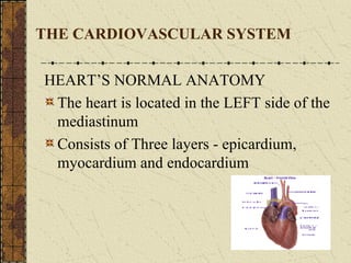
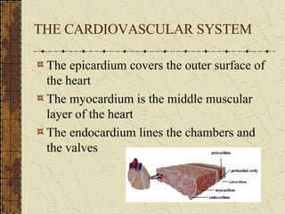
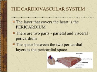
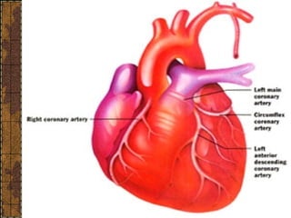
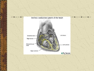
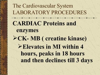
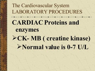
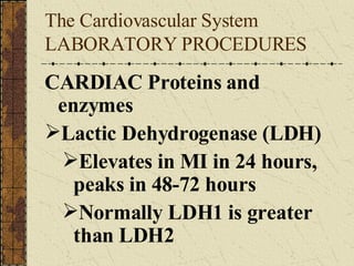
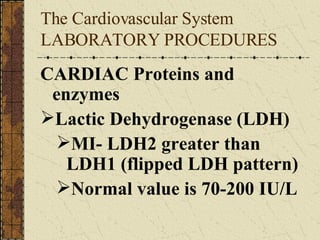
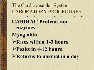
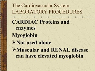
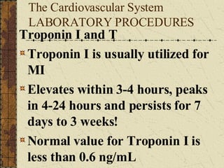
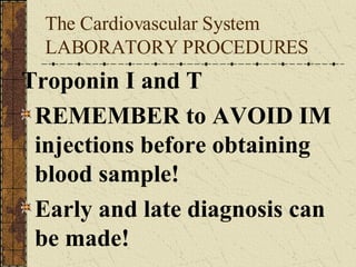
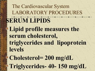
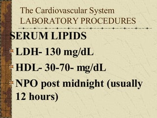
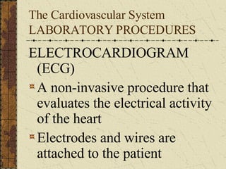
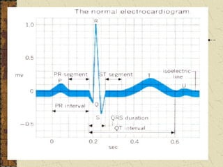
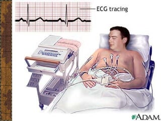
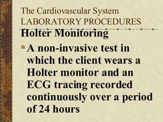
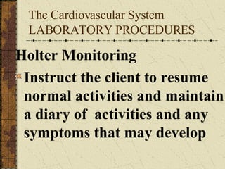
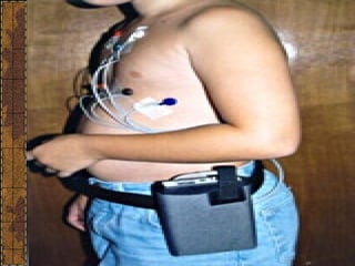
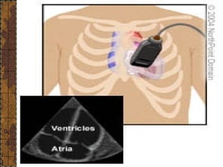
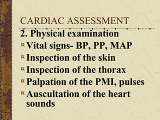
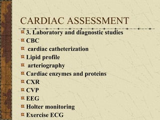
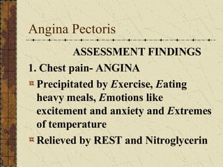
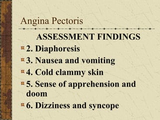
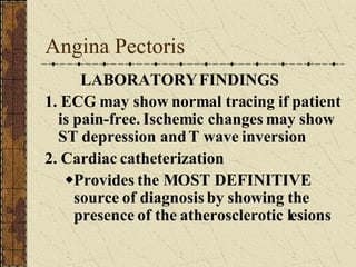
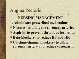
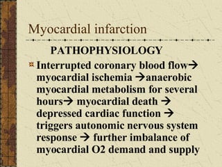
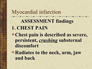
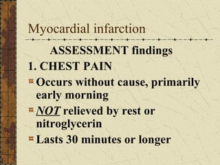
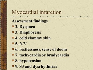
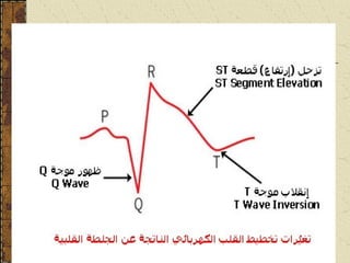
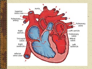
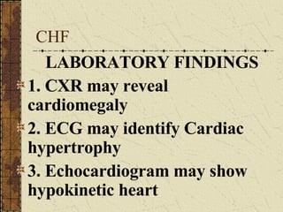
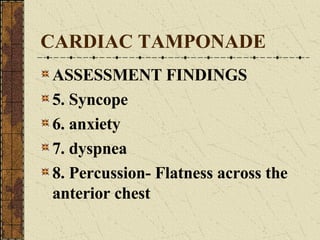
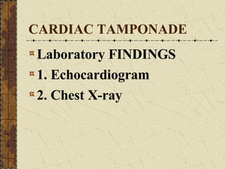
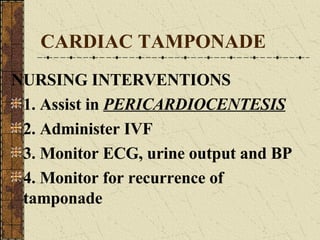
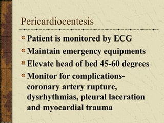
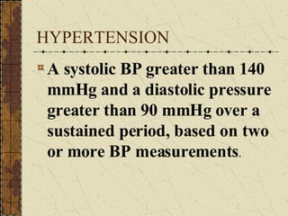
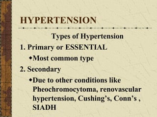
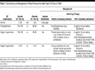
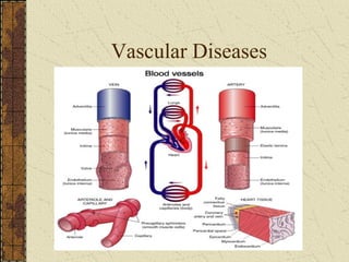
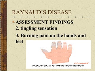
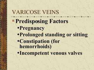
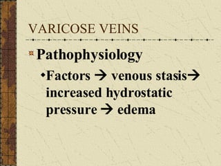
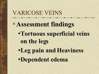
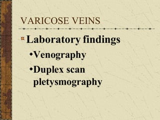
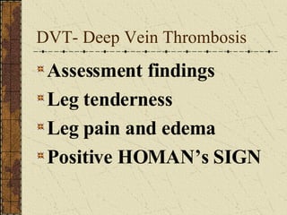
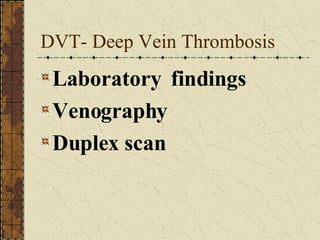
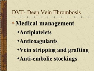
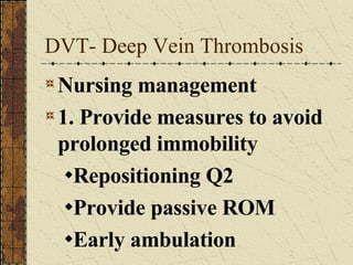
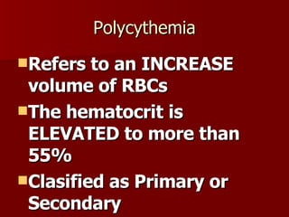
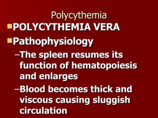
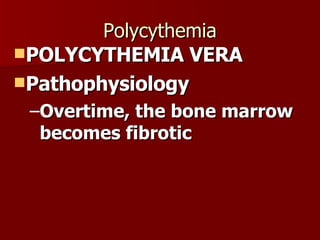
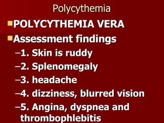
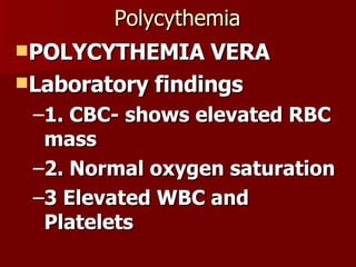
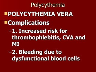
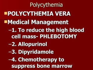
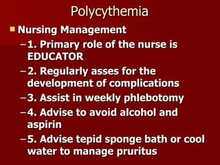
CARDIOLOGY  NURSING
THE CARDIOVASCULAR SYSTEM HEART’S NORMAL ANATOMY The heart is located in the LEFT side of the mediastinum Consists of Three layers - epicardium, myocardium and endocardium
THE CARDIOVASCULAR SYSTEM The epicardium covers the outer surface of the heart The myocardium is the middle muscular layer of the heart The endocardium lines the chambers and the valves
THE CARDIOVASCULAR SYSTEM The layer that covers the heart is the PERICARDIUM There are two parts - parietal and visceral pericardium The space between the two pericardial layers is the pericardial space
THE CARDIOVASCULAR SYSTEM The heart also has four chambers - two atria and two ventricles The Left atrium and the right atrium The left ventricle and the right ventricle
The Cardiovascular System The heart chambers are guarded by valves The atrio-ventricular valves - tricuspid and bicuspid The semi-lunar valves - pulmonic and aortic valves
The Cardiovascular System The Blood supply of the heart comes from the Coronary arteries 1. Right coronary artery supplies the RIGHT atrium and RIGHT ventricle, inferior portion of the LEFT ventricle, the POSTERIOR septal wall and the two nodes - AV (90%) and SA node (55%)
The Cardiovascular System 2. Left coronary artery- branches into the LAD and the circumflex branch The LAD supplies blood to the anterior wall of the LEFT ventricle, the anterior septum and the Apex of the left ventricle The CIRCUMFLEX branch supplies the left atrium and the posterior LEFT ventricle
 
The Cardiovascular System The CONDUCTING SYSTEM OF THE HEART Consists of the 1. SA node- the pacemaker 2. AV node- slowest conduction 3. Bundle of His – branches into the Right and the Left bundle branch 4. Purkinje fibers- fastest conduction
 
The Heart sounds 1. S1- due to closure of the AV valves 2. S2- due to the closure of the semi-lunar valves 3. S3-  due to increased ventricular filling 4. S4- due to forceful atrial contraction  The Cardiovascular System
The Cardiovascular System Heart rate Normal range is 60-100 beats per minute Tachycardia is greater than 100 bpm Bradycardia is less than 60 bpm Sympathetic system INCREASES HR Parasympathetic system (Vagus) DECREASES HR
The Cardiovascular System Blood pressure Cardiac output X peripheral resistance Control is neural (central and peripheral) and hormonal Baroreceptors in the carotid and aorta Hormones- ADH, aldosterone, epinephrine can increase BP; ANF can decrease BP
The Cardiovascular System The vascular system consists of the arteries, veins and capillaries The arteries are vessels that carry blood away from the heart to the periphery The veins are the vessels that carry blood to the heart The capillaries are lined with squamos cells, they connect the veins and arteries
The Cardiovascular System The lymphatic system also is part of the vascular system and the function of this system is to collect the extravasated fluid from the tissues and returns it to the blood
The Cardiovascular System Cardiac Assessment
The Cardiovascular System Laboratory Test Rationale 1. To assist in diagnosing MI 2. To identify abnormalities 3. To assess inflammation
The Cardiovascular System Laboratory Test Rationale 4. To determine baseline value 5. To monitor serum level of medications 6. To assess the effects of medications
The Cardiovascular System LABORATORY PROCEDURES  CARDIAC Proteins and enzymes CK- MB ( creatine kinase) Elevates in MI within 4 hours, peaks in 18 hours and then declines till 3 days
The Cardiovascular System LABORATORY PROCEDURES  CARDIAC Proteins and enzymes CK- MB ( creatine kinase) Normal value is 0-7 U/L
The Cardiovascular System LABORATORY PROCEDURES  CARDIAC Proteins and enzymes Lactic Dehydrogenase (LDH) Elevates in MI in 24 hours, peaks in 48-72 hours Normally LDH1 is greater than LDH2
The Cardiovascular System LABORATORY PROCEDURES  CARDIAC Proteins and enzymes Lactic Dehydrogenase (LDH) MI- LDH2 greater than LDH1 (flipped LDH pattern) Normal value is 70-200 IU/L
The Cardiovascular System LABORATORY PROCEDURES  CARDIAC Proteins and enzymes Myoglobin Rises within 1-3 hours Peaks in 4-12 hours Returns to normal in a day
The Cardiovascular System LABORATORY PROCEDURES  CARDIAC Proteins and enzymes Myoglobin Not used alone Muscular and RENAL disease can have elevated myoglobin
The Cardiovascular System LABORATORY PROCEDURES Troponin I and T Troponin I is usually utilized for MI Elevates within 3-4 hours, peaks in 4-24 hours and persists for 7 days to 3 weeks! Normal value for Troponin I is less than 0.6 ng/mL
The Cardiovascular System LABORATORY PROCEDURES Troponin I and T REMEMBER to AVOID IM injections before obtaining blood sample! Early and late diagnosis can be made!
The Cardiovascular System LABORATORY PROCEDURES SERUM LIPIDS Lipid profile measures the serum cholesterol, triglycerides and  lipoprotein levels Cholesterol= 200 mg/dL Triglycerides- 40- 150 mg/dL
The Cardiovascular System LABORATORY PROCEDURES SERUM LIPIDS LDH- 130 mg/dL HDL- 30-70- mg/dL NPO post midnight (usually 12 hours)
The Cardiovascular System LABORATORY PROCEDURES ELECTROCARDIOGRAM (ECG) A non-invasive procedure that evaluates the electrical activity of the heart Electrodes and wires are attached to the patient
 
 
The Cardiovascular System LABORATORY PROCEDURES Holter Monitoring A non-invasive test in which the client wears a Holter monitor and an ECG tracing recorded continuously over a period of 24 hours
The Cardiovascular System LABORATORY PROCEDURES Holter Monitoring Instruct the client to resume normal activities and maintain a diary of  activities and any symptoms that may develop
 
The Cardiovascular System LABORATORY PROCEDURES ECHOCARDIOGRAM Non-invasive test that studies the structural and functional changes of the heart with the use of ultrasound No special preparation is needed
 
The Cardiovascular System LABORATORY PROCEDURES Stress Test A non-invasive test that studies the heart during activity and detects and evaluates CAD Exercise test, pharmacologic test and emotional test
The Cardiovascular System LABORATORY PROCEDURES Stress Test Treadmill testing is the most commonly used stress test Used to determine CAD, Chest pain causes, drug effects and dysrhythmias in exercise
The Cardiovascular System LABORATORY PROCEDURES Stress Test Pre-test: consent may be required, adequate rest , eat a light meal or fast for 4 hours and avoid smoking, alcohol and caffeine
The Cardiovascular System LABORATORY PROCEDURES Post-test: instruct client to notify the physician if any chest pain, dizziness or shortness of breath . Instruct client to avoid taking a hot shower for 10-12 hours after the test
The Cardiovascular System LABORATORY PROCEDURES Pharmacological stress test Use of dipyridamole Maximally dilates coronary artery Side-effect: flushing of face
The Cardiovascular System LABORATORY PROCEDURES Pharmacological stress test Pre-test: 4 hours fasting, avoid alcohol, caffeine Post test: report symptoms of chest pain
The Cardiovascular System LABORATORY PROCEDURES CARDIAC catheterization Insertion of a catheter into the heart and surrounding vessels Determines the structure and performance of the heart valves and surrounding vessels
The Cardiovascular System LABORATORY PROCEDURES CARDIAC catheterization Used to diagnose CAD, assess coronary atery patency and determine extent of atherosclerosis
The Cardiovascular System LABORATORY PROCEDURES Pretest: Ensure Consent, assess for allergy to seafood and iodine, NPO, document weight and height, baseline VS, blood tests and document the peripheral pulses
The Cardiovascular System LABORATORY PROCEDURES Pretest: Fast for 8-12 hours, teachings, medications to allay anxiety
The Cardiovascular System LABORATORY PROCEDURES Intra-test: inform patient of a fluttery feeling as the catheter passes through the heart; inform the patient that a feeling of warmth and metallic taste may occur when dye is administered
The Cardiovascular System LABORATORY PROCEDURES Post-test: Monitor VS and cardiac rhythm Monitor peripheral pulses, color and warmth and sensation of the extremity distal to insertion site  Maintain sandbag to the insertion site if required to maintain pressure Monitor for bleeding and hematoma formation
The Cardiovascular System LABORATORY PROCEDURES Maintain strict bed rest for 6-12 hours Client may turn from side to side but bed should not be elevated more than 30 degrees and legs always straight Encourage fluid intake to flush out the dye Immobilize the arm if the antecubital vein is used Monitor for dye allergy
The Cardiovascular System LABORATORY PROCEDURES CVP The CVP is the pressure within the SVC Reflects the pressure under which blood is returned to the SVC and right atrium
The Cardiovascular System LABORATORY PROCEDURES CVP Normal CVP is 0 to 8 mmHg/  4-10 cm H2O Elevated CVP indicates increase in blood volume, excessive IVF or heart/renal failure Low CVP may indicated hypovolemia, hemorrhage and severe vasodilatation
The Cardiovascular System LABORATORY PROCEDURES Measuring CVP 1. Position the client supine with bed elevated at  45 degrees 2. Position the zero point of the CVP line at the level of the right atrium. Usually this is at the  MAL, 4 th  ICS  3. Instruct the client to be relaxed and avoid coughing and straining.
 
CARDIAC ASSESSMENT ASSESSMENT 1. Health History Obtain description of present illness and the chief complaint Chest pain, SOB, Edema, etc. Assess risk factors
CARDIAC ASSESSMENT 2. Physical examination  Vital signs- BP, PP, MAP Inspection of the skin Inspection of the thorax Palpation of the PMI, pulses Auscultation of the heart sounds
 
CARDIAC ASSESSMENT 3. Laboratory and diagnostic studies CBC  cardiac catheterization Lipid profile   arteriography Cardiac enzymes and proteins CXR   CVP EEG Holter monitoring Exercise ECG
CARDIAC IMPLEMENTATION Assess the cardio-pulmonary status VS, BP, Cardiac assessment  2. Enhance cardiac output Establish IV line to administer fluids
CARDIAC IMPLEMENTATION 3. Promote gas exchange Administer O2 Position client in  SEMI-Fowler’s Encourage coughing and deep breathing exercises
CARDIAC IMPLEMENTATION 4. Increase client activity tolerance Balance rest and activity periods Assist in daily activities 5. Promote client comfort Assess the client’s description of pain and chest discomfort Administer medication as prescribed
CARDIAC IMPLEMENTATION 6. Promote adequate sleep 7. Prevent infection Monitor skin integrity of lower extremities Assess skin site for edema, redness and warmth Monitor for fever Change position frequently
CARDIAC IMPLEMENTATION 8. Minimize patient anxiety Encourage verbalization of feelings, fears and concerns Answer client questions. Provide information about procedures and medications
CARDIAC DISEASES Coronary Artery Disease Myocardial Infarction Congestive Heart Failure Infective Endocarditis Cardiac Tamponade Cardiogenic Shock
VASCULAR DISEASES Hypertension Buerger’s disease Varicose veins Deep vein thrombosis Aneurysm
CAD CAD results from the focal narrowing of the large and medium-sized coronary arteries due to deposition of atheromatous plaque in the vessel wall
CAD RISK FACTORS 1. Age above 45/55 and Sex- Males and post-menopausal females 2. Family History 3. Hypertension 4. DM 5. Smoking 6. Obesity 7. Sedentary lifestyle 8. Hyperlipedimia
CAD RISK FACTORS Most important MODIFIABLE factors: Smoking Hypertension Diabetes Cholesterol abnormalities
CAD Pathophysiology Fatty streak formation in the vascular intima    T-cells and monocytes ingest lipids in the area of deposition   atheroma    narrowing of the arterial lumen    reduced coronary blood flow    myocardial ischemia
CAD Pathophysiology There is decreased perfusion of myocardial tissue and inadequate myocardial oxygen supply If 50% of the left coronary arterial lumen is reduced or 75% of the other coronary artery, this becomes significant Potential for Thrombosis and embolism
Angina Pectoris Chest pain resulting from coronary atherosclerosis or myocardial ischemia
Angina Pectoris: Clinical Syndromes Three Common Types of ANGINA 1. STABLE ANGINA The typical angina that occurs during exertion, relieved by rest and drugs and the  severity does not change
Angina Pectoris: Clinical Syndromes Three Common Types of ANGINA 2. Unstable angina Occurs unpredictably during exertion and emotion,  severity increases with time  and pain may not be relieved by rest and drug
Angina Pectoris: Clinical Syndromes Three Common Types of ANGINA 3. Variant angina Prinzmetal angina, results from coronary artery VASOSPASMS,  may occur at rest
Angina Pectoris ASSESSMENT FINDINGS 1. Chest pain- ANGINA The most characteristic symptom PAIN is described as mild to severe retrosternal pain,  squeezing , tightness or burning sensation Radiates to the jaw  and left arm
Angina Pectoris ASSESSMENT FINDINGS 1. Chest pain- ANGINA Precipitated by  E xercise,  E ating heavy meals,  E motions like excitement and anxiety and  E xtremes of temperature Relieved by REST and Nitroglycerin
Angina Pectoris ASSESSMENT FINDINGS 2. Diaphoresis 3. Nausea and vomiting 4. Cold clammy skin 5. Sense of apprehension and doom 6. Dizziness and syncope
Angina Pectoris LABORATORY FINDINGS 1. ECG may show normal tracing if patient is pain-free. Ischemic changes may show ST depression and T wave inversion 2. Cardiac catheterization Provides the MOST DEFINITIVE source of diagnosis by showing the presence of the atherosclerotic lesions
Angina Pectoris NURSING MANAGEMENT 1. Administer prescribed medications Nitrates- to dilate the coronary arteries Aspirin- to prevent thrombus formation Beta-blockers- to reduce BP and HR Calcium-channel blockers- to dilate coronary artery and reduce vasospasm
2. Teach the patient management of anginal attacks Advise patient to stop all activities  Put one nitroglycerin tablet under the tongue Wait for 5 minutes If not relieved, take another tablet and wait for 5 minutes Another tablet can be taken (third tablet) If unrelieved after THREE tablets   seek medical attention
Angina Pectoris 3. Obtain a 12-lead ECG 4. Promote myocardial perfusion Instruct patient to maintain bed rest Administer O2 @ 3 lpm Advise to avoid valsalva maneuvers Provide laxatives or high fiber diet to lessen constipation Encourage to avoid increased physical activities
Angina Pectoris 5. Assist in possible treatment modalities PTCA- percutaneous transluminal coronary angioplasty To compress the plaque against the vessel wall, increasing the arterial lumen CABG- coronary artery bypass graft To improve the blood flow to the myocardial tissue
 
Angina Pectoris 6. Provide information to family members to minimize anxiety and promote family cooperation 7. Assist client to identify risk factors that can be modified 8. Refer patient to proper agencies
Myocardial infarction Death of myocardial tissue in regions of the heart with abrupt interruption of coronary blood supply
 
Myocardial infarction ETIOLOGY and Risk factors 1. CAD 2. Coronary vasospasm 3. Coronary artery occlusion by embolus and thrombus 4. Conditions that decrease perfusion- hemorrhage, shock
Myocardial infarction Risk factors 1. Hypercholesterolemia 2. Smoking 3. Hypertension 4. Obesity 5. Stress 6. Sedentary lifestyle
Myocardial infarction PATHOPHYSIOLOGY Interrupted coronary blood flow   myocardial ischemia   anaerobic myocardial metabolism for several hours   myocardial death    depressed cardiac function    triggers autonomic nervous system response    further imbalance of myocardial O2 demand and supply
Myocardial infarction ASSESSMENT findings 1. CHEST PAIN Chest pain is described as severe, persistent,  crushing  substernal discomfort Radiates to the neck, arm, jaw and back
Myocardial infarction ASSESSMENT findings 1. CHEST PAIN Occurs without cause, primarily early morning NOT  relieved by rest or nitroglycerin Lasts 30 minutes or longer
Myocardial infarction Assessment findings 2. Dyspnea 3. Diaphoresis 4. cold clammy skin 5. N/V 6. restlessness, sense of doom 7. tachycardia or bradycardia 8. hypotension 9. S3 and dysrhythmias
Myocardial infarction Laboratory findings 1. ECG- the ST segment is ELEVATED. T wave inversion, presence of Q wave 2. Myocardial enzymes- elevated CK-MB, LDH and Troponin levels 3. CBC- may show elevated WBC count  4. Test after the acute stage- Exercise tolerance test, thallium scans, cardiac catheterization
 
Myocardial infarction Nursing Interventions 1. Provide Oxygen at 2 lpm, Semi-fowler’s 2. Administer medications Morphine to relieve pain nitrates, thrombolytics, aspirin and anticoagulants Stool softener and hypolipidemics 3. Minimize patient anxiety Provide information as to procedures and drug therapy
Myocardial infarction 4. Provide adequate rest periods 5. Minimize metabolic demands Provide soft diet Provide a low-sodium, low cholesterol and low fat diet 6. Minimize anxiety Reassure client and provide information as needed
Myocardial infarction 7. Assist in treatment modalities such as PTCA and CABG 8. Monitor for complications of MI- especially dysrhythmias, since ventricular tachycardia can happen in the first few hours after MI 9. Provide client teaching
 
MI Medical Management 1. ANALGESIC The choice is  MORPHINE It reduces pain and anxiety Relaxes bronchioles to enhance oxygenation
MI Medical Management 2. ACE Prevents formation of angiotensin II Limits the area of infarction
MI Medical Management 3. Thrombolytics Streptokinase, Alteplase Dissolve clots in the coronary artery allowing blood to flow
Myocardial infarction NURSING INTERVENTIONS AFTER ACUTE EPISODE 1. Maintain bed rest for the first 3 days 2. Provide passive ROM exercises 3. Progress with dangling of the feet at side of bed
Myocardial infarction NURSING INTERVENTIONS AFTER ACUTE EPISODE 4. Proceed with sitting out of bed, on the chair for 30 minutes TID 5. Proceed with ambulation in the room   toilet   hallway TID
Myocardial infarction NURSING INTERVENTIONS AFTER ACUTE EPISODE Cardiac rehabilitation To extend and improve quality of life Physical conditioning Patients who are able to walk 3-4 mph are usually ready to resume sexual activities
CARDIOMYOPATHIES Heart muscle disease associated with cardiac dysfunction
CARDIOMYOPATHIES 1. Dilated Cardiomyopathy 2. Hypertrophic Cardiomyopathy 3. Restrictive cardiomyopathy
DILATED CARDIOMYOPATHY ASSOCIATED FACTORS 1. Heavy alcohol intake 2. Pregnancy 3. Viral infection 4. Idiopathic
DILATED CARDIOMYOPATHY PATHOPHYSIOLOGY Diminished contractile proteins   poor contraction   decreased blood ejection   increased blood remaining in the ventricle   ventricular stretching and dilatation.  SYSTOLIC DYSFUNCTION
HYPERTROPHIC CARDIOMYOPATHY Associated factors: 1. Genetic 2. Idiopathic
HYPERTROPHIC CARDIOMYOPATHY Pathophysiology Increased size of myocardium   reduced ventricular volume   increased resistance to ventricular filling    diastolic dysfunction
RESTRICTIVE CARDIOMYOPATHY Associated factors 1. Infiltrative diseases like AMYLOIDOSIS 2. Idiopathic
RESTRICTIVE CARDIOMYOPATHY Pathophysiology Rigid ventricular wall   impaired stretch and diastolic filling   decreased output Diastolic dysfunction
CARDIOMYOPATHIES Assessment findings 1. PND 2. Orthopnea 3. Edema 4. Chest pain 5. Palpitations 6. dizziness 7. Syncope with exertion
CARDIOMYOPATHIES Laboratory Findings 1. CXR- may reveal cardiomegaly 2. ECHOCARDIOGRAM 3. ECG 4. Myocardial Biopsy
CARDIOMYOPATHIES Medical Management 1. Surgery 2. pacemaker insertion 3. Pharmacological drugs for symptom relief
CARDIOMYOPATHIES Nursing  Management 1.Improve cardiac output Adequate rest Oxygen therapy Low sodium diet
CARDIOMYOPATHIES Nursing  Management 2. Increase patient tolerance Schedule activities with rest periods in between
CARDIOMYOPATHIES Nursing  Management 3. Reduce patient anxiety Support Offer information about transplantations Support family in anticipatory grieving
Infective endocarditis Infection of the heart valves and the endothelial surface  of the heart Can be acute or chronic
Infective endocarditis Etiologic factors 1. Bacteria- Organism depends on several factors 2. Fungi
Infective endocarditis Risk factors 1. Prosthetic valves 2. Congenital malformation 3. Cardiomyopathy 4. IV drug users 5. Valvular dysfunctions
Infective endocarditis Pathophysiology Direct invasion of microbes   microbes adhere to damaged valve surface and proliferate   damage attracts platelets causing clot formation   erosion of valvular leaflets and vegetation can embolize
Infective endocarditis Assessment findings 1. Intermittent HIGH fever 2. anorexia, weight loss 3. cough, back pain and joint pain 4. splinter hemorrhages under nails
Infective endocarditis Assessment findings 5. Osler’s nodes- painful nodules on fingerpads 6. Roth’s spots- pale hemorrhages in the retina
Infective endocarditis Assessment findings 7. Heart murmurs 8. Heart failure
Infective endocarditis Prevention Antibiotic prophylaxis if patient is undergoing procedures like dental extractions, bronchoscopy, surgery, etc.
Infective endocarditis LABORATORY EXAM Blood Cultures to determine the exact organism
Infective endocarditis Nursing management 1. regular monitoring of temperature, heart sounds  2. manage infection 3. long-term antibiotic therapy
Infective endocarditis Medical management 1. Pharmacotherapy IV antibiotic for 2-6 weeks Antifungal agents are given – amphotericin B
Infective endocarditis Medical management 2. Surgery Valvular replacement
CHF A syndrome of congestion of both pulmonary and systemic circulation caused by inadequate cardiac function and inadequate cardiac output to meet the metabolic demands of tissues
CHF Inability of the heart to pump sufficiently The heart is unable to maintain adequate circulation to meet the metabolic needs of the body Classified according to the major ventricular dysfunction- Left or Right
 
CHF Etiology of CHF 1. CAD 2. Valvular heart diseases 3. Hypertension 4. MI 5. Cardiomyopathy 6. Lung diseases 7. Post-partum 8. Pericarditis and cardiac tamponade
New York Heart Association Class 1 Ordinary physical activity does NOT cause chest pain and fatigue No pulmonary congestion Asymptomatic NO limitation of ADLs
New York Heart Association Class 2 SLIGHT limitation of ADLs NO symptom at rest Symptom with INCREASED activity Basilar crackles and S3
New York Heart Association Class 3 Markedly limitation on ADLs Comfortable at rest BUT symptoms present in LESS than ordinary activity
New York Heart Association Class 4 SYMPTOMS are present at rest
CHF PATHOPHYSIOLOGY LEFT Ventricular pump failure   back up of blood into the pulmonary veins   increased pulmonary capillary pressure   pulmonary congestion
CHF PATHOPHYSIOLOGY LEFT ventricular failure   decreased cardiac output   decreased perfusion to the brain, kidney and other tissues    oliguria, dizziness
CHF PATHOPHYSIOLOGY RIGHT ventricular failure    blood pooling in the venous circulation    increased hydrostatic pressure   peripheral edema
CHF PATHOPHYSIOLOGY RIGHT ventricular failure   blood pooling   venous congestion in the kidney, liver and GIT
LEFT SIDED CHF ASSESSMENT FINDINGS 1. Dyspnea on exertion 2. PND 3. Orthopnea 4. Pulmonary crackles/rales 5. cough with Pinkish, frothy sputum 6. Tachycardia
LEFT SIDED CHF ASSESSMENT FINDINGS 7. Cool extremities 8. Cyanosis 9. decreased peripheral pulses 10. Fatigue 11. Oliguria 12. signs of cerebral anoxia
RIGHT SIDED CHF ASSESSMENT FINDINGS 1. Peripheral dependent, pitting edema 2. Weight gain 3. Distended neck vein 4. hepatomegaly 5. Ascites
RIGHT SIDED CHF ASSESSMENT FINDINGS 6. Body weakness 7. Anorexia, nausea 8. Pulsus alternans
CHF LABORATORY FINDINGS 1. CXR may reveal cardiomegaly 2. ECG may identify Cardiac hypertrophy 3. Echocardiogram may show hypokinetic heart
CHF LABORATORY FINDINGS 4. ABG and Pulse oximetry may show decreased O2 saturation 5. PCWP is increased in LEFT sided CHF and CVP is increased in RIGHT sided CHF
CHF NURSING INTERVENTIONS 1. Assess patient's cardio-pulmonary status 2. Assess VS, CVP and PCWP. Weigh patient daily to monitor fluid retention
CHF NURSING INTERVENTIONS 3. Administer medications- usually cardiac glycosides are given- DIGOXIN or DIGITOXIN, Diuretics, vasodilators and hypolipidemics are prescribed
CHF NURSING INTERVENTIONS 4. Provide a LOW sodium diet. Limit fluid intake as necessary 5. Provide adequate rest periods to prevent fatigue
CHF NURSING INTERVENTIONS 6. Position on semi-fowler’s to fowler’s for adequate chest expansion 7. Prevent complications of immobility
CHF NURSING INTERVENTION AFTER THE ACUTE STAGE 1. Provide opportunities for verbalization of feelings 2. Instruct the patient about the medication regimen- digitalis, vasodilators and diuretics 3. Instruct to avoid OTC drugs, Stimulants, smoking and alcohol
CHF NURSING INTERVENTION AFTER THE ACUTE STAGE 4. Provide a LOW fat and LOW sodium diet 5. Provide potassium supplements 6. Instruct about fluid restriction
CHF NURSING INTERVENTION AFTER THE ACUTE STAGE 7. Provide adequate rest periods and schedule activities 8. Monitor daily weight and report signs of fluid retention
CARDIOGENIC SHOCK Heart fails to pump adequately  resulting to a decreased cardiac output and decreased tissue perfusion ETIOLOGY 1. Massive MI 2. Severe CHF 3. Cardiomyopathy 4. Cardiac trauma 5. Cardiac tamponade
CARDIOGENIC SHOCK ASSESSMENT FINDINGS 1. HYPOTENSION 2. oliguria (less than 30 ml/hour) 3. tachycardia 4. narrow pulse pressure 5. weak peripheral pulses 6. cold clammy skin 7. changes in sensorium/LOC 8. pulmonary congestion
CARDIOGENIC SHOCK LABORATORY FINDINGS Increased CVP Normal is 4-10 cmH2O
CARDIOGENIC SHOCK NURSING INTERVENTIONS 1. Place patient in a modified Trendelenburg (shock ) position 2. Administer IVF, vasopressors and inotropics such as DOPAMINE and DOBUTAMINE 3. Administer O2 4. Morphine is administered to decreased pulmonary congestion and to relieve pain
CARDIOGENIC SHOCK 5. Assist in intubation, mechanical ventilation, PTCA, CABG, insertion of Swan-Ganz cath and IABP 6. Monitor urinary output, BP and pulses 7. cautiously administer diuretics and nitrates
CARDIAC TAMPONADE A condition where the heart is unable to pump blood due to accumulation of fluid in the pericardial sac (pericardial effusion)
CARDIAC TAMPONADE This condition restricts ventricular filling resulting to decreased cardiac output Acute tamponade may happen when there is a sudden accumulation of more than  50 ml fluid in the pericardial sac
CARDIAC TAMPONADE Causative factors 1. Cardiac trauma 2. Complication of Myocardial infarction 3. Pericarditis 4. Cancer metastasis
CARDIAC TAMPONADE ASSESSMENT FINDINGS 1. BECK’s Triad- Jugular vein distention, hypotension and distant/muffled heart sound 2. Pulsus paradoxus 3. Increased CVP 4. decreased cardiac output
CARDIAC TAMPONADE ASSESSMENT FINDINGS 5. Syncope 6. anxiety 7. dyspnea  8. Percussion- Flatness across the anterior chest
CARDIAC TAMPONADE Laboratory FINDINGS 1. Echocardiogram 2. Chest X-ray
CARDIAC TAMPONADE NURSING INTERVENTIONS 1. Assist in  PERICARDIOCENTESIS 2. Administer IVF 3. Monitor ECG, urine output and BP 4. Monitor for recurrence of tamponade
Pericardiocentesis Patient is monitored by ECG  Maintain emergency equipments Elevate head of bed 45-60 degrees Monitor for complications- coronary artery rupture, dysrhythmias, pleural laceration and myocardial trauma
HYPERTENSION A systolic BP greater than 140 mmHg and a diastolic pressure greater than 90 mmHg over a sustained period, based on two or more BP measurements .
HYPERTENSION Types of Hypertension 1. Primary or ESSENTIAL Most common type 2. Secondary  Due to other conditions like Pheochromocytoma, renovascular hypertension, Cushing’s, Conn’s , SIADH
HYPERTENSION CLASSIFICATION OF HYPERTENSION by JNC-VII
 
HYPERTENSION PATHOPHYSIOLOGY Multi-factorial etiology BP= CO (SV X HR)  x TPR Any increase in the above parameters will increase BP 1. Increased sympathetic activity 2. Increased absorption of Sodium, and water in the kidney
HYPERTENSION PATHOPHYSIOLOGY Multifactorial etiology BP= CO (SV X HR)  x TPR Any increase in the above parameters will increase BP 3. Increased activity of the RAAS 4. Increased vasoconstriction of the peripheral vessels 5. insulin resistance
HYPERTENSION ASSESSMENT FINDINGS 1. Headache 2. Visual changes 3. chest pain 4. dizziness 5. N/V
HYPERTENSION Risk factors for Cardiovascular Problems in Hypertensive patients Major Risk factors 1. Smoking 2. Hyperlipidemia 3. DM 4. Age older than 60  5. Gender- Male and post menopausal W 6. Family History
HYPERTENSION DIAGNOSTIC STUDIES 1. Health history and PE 2. Routine laboratory- urinalysis, ECG, lipid profile, BUN, serum creatinine , FBS 3. Other lab- CXR, creatinine clearance, 24-huour urine protein
HYPERTENSION MEDICAL MANAGEMENT 1. Lifestyle modification 2. Drug therapy 3. Diet therapy
HYPERTENSION MEDICAL MANAGEMENT Drug therapy Diuretics Beta blockers Calcium channel blockers ACE inhibitors A2 Receptor blockers Vasodilators
HYPERTENSION NURSING INTERVENTIONS 1. Provide health teaching to patient Teach about the disease process Elaborate on lifestyle changes Assist in meal planning to lose weight
HYPERTENSION NURSING INTERVENTIONS 1. Provide health teaching to the patient Provide list of LOW fat ,  LOW sodium diet of less than 2-3 grams of Na/day Limit alcohol intake to 30 ml/day Regular aerobic exercise Advise to completely Stop smoking
HYPERTENSION Nursing Interventions 2. Provide information about anti-hypertensive drugs Instruct proper compliance and not abrupt cessation of drugs even if pt becomes asymptomatic/ improved condition Instruct to avoid over-the-counter drugs that may interfere with the current medication
HYPERTENSION Nursing Intervention 3. Promote Home care management Instruct regular monitoring of BP Involve family members in care Instruct regular follow-up 4. Manage hypertensive emergency and urgency properly
Vascular Diseases
ANEURYSM Dilation involving an artery formed at a weak point in the vessel wall
ANEURYSM Saccular= when one side of the vessel is affected  Fusiform= when the entire segment becomes dilated
ANEURYSM RISK FACTORS Atherosclerosis Infection= syphilis Connective tissue disorder Genetic disorder= Marfan’s Syndrome
ANEURYSM PATHOPHYSIOLOGY Damage to the intima and media   weakness   outpouching Dissecting aneurysm   tear in the intima and media with dissection of blood through the layers
ANEURYSM ASSESSMENT Asymptomatic Pulsatile sensation on the abdomen Palpable bruit
ANEURYSM LABORATORY: CT scan Ultrasound X-ray Aortography
ANEURYSM Medical Management: Anti-hypertensives Synthetic graft
ANEURYSM Nursing Management: Administer medications Emphasize the need to avoid increased abdominal pressure No deep abdominal palpation Remind patient the need for serial ultrasound to detect diameter changes
PERIPHERAL ARTERIAL OCCLUSIVE DISEASE Refers to arterial insufficiency of the extremities  usually secondary to peripheral atherosclerosis. Usually found in males age 50 and above The legs are most often affected
PERIPHERAL ARTERIAL OCCLUSIVE DISEASE Risk factors for Peripheral Arterial occlusive disease Non-Modifiable 1. Age 2. gender 3. family predisposition
PERIPHERAL ARTERIAL OCCLUSIVE DISEASE Risk factors for Peripheral Arterial occlusive disease Modifiable 1. Smoking   2. HPN 3. Obesity 4. Sedentary lifestyle 5. DM 6. Stress
PERIPHERAL ARTERIAL OCCLUSIVE DISEASE ASSESSMENT FINDINGS 1.  INTERMITTENT CLAUDICATION-  the hallmark of PAOD This is PAIN described as aching, cramping or fatiguing discomfort consistently reproduced with the same degree of exercise or activity
PERIPHERAL ARTERIAL OCCLUSIVE DISEASE ASSESSMENT FINDINGS 1. INTERMITTENT CLAUDICATION- the hallmark of PAOD This pain is RELIEVED by REST This commonly affects the muscle group below the arterial occlusion
PERIPHERAL ARTERIAL OCCLUSIVE DISEASE Assessment Findings 2. Progressive pain on the extremity as the disease advances 3. Sensation of cold and numbness of the extremities
PERIPHERAL ARTERIAL OCCLUSIVE DISEASE Assessment Findings 4. Skin is pale when elevated and cyanotic/ruddy when placed on a dependent position 5. Muscle atrophy, leg ulceration and gangrene
PERIPHERAL ARTERIAL OCCLUSIVE DISEASE Diagnostic Findings 1. Unequal pulses between the extremities 2. Duplex ultrasonography 3. Doppler flow studies
PAOD Medical Management 1. Drug therapy Pentoxyfylline (Trental) reduces blood viscosity and improves supply of O2 blood to muscles Cilostazol (Pletaal) inhibits platelet aggregation and increases vasodilatation 2. Surgery- Bypass graft and anastomoses
PERIPHERAL ARTERIAL OCCLUSIVE DISEASE Nursing Interventions 1. Maintain Circulation to the extremity Evaluate regularly peripheral pulses, temperature, sensation, motor function and capillary refill time Administer post-operative care to patient who underwent surgery
PERIPHERAL ARTERIAL OCCLUSIVE DISEASE Nursing Interventions 2. Monitor and manage complications Note for bleeding, hematoma, decreased urine output Elevate the legs to diminish edema Encourage exercise of the extremity while on bed Teach patient to avoid leg-crossing
PERIPHERAL ARTERIAL OCCLUSIVE DISEASE Nursing Interventions 3. Promote Home management Encourage lifestyle changes Instruct to AVOID smoking Instruct to avoid leg crossing
BUERGER’S DISEASE Thromboangiitis obliterans A disease characterized by recurring inflammation of the medium and small arteries and veins of the lower extremities Occurs in MEN ages 20-35  RISK FACTOR: SMOKING!
BUERGER’S DISEASE PATHOPHYSIOLOGY Cause is UNKNOWN Probably an Autoimmune disease Inflammation of the arteries   thrombus formation   occlusion of the vessels
BUERGER’S DISEASE ASSESSMENT FINDINGS 1. Leg PAIN Foot cramps in the arch (instep claudication) after exercise Relieved by rest Aggravated by smoking, emotional disturbance and cold chilling 2. Digital rest pain not changed by activity or rest
BUERGER’S DISEASE ASSESSMENT FINDINGS 3. Intense RUBOR (reddish-blue discoloration), progresses to CYANOSIS as disease advances 4. Paresthesia
BUERGER’S DISEASE Diagnostic Studies 1. Duplex ultrasonography 2. Contrast angiography
BUERGER’S DISEASE Nursing Interventions 1. Assist in the medical and surgical management Bypass graft amputation 2. Strongly advise to AVOID smoking 3. Manage complications appropriately
Medical Management 1. Drug therapy Pentoxyfylline (Trental) reduces blood viscosity and improves supply of O2 blood to muscles Cilostazol (Pletaal) inhibits platelet aggregation and increases vasodilatation 2. Surgery- Bypass graft and anastomoses
BUERGER’S DISEASE Nursing Interventions Post-operative care: after amputation Elevate stump for the FIRST 24 HOURS to minimize edema and promote venous return Place patient on PRONE position after 24 hours Assess skin for bleeding and hematoma Wrap the extremity with elastic bandage
RAYNAUD’S DISEASE A form of intermittent arteriolar VASOCONSTRICTION that results in coldness, pain and pallor of the fingertips or toes Cause : UNKNOWN Most commonly affects WOMEN, 16- 40 years old
RAYNAUD’S DISEASE ASSESSMENT FINDINGS 1. Raynaud’s phenomenon A localized episode of vasoconstriction of the small arteries of the hands and feet that causes color and temperature changes
RAYNAUD’S DISEASE W-B-R Pallor- due to vasoconstriction, then Blue- due to pooling of Deoxygenated blood Red- due to exaggerated reflow/hyperemia
RAYNAUD’S DISEASE ASSESSMENT FINDINGS 2. tingling sensation 3. Burning pain on the hands and feet
RAYNAUD’S DISEASE Medical management Drug therapy with the use of CALCIUM channel blockers To prevent vasospasms
RAYNAUD’S DISEASE Nursing Interventions 1. instruct patient to avoid situations that may be stressful 2. instruct to avoid exposure to cold and remain indoors when the climate is cold 3. instruct to avoid all kinds of nicotine 4. instruct about safety. Careful handling of sharp objects
Venous diseases
VARICOSE VEINS THESE are dilated veins usually in the lower extremities
VARICOSE VEINS Predisposing Factors Pregnancy Prolonged standing or sitting Constipation (for hemorrhoids) Incompetent venous valves
VARICOSE VEINS Pathophysiology Factors    venous stasis   increased hydrostatic pressure    edema
VARICOSE VEINS Assessment findings Tortuous superficial veins on the legs Leg pain and Heaviness Dependent edema
VARICOSE VEINS Laboratory findings Venography Duplex scan pletysmography
VARICOSE VEINS Medical management Pharmacological therapy Leg vein stripping Anti-embolic stockings
VARICOSE VEINS Nursing management 1. Advise patient to elevate the legs 2. Caution patient to avoid prolonged standing or sitting
VARICOSE VEINS Nursing management 3. Provide high-fiber foods to prevent constipation 4. Teach simple exercise to promote venous return
VARICOSE VEINS Nursing management 5. Caution patient to avoid knee-length stockings and constrictive clothings
VARICOSE VEINS Nursing management 6. Apply anti-embolic stockings as directed 7. Avoid massage on the affected area
DVT- Deep Vein Thrombosis Inflammation of the deep veins of the lower extremities and the pelvic veins The inflammation results to formation of blood clots in the area
DVT- Deep Vein Thrombosis Predisposing factors Prolonged immobility Varicosities Traumatic procedures
DVT- Deep Vein Thrombosis Complication PULMONARY thromboembolism
DVT- Deep Vein Thrombosis Assessment findings Leg tenderness Leg pain and edema Positive HOMAN’s SIGN
DVT- Deep Vein Thrombosis Laboratory  findings Venography Duplex scan
DVT- Deep Vein Thrombosis Medical management Antiplatelets Anticoagulants Vein stripping and grafting Anti-embolic stockings
DVT- Deep Vein Thrombosis Nursing management 1. Provide measures to avoid prolonged immobility Repositioning Q2 Provide passive ROM Early ambulation
DVT- Deep Vein Thrombosis Nursing management 2. Provide skin care to prevent the complication of leg ulcers 3. Provide anti-embolic stockings
DVT- Deep Vein Thrombosis Nursing management 4. Administer anticoagulants as prescribed 5. Monitor for signs of pulmonary embolism
 
Blood disorders Anemia Nutritional anemia Hemolytic anemia Aplastic anemia Sickle cell anemia
ANEMIA A condition in which the hemoglobin concentration is lower than normal
ANEMIA Three broad categories 1. Loss of RBC- occurs with bleeding 2. Decreased RBC production 3. Increased RBC destruction
Hypoproliferative Anemia Iron Deficiency Anemia Results when the dietary intake of iron is inadequate to produce hemoglobin
Hypoproliferative Anemia Iron Deficiency Anemia Etiologic Factors 1. Bleeding- the most common cause 2. Mal-absorption 3. Malnutrition 4. Alcoholism
Hypoproliferative Anemia Iron Deficiency Anemia Pathophysiology The body stores of iron decrease, leading to depletion of hemoglobin synthesis
Hypoproliferative Anemia Iron Deficiency Anemia Pathophysiology The oxygen carrying capacity of hemoglobin is reduced   tissue hypoxia
Hypoproliferative Anemia Iron Deficiency Anemia Assessment Findings 1. Pallor of the skin and mucous membrane 2. Weakness and fatigue 3. General malaise 4. Pica
Hypoproliferative Anemia Iron Deficiency Anemia Assessment Findings 5. Brittle nails 6. Smooth and sore tongue 7. Angular cheilosis
Hypoproliferative Anemia Iron Deficiency Anemia Laboratory findings 1. CBC- Low levels of Hct, Hgb and RBC count 2. low serum iron, low ferritin 3. Bone marrow aspiration- MOST definitive
Hypoproliferative Anemia Iron Deficiency Anemia Medical management 1. Hematinics 2. Blood transfusion
Hypoproliferative Anemia Iron Deficiency Anemia Nursing Management 1. Provide iron rich-foods Organ meats (liver) Beans Leafy green vegetables Raisins and molasses
Hypoproliferative Anemia Nursing Management 2. Administer iron  Oral preparations tablets- Fe fumarate, sulfate and gluconate Advise to take iron ONE hour before meals Take it with vitamin C Continue taking it for several months
Hypoproliferative Anemia Nursing Management 2. Administer iron  Oral preparations- liquid It stains teeth Drink it with a straw Stool may turn blackish- dark in color Advise to eat high-fiber diet to counteract constipation
Hypoproliferative Anemia Nursing Management 2. Administer iron  IM preparation Administer DEEP IM using the Z-track method Avoid vigorous rubbing Can cause local pain and staining
APLASTIC ANEMIA A condition characterized by decreased number of RBC as well as WBC and platelets
APLASTIC ANEMIA CAUSATIVE FACTORS 1. Environmental toxins- pesticides, benzene 2. Certain drugs- Chemotherapeutic agents, chloramphenicol, phenothiazines, Sulfonamides 3. Heavy metals 4. Radiation
APLASTIC ANEMIA Pathophysiology Toxins cause a direct bone marrow depression   acellualr bone marrow   decreased production of blood elements
APLASTIC ANEMIA ASSESSMENT FINDINGS 1. fatigue 2. pallor 3. dyspnea 4. bruising 5. splenomegaly 6. retinal hemorrhages
APLASTIC ANEMIA LABORATORY FINDINGS 1. CBC- decreased blood cell numbers 2. Bone marrow aspiration confirms the anemia- hypoplastic or acellular marrow replaced by fats
APLASTIC ANEMIA Medical Management 1. Bone marrow transplantation 2. Immunosupressant drugs 3. Rarely, steroids 4. Blood transfusion
APLASTIC ANEMIA Nursing management 1. Assess for signs of bleeding and infection 2. Instruct to avoid exposure to offending agents
Megaloblastic Anemias Anemias characterized by abnormally large RBC secondary to impaired DNA synthesis due to deficiency of Folic acid and/or vitamin B12
Megaloblastic Anemias Folic Acid deficiency Causative factors 1. Alcoholism 2. Mal-absorption 3. Diet deficient in uncooked vegetables
Megaloblastic Anemias Pathophysiology of Folic acid deficiency Decreased folic acid   impaired DNA synthesis in the bone marrow   impaired RBC development, impaired nuclear maturation but CYTOplasmic maturation continues   large size
Megaloblastic Anemias Vitamin B12 deficiency Causative factors 1. Strict vegetarian diet 2. Gastrointestinal malabsorption 3. Crohn's disease 4. gastrectomy
Megaloblastic Anemias Vitamin B12 deficiency Pernicious Anemia Due to the absence of intrinsic factor secreted by the parietal cells  Intrinsic factor binds with Vit. B12 to promote absorption
Megaloblastic Anemias Assessment findings 1. weakness 2. fatigue 3. listless 4. neurologic manifestations are present only in Vit. B12 deficiency
Megaloblastic Anemias Assessment findings Pernicious Anemia Beefy, red, swollen tongue Mild diarrhea Extreme pallor Paresthesias in the extremities
Megaloblastic Anemias Laboratory findings 1. Peripheral blood smear- shows giant RBCs, WBCs with giant hypersegmented nuclei 2. Very high MCV 3. Schilling’s test 4. Intrinsic factor antibody test
Megaloblastic Anemias Medical Management 1. Vitamin supplementation Folic acid 1 mg daily 2. Diet supplementation Vegetarians should have vitamin intake 3. Lifetime monthly injection of IM Vit B12
Megaloblastic Anemias Nursing Management 1. Monitor patient 2. Provide assistance in ambulation 3. Oral care for tongue sore 4. Explain the need for lifetime IM injection of vit B12
Hemolytic Anemia: Sickle Cell A severe chronic incurable hemolytic anemia that results from heritance of the sickle hemoglobin gene.
Hemolytic Anemia: Sickle Cell Causative factor Genetic inheritance of the sickle gene-  HbS gene
Hemolytic Anemia: Sickle Cell Pathophysiology Decreased O2, Cold, Vasoconstriction can precipitate sickling process
Hemolytic Anemia: Sickle Cell Pathophysiology Factors   cause defective hemoglobin to acquire a rigid, crystal-like C-shaped configuration   Sickled RBCs will adhere to endothelium   pile up and plug the vessels   ischemia results   pain, swelling and fever
Hemolytic Anemia: Sickle Cell Assessment Findings 1. jaundice 2. enlarged skull and facial bones 3. tachycardia, murmurs and cardiomegaly
Hemolytic Anemia: Sickle Cell Assessment Findings Primary sites of thrombotic occlusion: spleen, lungs and CNS Chest pain, dyspnea
Hemolytic Anemia: Sickle Cell Assessment Findings 1. Sickle cell crises Results from tissue hypoxia and necrosis 2. Acute chest syndrome Manifested by a rapidly falling hemoglobin level, tachycardia, fever and chest infiltrates in the CXR
Hemolytic Anemia: Sickle Cell Medical Management 1. Bone marrow transplant 2. Hydroxyurea Increases the HbF 3. Long term RBC trnasfusion
Hemolytic Anemia: Sickle Cell Nursing Management 1. manage the pain Support and elevate acutely inflamed joint Relaxation techniques analgesics
Hemolytic Anemia: Sickle Cell Nursing Management 2. Prevent and manage infection Monitor status of patient Initiate prompt antibiotic therapy
Hemolytic Anemia: Sickle Cell Nursing Management 3. Promote coping skills Provide accurate information Allow patient to verbalize her concerns about medication, prognosis and future pregnancy
Hemolytic Anemia: Sickle Cell Nursing Management 4. Monitor and prevent potential complications Provide always adequate hydration Avoid cold, temperature that may cause vasoconstriction
Hemolytic Anemia: Sickle Cell Nursing Management 4. Monitor and prevent potential complications Leg ulcer Aseptic technique
Hemolytic Anemia: Sickle Cell Nursing Management 4. Monitor and prevent potential complications Priapism Sudden painful erection Instruct patient to empty bladder, then take a warm bath
Polycythemia Refers to an INCREASE volume of RBCs The hematocrit is ELEVATED to more than 55% Clasified as Primary or Secondary
Polycythemia POLYCYTHEMIA VERA Primary Polycythemia A proliferative disorder in which the myeloid stem cells become uncontrolled
Polycythemia POLYCYTHEMIA VERA Causative factor unknown
Polycythemia POLYCYTHEMIA VERA Pathophysiology The stem cells grow uncontrollably The bone marrow becomes HYPERcellular and all the blood cells are increased in number
Polycythemia POLYCYTHEMIA VERA Pathophysiology The spleen resumes its function of hematopoiesis and enlarges Blood becomes thick and viscous causing sluggish circulation
Polycythemia POLYCYTHEMIA VERA Pathophysiology Overtime, the bone marrow becomes fibrotic
Polycythemia POLYCYTHEMIA VERA Assessment findings 1. Skin is ruddy 2. Splenomegaly 3. headache 4. dizziness, blurred vision 5. Angina, dyspnea and thrombophlebitis
Polycythemia POLYCYTHEMIA VERA Laboratory findings 1. CBC- shows elevated RBC mass 2. Normal oxygen saturation 3 Elevated WBC and Platelets
Polycythemia POLYCYTHEMIA VERA Complications 1. Increased risk for thrombophlebitis, CVA and MI 2. Bleeding due to dysfunctional blood cells
Polycythemia POLYCYTHEMIA VERA Medical Management 1. To reduce the high blood cell mass- PHLEBOTOMY 2. Allopurinol 3. Dipyridamole 4. Chemotherapy to suppress bone marrow
Polycythemia Nursing Management 1. Primary role of the nurse is EDUCATOR 2. Regularly asses for the development of complications 3. Assist in weekly phlebotomy 4. Advise to avoid alcohol and aspirin 5. Advise tepid sponge bath or cool water to manage pruritus
Leukemia Malignant disorders of blood forming cells characterized by UNCONTROLLED proliferation of WHITE BLOOD CELLS in the bone marrow- replacing marrow elements . The WBC can also proliferate in the liver, spleen and lymph nodes.
Leukemia The leukemias are named after the specific lines of blood cells afffected primarily Myeloid Lymphoid Monocytic
Leukemia The leukemias are named also according to the maturation of cells ACUTE The cells are primarily immature CHRONIC The cells are primarily mature or diferentiated
Leukemia ACUTE myelocytic leukemia ACUTE lymphocytic leukemia CHRONIC myelocytic leukemia CHRONIC lymphocytic leukemia
Leukemia ETIOLOGIC FACTORS UNKNOWM Probably exposure to radiation Chemical agents Infectious agents Genetic
Leukemia PATHOPHYSIOLOGY of ACUTE Leukemia Uncontrolled proliferation of immature cells   suppresses bone marrow function   severe anemia, thrombocytopenia and granulocytopenia
Leukemia PATHOPHYSIOLOGY of CHRONIC Leukemia Uncontrolled proliferation of DIFFERENTIATED cells   slow suppression of  bone marrow function   milder symptoms
Leukemia ASSESSMENT FINDINGS ACUTE LEUKEMIA Pallor Fatigue Dyspnea Hemorrhages Organomegaly Headache vomiting
Leukemia ASSESSMENT FINDINGS CHRONIC LEUKEMIA Less severe symptoms organomegaly
Leukemia LABORATORY FINDINGS Peripheral WBC count varies widely  Bone marrow aspiration biopsy reveals a large percentage of immature cells- BLASTS Erythrocytes and platelets are decreased
Leukemia Medical Management Chemotherapy Bone marrow transplantation
Leukemia Nursing Management 1. Manage AND prevent infection Monitor temperature Assess for signs of infection Be alert if the neutrophil count drops below 1,000 cells/mm3
Leukemia Nursing Management 2. Maintain skin integrity 3. Provide pain relief 4. Provide information as to therapy- chemo and bone marrow transplantation

Cardiovascular & Hematologic System

  • 1.
  • 2.
    THE CARDIOVASCULAR SYSTEMHEART’S NORMAL ANATOMY The heart is located in the LEFT side of the mediastinum Consists of Three layers - epicardium, myocardium and endocardium
  • 3.
    THE CARDIOVASCULAR SYSTEMThe epicardium covers the outer surface of the heart The myocardium is the middle muscular layer of the heart The endocardium lines the chambers and the valves
  • 4.
    THE CARDIOVASCULAR SYSTEMThe layer that covers the heart is the PERICARDIUM There are two parts - parietal and visceral pericardium The space between the two pericardial layers is the pericardial space
  • 5.
    THE CARDIOVASCULAR SYSTEMThe heart also has four chambers - two atria and two ventricles The Left atrium and the right atrium The left ventricle and the right ventricle
  • 6.
    The Cardiovascular SystemThe heart chambers are guarded by valves The atrio-ventricular valves - tricuspid and bicuspid The semi-lunar valves - pulmonic and aortic valves
  • 7.
    The Cardiovascular SystemThe Blood supply of the heart comes from the Coronary arteries 1. Right coronary artery supplies the RIGHT atrium and RIGHT ventricle, inferior portion of the LEFT ventricle, the POSTERIOR septal wall and the two nodes - AV (90%) and SA node (55%)
  • 8.
    The Cardiovascular System2. Left coronary artery- branches into the LAD and the circumflex branch The LAD supplies blood to the anterior wall of the LEFT ventricle, the anterior septum and the Apex of the left ventricle The CIRCUMFLEX branch supplies the left atrium and the posterior LEFT ventricle
  • 9.
  • 10.
    The Cardiovascular SystemThe CONDUCTING SYSTEM OF THE HEART Consists of the 1. SA node- the pacemaker 2. AV node- slowest conduction 3. Bundle of His – branches into the Right and the Left bundle branch 4. Purkinje fibers- fastest conduction
  • 11.
  • 12.
    The Heart sounds1. S1- due to closure of the AV valves 2. S2- due to the closure of the semi-lunar valves 3. S3- due to increased ventricular filling 4. S4- due to forceful atrial contraction The Cardiovascular System
  • 13.
    The Cardiovascular SystemHeart rate Normal range is 60-100 beats per minute Tachycardia is greater than 100 bpm Bradycardia is less than 60 bpm Sympathetic system INCREASES HR Parasympathetic system (Vagus) DECREASES HR
  • 14.
    The Cardiovascular SystemBlood pressure Cardiac output X peripheral resistance Control is neural (central and peripheral) and hormonal Baroreceptors in the carotid and aorta Hormones- ADH, aldosterone, epinephrine can increase BP; ANF can decrease BP
  • 15.
    The Cardiovascular SystemThe vascular system consists of the arteries, veins and capillaries The arteries are vessels that carry blood away from the heart to the periphery The veins are the vessels that carry blood to the heart The capillaries are lined with squamos cells, they connect the veins and arteries
  • 16.
    The Cardiovascular SystemThe lymphatic system also is part of the vascular system and the function of this system is to collect the extravasated fluid from the tissues and returns it to the blood
  • 17.
    The Cardiovascular SystemCardiac Assessment
  • 18.
    The Cardiovascular SystemLaboratory Test Rationale 1. To assist in diagnosing MI 2. To identify abnormalities 3. To assess inflammation
  • 19.
    The Cardiovascular SystemLaboratory Test Rationale 4. To determine baseline value 5. To monitor serum level of medications 6. To assess the effects of medications
  • 20.
    The Cardiovascular SystemLABORATORY PROCEDURES CARDIAC Proteins and enzymes CK- MB ( creatine kinase) Elevates in MI within 4 hours, peaks in 18 hours and then declines till 3 days
  • 21.
    The Cardiovascular SystemLABORATORY PROCEDURES CARDIAC Proteins and enzymes CK- MB ( creatine kinase) Normal value is 0-7 U/L
  • 22.
    The Cardiovascular SystemLABORATORY PROCEDURES CARDIAC Proteins and enzymes Lactic Dehydrogenase (LDH) Elevates in MI in 24 hours, peaks in 48-72 hours Normally LDH1 is greater than LDH2
  • 23.
    The Cardiovascular SystemLABORATORY PROCEDURES CARDIAC Proteins and enzymes Lactic Dehydrogenase (LDH) MI- LDH2 greater than LDH1 (flipped LDH pattern) Normal value is 70-200 IU/L
  • 24.
    The Cardiovascular SystemLABORATORY PROCEDURES CARDIAC Proteins and enzymes Myoglobin Rises within 1-3 hours Peaks in 4-12 hours Returns to normal in a day
  • 25.
    The Cardiovascular SystemLABORATORY PROCEDURES CARDIAC Proteins and enzymes Myoglobin Not used alone Muscular and RENAL disease can have elevated myoglobin
  • 26.
    The Cardiovascular SystemLABORATORY PROCEDURES Troponin I and T Troponin I is usually utilized for MI Elevates within 3-4 hours, peaks in 4-24 hours and persists for 7 days to 3 weeks! Normal value for Troponin I is less than 0.6 ng/mL
  • 27.
    The Cardiovascular SystemLABORATORY PROCEDURES Troponin I and T REMEMBER to AVOID IM injections before obtaining blood sample! Early and late diagnosis can be made!
  • 28.
    The Cardiovascular SystemLABORATORY PROCEDURES SERUM LIPIDS Lipid profile measures the serum cholesterol, triglycerides and lipoprotein levels Cholesterol= 200 mg/dL Triglycerides- 40- 150 mg/dL
  • 29.
    The Cardiovascular SystemLABORATORY PROCEDURES SERUM LIPIDS LDH- 130 mg/dL HDL- 30-70- mg/dL NPO post midnight (usually 12 hours)
  • 30.
    The Cardiovascular SystemLABORATORY PROCEDURES ELECTROCARDIOGRAM (ECG) A non-invasive procedure that evaluates the electrical activity of the heart Electrodes and wires are attached to the patient
  • 31.
  • 32.
  • 33.
    The Cardiovascular SystemLABORATORY PROCEDURES Holter Monitoring A non-invasive test in which the client wears a Holter monitor and an ECG tracing recorded continuously over a period of 24 hours
  • 34.
    The Cardiovascular SystemLABORATORY PROCEDURES Holter Monitoring Instruct the client to resume normal activities and maintain a diary of activities and any symptoms that may develop
  • 35.
  • 36.
    The Cardiovascular SystemLABORATORY PROCEDURES ECHOCARDIOGRAM Non-invasive test that studies the structural and functional changes of the heart with the use of ultrasound No special preparation is needed
  • 37.
  • 38.
    The Cardiovascular SystemLABORATORY PROCEDURES Stress Test A non-invasive test that studies the heart during activity and detects and evaluates CAD Exercise test, pharmacologic test and emotional test
  • 39.
    The Cardiovascular SystemLABORATORY PROCEDURES Stress Test Treadmill testing is the most commonly used stress test Used to determine CAD, Chest pain causes, drug effects and dysrhythmias in exercise
  • 40.
    The Cardiovascular SystemLABORATORY PROCEDURES Stress Test Pre-test: consent may be required, adequate rest , eat a light meal or fast for 4 hours and avoid smoking, alcohol and caffeine
  • 41.
    The Cardiovascular SystemLABORATORY PROCEDURES Post-test: instruct client to notify the physician if any chest pain, dizziness or shortness of breath . Instruct client to avoid taking a hot shower for 10-12 hours after the test
  • 42.
    The Cardiovascular SystemLABORATORY PROCEDURES Pharmacological stress test Use of dipyridamole Maximally dilates coronary artery Side-effect: flushing of face
  • 43.
    The Cardiovascular SystemLABORATORY PROCEDURES Pharmacological stress test Pre-test: 4 hours fasting, avoid alcohol, caffeine Post test: report symptoms of chest pain
  • 44.
    The Cardiovascular SystemLABORATORY PROCEDURES CARDIAC catheterization Insertion of a catheter into the heart and surrounding vessels Determines the structure and performance of the heart valves and surrounding vessels
  • 45.
    The Cardiovascular SystemLABORATORY PROCEDURES CARDIAC catheterization Used to diagnose CAD, assess coronary atery patency and determine extent of atherosclerosis
  • 46.
    The Cardiovascular SystemLABORATORY PROCEDURES Pretest: Ensure Consent, assess for allergy to seafood and iodine, NPO, document weight and height, baseline VS, blood tests and document the peripheral pulses
  • 47.
    The Cardiovascular SystemLABORATORY PROCEDURES Pretest: Fast for 8-12 hours, teachings, medications to allay anxiety
  • 48.
    The Cardiovascular SystemLABORATORY PROCEDURES Intra-test: inform patient of a fluttery feeling as the catheter passes through the heart; inform the patient that a feeling of warmth and metallic taste may occur when dye is administered
  • 49.
    The Cardiovascular SystemLABORATORY PROCEDURES Post-test: Monitor VS and cardiac rhythm Monitor peripheral pulses, color and warmth and sensation of the extremity distal to insertion site Maintain sandbag to the insertion site if required to maintain pressure Monitor for bleeding and hematoma formation
  • 50.
    The Cardiovascular SystemLABORATORY PROCEDURES Maintain strict bed rest for 6-12 hours Client may turn from side to side but bed should not be elevated more than 30 degrees and legs always straight Encourage fluid intake to flush out the dye Immobilize the arm if the antecubital vein is used Monitor for dye allergy
  • 51.
    The Cardiovascular SystemLABORATORY PROCEDURES CVP The CVP is the pressure within the SVC Reflects the pressure under which blood is returned to the SVC and right atrium
  • 52.
    The Cardiovascular SystemLABORATORY PROCEDURES CVP Normal CVP is 0 to 8 mmHg/ 4-10 cm H2O Elevated CVP indicates increase in blood volume, excessive IVF or heart/renal failure Low CVP may indicated hypovolemia, hemorrhage and severe vasodilatation
  • 53.
    The Cardiovascular SystemLABORATORY PROCEDURES Measuring CVP 1. Position the client supine with bed elevated at 45 degrees 2. Position the zero point of the CVP line at the level of the right atrium. Usually this is at the MAL, 4 th ICS 3. Instruct the client to be relaxed and avoid coughing and straining.
  • 54.
  • 55.
    CARDIAC ASSESSMENT ASSESSMENT1. Health History Obtain description of present illness and the chief complaint Chest pain, SOB, Edema, etc. Assess risk factors
  • 56.
    CARDIAC ASSESSMENT 2.Physical examination Vital signs- BP, PP, MAP Inspection of the skin Inspection of the thorax Palpation of the PMI, pulses Auscultation of the heart sounds
  • 57.
  • 58.
    CARDIAC ASSESSMENT 3.Laboratory and diagnostic studies CBC cardiac catheterization Lipid profile arteriography Cardiac enzymes and proteins CXR CVP EEG Holter monitoring Exercise ECG
  • 59.
    CARDIAC IMPLEMENTATION Assessthe cardio-pulmonary status VS, BP, Cardiac assessment 2. Enhance cardiac output Establish IV line to administer fluids
  • 60.
    CARDIAC IMPLEMENTATION 3.Promote gas exchange Administer O2 Position client in SEMI-Fowler’s Encourage coughing and deep breathing exercises
  • 61.
    CARDIAC IMPLEMENTATION 4.Increase client activity tolerance Balance rest and activity periods Assist in daily activities 5. Promote client comfort Assess the client’s description of pain and chest discomfort Administer medication as prescribed
  • 62.
    CARDIAC IMPLEMENTATION 6.Promote adequate sleep 7. Prevent infection Monitor skin integrity of lower extremities Assess skin site for edema, redness and warmth Monitor for fever Change position frequently
  • 63.
    CARDIAC IMPLEMENTATION 8.Minimize patient anxiety Encourage verbalization of feelings, fears and concerns Answer client questions. Provide information about procedures and medications
  • 64.
    CARDIAC DISEASES CoronaryArtery Disease Myocardial Infarction Congestive Heart Failure Infective Endocarditis Cardiac Tamponade Cardiogenic Shock
  • 65.
    VASCULAR DISEASES HypertensionBuerger’s disease Varicose veins Deep vein thrombosis Aneurysm
  • 66.
    CAD CAD resultsfrom the focal narrowing of the large and medium-sized coronary arteries due to deposition of atheromatous plaque in the vessel wall
  • 67.
    CAD RISK FACTORS1. Age above 45/55 and Sex- Males and post-menopausal females 2. Family History 3. Hypertension 4. DM 5. Smoking 6. Obesity 7. Sedentary lifestyle 8. Hyperlipedimia
  • 68.
    CAD RISK FACTORSMost important MODIFIABLE factors: Smoking Hypertension Diabetes Cholesterol abnormalities
  • 69.
    CAD Pathophysiology Fattystreak formation in the vascular intima  T-cells and monocytes ingest lipids in the area of deposition  atheroma  narrowing of the arterial lumen  reduced coronary blood flow  myocardial ischemia
  • 70.
    CAD Pathophysiology Thereis decreased perfusion of myocardial tissue and inadequate myocardial oxygen supply If 50% of the left coronary arterial lumen is reduced or 75% of the other coronary artery, this becomes significant Potential for Thrombosis and embolism
  • 71.
    Angina Pectoris Chestpain resulting from coronary atherosclerosis or myocardial ischemia
  • 72.
    Angina Pectoris: ClinicalSyndromes Three Common Types of ANGINA 1. STABLE ANGINA The typical angina that occurs during exertion, relieved by rest and drugs and the severity does not change
  • 73.
    Angina Pectoris: ClinicalSyndromes Three Common Types of ANGINA 2. Unstable angina Occurs unpredictably during exertion and emotion, severity increases with time and pain may not be relieved by rest and drug
  • 74.
    Angina Pectoris: ClinicalSyndromes Three Common Types of ANGINA 3. Variant angina Prinzmetal angina, results from coronary artery VASOSPASMS, may occur at rest
  • 75.
    Angina Pectoris ASSESSMENTFINDINGS 1. Chest pain- ANGINA The most characteristic symptom PAIN is described as mild to severe retrosternal pain, squeezing , tightness or burning sensation Radiates to the jaw and left arm
  • 76.
    Angina Pectoris ASSESSMENTFINDINGS 1. Chest pain- ANGINA Precipitated by E xercise, E ating heavy meals, E motions like excitement and anxiety and E xtremes of temperature Relieved by REST and Nitroglycerin
  • 77.
    Angina Pectoris ASSESSMENTFINDINGS 2. Diaphoresis 3. Nausea and vomiting 4. Cold clammy skin 5. Sense of apprehension and doom 6. Dizziness and syncope
  • 78.
    Angina Pectoris LABORATORYFINDINGS 1. ECG may show normal tracing if patient is pain-free. Ischemic changes may show ST depression and T wave inversion 2. Cardiac catheterization Provides the MOST DEFINITIVE source of diagnosis by showing the presence of the atherosclerotic lesions
  • 79.
    Angina Pectoris NURSINGMANAGEMENT 1. Administer prescribed medications Nitrates- to dilate the coronary arteries Aspirin- to prevent thrombus formation Beta-blockers- to reduce BP and HR Calcium-channel blockers- to dilate coronary artery and reduce vasospasm
  • 80.
    2. Teach thepatient management of anginal attacks Advise patient to stop all activities Put one nitroglycerin tablet under the tongue Wait for 5 minutes If not relieved, take another tablet and wait for 5 minutes Another tablet can be taken (third tablet) If unrelieved after THREE tablets  seek medical attention
  • 81.
    Angina Pectoris 3.Obtain a 12-lead ECG 4. Promote myocardial perfusion Instruct patient to maintain bed rest Administer O2 @ 3 lpm Advise to avoid valsalva maneuvers Provide laxatives or high fiber diet to lessen constipation Encourage to avoid increased physical activities
  • 82.
    Angina Pectoris 5.Assist in possible treatment modalities PTCA- percutaneous transluminal coronary angioplasty To compress the plaque against the vessel wall, increasing the arterial lumen CABG- coronary artery bypass graft To improve the blood flow to the myocardial tissue
  • 83.
  • 84.
    Angina Pectoris 6.Provide information to family members to minimize anxiety and promote family cooperation 7. Assist client to identify risk factors that can be modified 8. Refer patient to proper agencies
  • 85.
    Myocardial infarction Deathof myocardial tissue in regions of the heart with abrupt interruption of coronary blood supply
  • 86.
  • 87.
    Myocardial infarction ETIOLOGYand Risk factors 1. CAD 2. Coronary vasospasm 3. Coronary artery occlusion by embolus and thrombus 4. Conditions that decrease perfusion- hemorrhage, shock
  • 88.
    Myocardial infarction Riskfactors 1. Hypercholesterolemia 2. Smoking 3. Hypertension 4. Obesity 5. Stress 6. Sedentary lifestyle
  • 89.
    Myocardial infarction PATHOPHYSIOLOGYInterrupted coronary blood flow  myocardial ischemia  anaerobic myocardial metabolism for several hours  myocardial death  depressed cardiac function  triggers autonomic nervous system response  further imbalance of myocardial O2 demand and supply
  • 90.
    Myocardial infarction ASSESSMENTfindings 1. CHEST PAIN Chest pain is described as severe, persistent, crushing substernal discomfort Radiates to the neck, arm, jaw and back
  • 91.
    Myocardial infarction ASSESSMENTfindings 1. CHEST PAIN Occurs without cause, primarily early morning NOT relieved by rest or nitroglycerin Lasts 30 minutes or longer
  • 92.
    Myocardial infarction Assessmentfindings 2. Dyspnea 3. Diaphoresis 4. cold clammy skin 5. N/V 6. restlessness, sense of doom 7. tachycardia or bradycardia 8. hypotension 9. S3 and dysrhythmias
  • 93.
    Myocardial infarction Laboratoryfindings 1. ECG- the ST segment is ELEVATED. T wave inversion, presence of Q wave 2. Myocardial enzymes- elevated CK-MB, LDH and Troponin levels 3. CBC- may show elevated WBC count 4. Test after the acute stage- Exercise tolerance test, thallium scans, cardiac catheterization
  • 94.
  • 95.
    Myocardial infarction NursingInterventions 1. Provide Oxygen at 2 lpm, Semi-fowler’s 2. Administer medications Morphine to relieve pain nitrates, thrombolytics, aspirin and anticoagulants Stool softener and hypolipidemics 3. Minimize patient anxiety Provide information as to procedures and drug therapy
  • 96.
    Myocardial infarction 4.Provide adequate rest periods 5. Minimize metabolic demands Provide soft diet Provide a low-sodium, low cholesterol and low fat diet 6. Minimize anxiety Reassure client and provide information as needed
  • 97.
    Myocardial infarction 7.Assist in treatment modalities such as PTCA and CABG 8. Monitor for complications of MI- especially dysrhythmias, since ventricular tachycardia can happen in the first few hours after MI 9. Provide client teaching
  • 98.
  • 99.
    MI Medical Management1. ANALGESIC The choice is MORPHINE It reduces pain and anxiety Relaxes bronchioles to enhance oxygenation
  • 100.
    MI Medical Management2. ACE Prevents formation of angiotensin II Limits the area of infarction
  • 101.
    MI Medical Management3. Thrombolytics Streptokinase, Alteplase Dissolve clots in the coronary artery allowing blood to flow
  • 102.
    Myocardial infarction NURSINGINTERVENTIONS AFTER ACUTE EPISODE 1. Maintain bed rest for the first 3 days 2. Provide passive ROM exercises 3. Progress with dangling of the feet at side of bed
  • 103.
    Myocardial infarction NURSINGINTERVENTIONS AFTER ACUTE EPISODE 4. Proceed with sitting out of bed, on the chair for 30 minutes TID 5. Proceed with ambulation in the room  toilet  hallway TID
  • 104.
    Myocardial infarction NURSINGINTERVENTIONS AFTER ACUTE EPISODE Cardiac rehabilitation To extend and improve quality of life Physical conditioning Patients who are able to walk 3-4 mph are usually ready to resume sexual activities
  • 105.
    CARDIOMYOPATHIES Heart muscledisease associated with cardiac dysfunction
  • 106.
    CARDIOMYOPATHIES 1. DilatedCardiomyopathy 2. Hypertrophic Cardiomyopathy 3. Restrictive cardiomyopathy
  • 107.
    DILATED CARDIOMYOPATHY ASSOCIATEDFACTORS 1. Heavy alcohol intake 2. Pregnancy 3. Viral infection 4. Idiopathic
  • 108.
    DILATED CARDIOMYOPATHY PATHOPHYSIOLOGYDiminished contractile proteins  poor contraction  decreased blood ejection  increased blood remaining in the ventricle  ventricular stretching and dilatation. SYSTOLIC DYSFUNCTION
  • 109.
    HYPERTROPHIC CARDIOMYOPATHY Associatedfactors: 1. Genetic 2. Idiopathic
  • 110.
    HYPERTROPHIC CARDIOMYOPATHY PathophysiologyIncreased size of myocardium  reduced ventricular volume  increased resistance to ventricular filling  diastolic dysfunction
  • 111.
    RESTRICTIVE CARDIOMYOPATHY Associatedfactors 1. Infiltrative diseases like AMYLOIDOSIS 2. Idiopathic
  • 112.
    RESTRICTIVE CARDIOMYOPATHY PathophysiologyRigid ventricular wall  impaired stretch and diastolic filling  decreased output Diastolic dysfunction
  • 113.
    CARDIOMYOPATHIES Assessment findings1. PND 2. Orthopnea 3. Edema 4. Chest pain 5. Palpitations 6. dizziness 7. Syncope with exertion
  • 114.
    CARDIOMYOPATHIES Laboratory Findings1. CXR- may reveal cardiomegaly 2. ECHOCARDIOGRAM 3. ECG 4. Myocardial Biopsy
  • 115.
    CARDIOMYOPATHIES Medical Management1. Surgery 2. pacemaker insertion 3. Pharmacological drugs for symptom relief
  • 116.
    CARDIOMYOPATHIES Nursing Management 1.Improve cardiac output Adequate rest Oxygen therapy Low sodium diet
  • 117.
    CARDIOMYOPATHIES Nursing Management 2. Increase patient tolerance Schedule activities with rest periods in between
  • 118.
    CARDIOMYOPATHIES Nursing Management 3. Reduce patient anxiety Support Offer information about transplantations Support family in anticipatory grieving
  • 119.
    Infective endocarditis Infectionof the heart valves and the endothelial surface of the heart Can be acute or chronic
  • 120.
    Infective endocarditis Etiologicfactors 1. Bacteria- Organism depends on several factors 2. Fungi
  • 121.
    Infective endocarditis Riskfactors 1. Prosthetic valves 2. Congenital malformation 3. Cardiomyopathy 4. IV drug users 5. Valvular dysfunctions
  • 122.
    Infective endocarditis PathophysiologyDirect invasion of microbes  microbes adhere to damaged valve surface and proliferate  damage attracts platelets causing clot formation  erosion of valvular leaflets and vegetation can embolize
  • 123.
    Infective endocarditis Assessmentfindings 1. Intermittent HIGH fever 2. anorexia, weight loss 3. cough, back pain and joint pain 4. splinter hemorrhages under nails
  • 124.
    Infective endocarditis Assessmentfindings 5. Osler’s nodes- painful nodules on fingerpads 6. Roth’s spots- pale hemorrhages in the retina
  • 125.
    Infective endocarditis Assessmentfindings 7. Heart murmurs 8. Heart failure
  • 126.
    Infective endocarditis PreventionAntibiotic prophylaxis if patient is undergoing procedures like dental extractions, bronchoscopy, surgery, etc.
  • 127.
    Infective endocarditis LABORATORYEXAM Blood Cultures to determine the exact organism
  • 128.
    Infective endocarditis Nursingmanagement 1. regular monitoring of temperature, heart sounds 2. manage infection 3. long-term antibiotic therapy
  • 129.
    Infective endocarditis Medicalmanagement 1. Pharmacotherapy IV antibiotic for 2-6 weeks Antifungal agents are given – amphotericin B
  • 130.
    Infective endocarditis Medicalmanagement 2. Surgery Valvular replacement
  • 131.
    CHF A syndromeof congestion of both pulmonary and systemic circulation caused by inadequate cardiac function and inadequate cardiac output to meet the metabolic demands of tissues
  • 132.
    CHF Inability ofthe heart to pump sufficiently The heart is unable to maintain adequate circulation to meet the metabolic needs of the body Classified according to the major ventricular dysfunction- Left or Right
  • 133.
  • 134.
    CHF Etiology ofCHF 1. CAD 2. Valvular heart diseases 3. Hypertension 4. MI 5. Cardiomyopathy 6. Lung diseases 7. Post-partum 8. Pericarditis and cardiac tamponade
  • 135.
    New York HeartAssociation Class 1 Ordinary physical activity does NOT cause chest pain and fatigue No pulmonary congestion Asymptomatic NO limitation of ADLs
  • 136.
    New York HeartAssociation Class 2 SLIGHT limitation of ADLs NO symptom at rest Symptom with INCREASED activity Basilar crackles and S3
  • 137.
    New York HeartAssociation Class 3 Markedly limitation on ADLs Comfortable at rest BUT symptoms present in LESS than ordinary activity
  • 138.
    New York HeartAssociation Class 4 SYMPTOMS are present at rest
  • 139.
    CHF PATHOPHYSIOLOGY LEFTVentricular pump failure  back up of blood into the pulmonary veins  increased pulmonary capillary pressure  pulmonary congestion
  • 140.
    CHF PATHOPHYSIOLOGY LEFTventricular failure  decreased cardiac output  decreased perfusion to the brain, kidney and other tissues  oliguria, dizziness
  • 141.
    CHF PATHOPHYSIOLOGY RIGHTventricular failure  blood pooling in the venous circulation  increased hydrostatic pressure  peripheral edema
  • 142.
    CHF PATHOPHYSIOLOGY RIGHTventricular failure  blood pooling  venous congestion in the kidney, liver and GIT
  • 143.
    LEFT SIDED CHFASSESSMENT FINDINGS 1. Dyspnea on exertion 2. PND 3. Orthopnea 4. Pulmonary crackles/rales 5. cough with Pinkish, frothy sputum 6. Tachycardia
  • 144.
    LEFT SIDED CHFASSESSMENT FINDINGS 7. Cool extremities 8. Cyanosis 9. decreased peripheral pulses 10. Fatigue 11. Oliguria 12. signs of cerebral anoxia
  • 145.
    RIGHT SIDED CHFASSESSMENT FINDINGS 1. Peripheral dependent, pitting edema 2. Weight gain 3. Distended neck vein 4. hepatomegaly 5. Ascites
  • 146.
    RIGHT SIDED CHFASSESSMENT FINDINGS 6. Body weakness 7. Anorexia, nausea 8. Pulsus alternans
  • 147.
    CHF LABORATORY FINDINGS1. CXR may reveal cardiomegaly 2. ECG may identify Cardiac hypertrophy 3. Echocardiogram may show hypokinetic heart
  • 148.
    CHF LABORATORY FINDINGS4. ABG and Pulse oximetry may show decreased O2 saturation 5. PCWP is increased in LEFT sided CHF and CVP is increased in RIGHT sided CHF
  • 149.
    CHF NURSING INTERVENTIONS1. Assess patient's cardio-pulmonary status 2. Assess VS, CVP and PCWP. Weigh patient daily to monitor fluid retention
  • 150.
    CHF NURSING INTERVENTIONS3. Administer medications- usually cardiac glycosides are given- DIGOXIN or DIGITOXIN, Diuretics, vasodilators and hypolipidemics are prescribed
  • 151.
    CHF NURSING INTERVENTIONS4. Provide a LOW sodium diet. Limit fluid intake as necessary 5. Provide adequate rest periods to prevent fatigue
  • 152.
    CHF NURSING INTERVENTIONS6. Position on semi-fowler’s to fowler’s for adequate chest expansion 7. Prevent complications of immobility
  • 153.
    CHF NURSING INTERVENTIONAFTER THE ACUTE STAGE 1. Provide opportunities for verbalization of feelings 2. Instruct the patient about the medication regimen- digitalis, vasodilators and diuretics 3. Instruct to avoid OTC drugs, Stimulants, smoking and alcohol
  • 154.
    CHF NURSING INTERVENTIONAFTER THE ACUTE STAGE 4. Provide a LOW fat and LOW sodium diet 5. Provide potassium supplements 6. Instruct about fluid restriction
  • 155.
    CHF NURSING INTERVENTIONAFTER THE ACUTE STAGE 7. Provide adequate rest periods and schedule activities 8. Monitor daily weight and report signs of fluid retention
  • 156.
    CARDIOGENIC SHOCK Heartfails to pump adequately resulting to a decreased cardiac output and decreased tissue perfusion ETIOLOGY 1. Massive MI 2. Severe CHF 3. Cardiomyopathy 4. Cardiac trauma 5. Cardiac tamponade
  • 157.
    CARDIOGENIC SHOCK ASSESSMENTFINDINGS 1. HYPOTENSION 2. oliguria (less than 30 ml/hour) 3. tachycardia 4. narrow pulse pressure 5. weak peripheral pulses 6. cold clammy skin 7. changes in sensorium/LOC 8. pulmonary congestion
  • 158.
    CARDIOGENIC SHOCK LABORATORYFINDINGS Increased CVP Normal is 4-10 cmH2O
  • 159.
    CARDIOGENIC SHOCK NURSINGINTERVENTIONS 1. Place patient in a modified Trendelenburg (shock ) position 2. Administer IVF, vasopressors and inotropics such as DOPAMINE and DOBUTAMINE 3. Administer O2 4. Morphine is administered to decreased pulmonary congestion and to relieve pain
  • 160.
    CARDIOGENIC SHOCK 5.Assist in intubation, mechanical ventilation, PTCA, CABG, insertion of Swan-Ganz cath and IABP 6. Monitor urinary output, BP and pulses 7. cautiously administer diuretics and nitrates
  • 161.
    CARDIAC TAMPONADE Acondition where the heart is unable to pump blood due to accumulation of fluid in the pericardial sac (pericardial effusion)
  • 162.
    CARDIAC TAMPONADE Thiscondition restricts ventricular filling resulting to decreased cardiac output Acute tamponade may happen when there is a sudden accumulation of more than 50 ml fluid in the pericardial sac
  • 163.
    CARDIAC TAMPONADE Causativefactors 1. Cardiac trauma 2. Complication of Myocardial infarction 3. Pericarditis 4. Cancer metastasis
  • 164.
    CARDIAC TAMPONADE ASSESSMENTFINDINGS 1. BECK’s Triad- Jugular vein distention, hypotension and distant/muffled heart sound 2. Pulsus paradoxus 3. Increased CVP 4. decreased cardiac output
  • 165.
    CARDIAC TAMPONADE ASSESSMENTFINDINGS 5. Syncope 6. anxiety 7. dyspnea 8. Percussion- Flatness across the anterior chest
  • 166.
    CARDIAC TAMPONADE LaboratoryFINDINGS 1. Echocardiogram 2. Chest X-ray
  • 167.
    CARDIAC TAMPONADE NURSINGINTERVENTIONS 1. Assist in PERICARDIOCENTESIS 2. Administer IVF 3. Monitor ECG, urine output and BP 4. Monitor for recurrence of tamponade
  • 168.
    Pericardiocentesis Patient ismonitored by ECG Maintain emergency equipments Elevate head of bed 45-60 degrees Monitor for complications- coronary artery rupture, dysrhythmias, pleural laceration and myocardial trauma
  • 169.
    HYPERTENSION A systolicBP greater than 140 mmHg and a diastolic pressure greater than 90 mmHg over a sustained period, based on two or more BP measurements .
  • 170.
    HYPERTENSION Types ofHypertension 1. Primary or ESSENTIAL Most common type 2. Secondary Due to other conditions like Pheochromocytoma, renovascular hypertension, Cushing’s, Conn’s , SIADH
  • 171.
    HYPERTENSION CLASSIFICATION OFHYPERTENSION by JNC-VII
  • 172.
  • 173.
    HYPERTENSION PATHOPHYSIOLOGY Multi-factorialetiology BP= CO (SV X HR) x TPR Any increase in the above parameters will increase BP 1. Increased sympathetic activity 2. Increased absorption of Sodium, and water in the kidney
  • 174.
    HYPERTENSION PATHOPHYSIOLOGY Multifactorialetiology BP= CO (SV X HR) x TPR Any increase in the above parameters will increase BP 3. Increased activity of the RAAS 4. Increased vasoconstriction of the peripheral vessels 5. insulin resistance
  • 175.
    HYPERTENSION ASSESSMENT FINDINGS1. Headache 2. Visual changes 3. chest pain 4. dizziness 5. N/V
  • 176.
    HYPERTENSION Risk factorsfor Cardiovascular Problems in Hypertensive patients Major Risk factors 1. Smoking 2. Hyperlipidemia 3. DM 4. Age older than 60 5. Gender- Male and post menopausal W 6. Family History
  • 177.
    HYPERTENSION DIAGNOSTIC STUDIES1. Health history and PE 2. Routine laboratory- urinalysis, ECG, lipid profile, BUN, serum creatinine , FBS 3. Other lab- CXR, creatinine clearance, 24-huour urine protein
  • 178.
    HYPERTENSION MEDICAL MANAGEMENT1. Lifestyle modification 2. Drug therapy 3. Diet therapy
  • 179.
    HYPERTENSION MEDICAL MANAGEMENTDrug therapy Diuretics Beta blockers Calcium channel blockers ACE inhibitors A2 Receptor blockers Vasodilators
  • 180.
    HYPERTENSION NURSING INTERVENTIONS1. Provide health teaching to patient Teach about the disease process Elaborate on lifestyle changes Assist in meal planning to lose weight
  • 181.
    HYPERTENSION NURSING INTERVENTIONS1. Provide health teaching to the patient Provide list of LOW fat , LOW sodium diet of less than 2-3 grams of Na/day Limit alcohol intake to 30 ml/day Regular aerobic exercise Advise to completely Stop smoking
  • 182.
    HYPERTENSION Nursing Interventions2. Provide information about anti-hypertensive drugs Instruct proper compliance and not abrupt cessation of drugs even if pt becomes asymptomatic/ improved condition Instruct to avoid over-the-counter drugs that may interfere with the current medication
  • 183.
    HYPERTENSION Nursing Intervention3. Promote Home care management Instruct regular monitoring of BP Involve family members in care Instruct regular follow-up 4. Manage hypertensive emergency and urgency properly
  • 184.
  • 185.
    ANEURYSM Dilation involvingan artery formed at a weak point in the vessel wall
  • 186.
    ANEURYSM Saccular= whenone side of the vessel is affected Fusiform= when the entire segment becomes dilated
  • 187.
    ANEURYSM RISK FACTORSAtherosclerosis Infection= syphilis Connective tissue disorder Genetic disorder= Marfan’s Syndrome
  • 188.
    ANEURYSM PATHOPHYSIOLOGY Damageto the intima and media  weakness  outpouching Dissecting aneurysm  tear in the intima and media with dissection of blood through the layers
  • 189.
    ANEURYSM ASSESSMENT AsymptomaticPulsatile sensation on the abdomen Palpable bruit
  • 190.
    ANEURYSM LABORATORY: CTscan Ultrasound X-ray Aortography
  • 191.
    ANEURYSM Medical Management:Anti-hypertensives Synthetic graft
  • 192.
    ANEURYSM Nursing Management:Administer medications Emphasize the need to avoid increased abdominal pressure No deep abdominal palpation Remind patient the need for serial ultrasound to detect diameter changes
  • 193.
    PERIPHERAL ARTERIAL OCCLUSIVEDISEASE Refers to arterial insufficiency of the extremities usually secondary to peripheral atherosclerosis. Usually found in males age 50 and above The legs are most often affected
  • 194.
    PERIPHERAL ARTERIAL OCCLUSIVEDISEASE Risk factors for Peripheral Arterial occlusive disease Non-Modifiable 1. Age 2. gender 3. family predisposition
  • 195.
    PERIPHERAL ARTERIAL OCCLUSIVEDISEASE Risk factors for Peripheral Arterial occlusive disease Modifiable 1. Smoking 2. HPN 3. Obesity 4. Sedentary lifestyle 5. DM 6. Stress
  • 196.
    PERIPHERAL ARTERIAL OCCLUSIVEDISEASE ASSESSMENT FINDINGS 1. INTERMITTENT CLAUDICATION- the hallmark of PAOD This is PAIN described as aching, cramping or fatiguing discomfort consistently reproduced with the same degree of exercise or activity
  • 197.
    PERIPHERAL ARTERIAL OCCLUSIVEDISEASE ASSESSMENT FINDINGS 1. INTERMITTENT CLAUDICATION- the hallmark of PAOD This pain is RELIEVED by REST This commonly affects the muscle group below the arterial occlusion
  • 198.
    PERIPHERAL ARTERIAL OCCLUSIVEDISEASE Assessment Findings 2. Progressive pain on the extremity as the disease advances 3. Sensation of cold and numbness of the extremities
  • 199.
    PERIPHERAL ARTERIAL OCCLUSIVEDISEASE Assessment Findings 4. Skin is pale when elevated and cyanotic/ruddy when placed on a dependent position 5. Muscle atrophy, leg ulceration and gangrene
  • 200.
    PERIPHERAL ARTERIAL OCCLUSIVEDISEASE Diagnostic Findings 1. Unequal pulses between the extremities 2. Duplex ultrasonography 3. Doppler flow studies
  • 201.
    PAOD Medical Management1. Drug therapy Pentoxyfylline (Trental) reduces blood viscosity and improves supply of O2 blood to muscles Cilostazol (Pletaal) inhibits platelet aggregation and increases vasodilatation 2. Surgery- Bypass graft and anastomoses
  • 202.
    PERIPHERAL ARTERIAL OCCLUSIVEDISEASE Nursing Interventions 1. Maintain Circulation to the extremity Evaluate regularly peripheral pulses, temperature, sensation, motor function and capillary refill time Administer post-operative care to patient who underwent surgery
  • 203.
    PERIPHERAL ARTERIAL OCCLUSIVEDISEASE Nursing Interventions 2. Monitor and manage complications Note for bleeding, hematoma, decreased urine output Elevate the legs to diminish edema Encourage exercise of the extremity while on bed Teach patient to avoid leg-crossing
  • 204.
    PERIPHERAL ARTERIAL OCCLUSIVEDISEASE Nursing Interventions 3. Promote Home management Encourage lifestyle changes Instruct to AVOID smoking Instruct to avoid leg crossing
  • 205.
    BUERGER’S DISEASE Thromboangiitisobliterans A disease characterized by recurring inflammation of the medium and small arteries and veins of the lower extremities Occurs in MEN ages 20-35 RISK FACTOR: SMOKING!
  • 206.
    BUERGER’S DISEASE PATHOPHYSIOLOGYCause is UNKNOWN Probably an Autoimmune disease Inflammation of the arteries  thrombus formation  occlusion of the vessels
  • 207.
    BUERGER’S DISEASE ASSESSMENTFINDINGS 1. Leg PAIN Foot cramps in the arch (instep claudication) after exercise Relieved by rest Aggravated by smoking, emotional disturbance and cold chilling 2. Digital rest pain not changed by activity or rest
  • 208.
    BUERGER’S DISEASE ASSESSMENTFINDINGS 3. Intense RUBOR (reddish-blue discoloration), progresses to CYANOSIS as disease advances 4. Paresthesia
  • 209.
    BUERGER’S DISEASE DiagnosticStudies 1. Duplex ultrasonography 2. Contrast angiography
  • 210.
    BUERGER’S DISEASE NursingInterventions 1. Assist in the medical and surgical management Bypass graft amputation 2. Strongly advise to AVOID smoking 3. Manage complications appropriately
  • 211.
    Medical Management 1.Drug therapy Pentoxyfylline (Trental) reduces blood viscosity and improves supply of O2 blood to muscles Cilostazol (Pletaal) inhibits platelet aggregation and increases vasodilatation 2. Surgery- Bypass graft and anastomoses
  • 212.
    BUERGER’S DISEASE NursingInterventions Post-operative care: after amputation Elevate stump for the FIRST 24 HOURS to minimize edema and promote venous return Place patient on PRONE position after 24 hours Assess skin for bleeding and hematoma Wrap the extremity with elastic bandage
  • 213.
    RAYNAUD’S DISEASE Aform of intermittent arteriolar VASOCONSTRICTION that results in coldness, pain and pallor of the fingertips or toes Cause : UNKNOWN Most commonly affects WOMEN, 16- 40 years old
  • 214.
    RAYNAUD’S DISEASE ASSESSMENTFINDINGS 1. Raynaud’s phenomenon A localized episode of vasoconstriction of the small arteries of the hands and feet that causes color and temperature changes
  • 215.
    RAYNAUD’S DISEASE W-B-RPallor- due to vasoconstriction, then Blue- due to pooling of Deoxygenated blood Red- due to exaggerated reflow/hyperemia
  • 216.
    RAYNAUD’S DISEASE ASSESSMENTFINDINGS 2. tingling sensation 3. Burning pain on the hands and feet
  • 217.
    RAYNAUD’S DISEASE Medicalmanagement Drug therapy with the use of CALCIUM channel blockers To prevent vasospasms
  • 218.
    RAYNAUD’S DISEASE NursingInterventions 1. instruct patient to avoid situations that may be stressful 2. instruct to avoid exposure to cold and remain indoors when the climate is cold 3. instruct to avoid all kinds of nicotine 4. instruct about safety. Careful handling of sharp objects
  • 219.
  • 220.
    VARICOSE VEINS THESEare dilated veins usually in the lower extremities
  • 221.
    VARICOSE VEINS PredisposingFactors Pregnancy Prolonged standing or sitting Constipation (for hemorrhoids) Incompetent venous valves
  • 222.
    VARICOSE VEINS PathophysiologyFactors  venous stasis  increased hydrostatic pressure  edema
  • 223.
    VARICOSE VEINS Assessmentfindings Tortuous superficial veins on the legs Leg pain and Heaviness Dependent edema
  • 224.
    VARICOSE VEINS Laboratoryfindings Venography Duplex scan pletysmography
  • 225.
    VARICOSE VEINS Medicalmanagement Pharmacological therapy Leg vein stripping Anti-embolic stockings
  • 226.
    VARICOSE VEINS Nursingmanagement 1. Advise patient to elevate the legs 2. Caution patient to avoid prolonged standing or sitting
  • 227.
    VARICOSE VEINS Nursingmanagement 3. Provide high-fiber foods to prevent constipation 4. Teach simple exercise to promote venous return
  • 228.
    VARICOSE VEINS Nursingmanagement 5. Caution patient to avoid knee-length stockings and constrictive clothings
  • 229.
    VARICOSE VEINS Nursingmanagement 6. Apply anti-embolic stockings as directed 7. Avoid massage on the affected area
  • 230.
    DVT- Deep VeinThrombosis Inflammation of the deep veins of the lower extremities and the pelvic veins The inflammation results to formation of blood clots in the area
  • 231.
    DVT- Deep VeinThrombosis Predisposing factors Prolonged immobility Varicosities Traumatic procedures
  • 232.
    DVT- Deep VeinThrombosis Complication PULMONARY thromboembolism
  • 233.
    DVT- Deep VeinThrombosis Assessment findings Leg tenderness Leg pain and edema Positive HOMAN’s SIGN
  • 234.
    DVT- Deep VeinThrombosis Laboratory findings Venography Duplex scan
  • 235.
    DVT- Deep VeinThrombosis Medical management Antiplatelets Anticoagulants Vein stripping and grafting Anti-embolic stockings
  • 236.
    DVT- Deep VeinThrombosis Nursing management 1. Provide measures to avoid prolonged immobility Repositioning Q2 Provide passive ROM Early ambulation
  • 237.
    DVT- Deep VeinThrombosis Nursing management 2. Provide skin care to prevent the complication of leg ulcers 3. Provide anti-embolic stockings
  • 238.
    DVT- Deep VeinThrombosis Nursing management 4. Administer anticoagulants as prescribed 5. Monitor for signs of pulmonary embolism
  • 239.
  • 240.
    Blood disorders AnemiaNutritional anemia Hemolytic anemia Aplastic anemia Sickle cell anemia
  • 241.
    ANEMIA A conditionin which the hemoglobin concentration is lower than normal
  • 242.
    ANEMIA Three broadcategories 1. Loss of RBC- occurs with bleeding 2. Decreased RBC production 3. Increased RBC destruction
  • 243.
    Hypoproliferative Anemia IronDeficiency Anemia Results when the dietary intake of iron is inadequate to produce hemoglobin
  • 244.
    Hypoproliferative Anemia IronDeficiency Anemia Etiologic Factors 1. Bleeding- the most common cause 2. Mal-absorption 3. Malnutrition 4. Alcoholism
  • 245.
    Hypoproliferative Anemia IronDeficiency Anemia Pathophysiology The body stores of iron decrease, leading to depletion of hemoglobin synthesis
  • 246.
    Hypoproliferative Anemia IronDeficiency Anemia Pathophysiology The oxygen carrying capacity of hemoglobin is reduced  tissue hypoxia
  • 247.
    Hypoproliferative Anemia IronDeficiency Anemia Assessment Findings 1. Pallor of the skin and mucous membrane 2. Weakness and fatigue 3. General malaise 4. Pica
  • 248.
    Hypoproliferative Anemia IronDeficiency Anemia Assessment Findings 5. Brittle nails 6. Smooth and sore tongue 7. Angular cheilosis
  • 249.
    Hypoproliferative Anemia IronDeficiency Anemia Laboratory findings 1. CBC- Low levels of Hct, Hgb and RBC count 2. low serum iron, low ferritin 3. Bone marrow aspiration- MOST definitive
  • 250.
    Hypoproliferative Anemia IronDeficiency Anemia Medical management 1. Hematinics 2. Blood transfusion
  • 251.
    Hypoproliferative Anemia IronDeficiency Anemia Nursing Management 1. Provide iron rich-foods Organ meats (liver) Beans Leafy green vegetables Raisins and molasses
  • 252.
    Hypoproliferative Anemia NursingManagement 2. Administer iron Oral preparations tablets- Fe fumarate, sulfate and gluconate Advise to take iron ONE hour before meals Take it with vitamin C Continue taking it for several months
  • 253.
    Hypoproliferative Anemia NursingManagement 2. Administer iron Oral preparations- liquid It stains teeth Drink it with a straw Stool may turn blackish- dark in color Advise to eat high-fiber diet to counteract constipation
  • 254.
    Hypoproliferative Anemia NursingManagement 2. Administer iron IM preparation Administer DEEP IM using the Z-track method Avoid vigorous rubbing Can cause local pain and staining
  • 255.
    APLASTIC ANEMIA Acondition characterized by decreased number of RBC as well as WBC and platelets
  • 256.
    APLASTIC ANEMIA CAUSATIVEFACTORS 1. Environmental toxins- pesticides, benzene 2. Certain drugs- Chemotherapeutic agents, chloramphenicol, phenothiazines, Sulfonamides 3. Heavy metals 4. Radiation
  • 257.
    APLASTIC ANEMIA PathophysiologyToxins cause a direct bone marrow depression  acellualr bone marrow  decreased production of blood elements
  • 258.
    APLASTIC ANEMIA ASSESSMENTFINDINGS 1. fatigue 2. pallor 3. dyspnea 4. bruising 5. splenomegaly 6. retinal hemorrhages
  • 259.
    APLASTIC ANEMIA LABORATORYFINDINGS 1. CBC- decreased blood cell numbers 2. Bone marrow aspiration confirms the anemia- hypoplastic or acellular marrow replaced by fats
  • 260.
    APLASTIC ANEMIA MedicalManagement 1. Bone marrow transplantation 2. Immunosupressant drugs 3. Rarely, steroids 4. Blood transfusion
  • 261.
    APLASTIC ANEMIA Nursingmanagement 1. Assess for signs of bleeding and infection 2. Instruct to avoid exposure to offending agents
  • 262.
    Megaloblastic Anemias Anemiascharacterized by abnormally large RBC secondary to impaired DNA synthesis due to deficiency of Folic acid and/or vitamin B12
  • 263.
    Megaloblastic Anemias FolicAcid deficiency Causative factors 1. Alcoholism 2. Mal-absorption 3. Diet deficient in uncooked vegetables
  • 264.
    Megaloblastic Anemias Pathophysiologyof Folic acid deficiency Decreased folic acid  impaired DNA synthesis in the bone marrow  impaired RBC development, impaired nuclear maturation but CYTOplasmic maturation continues  large size
  • 265.
    Megaloblastic Anemias VitaminB12 deficiency Causative factors 1. Strict vegetarian diet 2. Gastrointestinal malabsorption 3. Crohn's disease 4. gastrectomy
  • 266.
    Megaloblastic Anemias VitaminB12 deficiency Pernicious Anemia Due to the absence of intrinsic factor secreted by the parietal cells Intrinsic factor binds with Vit. B12 to promote absorption
  • 267.
    Megaloblastic Anemias Assessmentfindings 1. weakness 2. fatigue 3. listless 4. neurologic manifestations are present only in Vit. B12 deficiency
  • 268.
    Megaloblastic Anemias Assessmentfindings Pernicious Anemia Beefy, red, swollen tongue Mild diarrhea Extreme pallor Paresthesias in the extremities
  • 269.
    Megaloblastic Anemias Laboratoryfindings 1. Peripheral blood smear- shows giant RBCs, WBCs with giant hypersegmented nuclei 2. Very high MCV 3. Schilling’s test 4. Intrinsic factor antibody test
  • 270.
    Megaloblastic Anemias MedicalManagement 1. Vitamin supplementation Folic acid 1 mg daily 2. Diet supplementation Vegetarians should have vitamin intake 3. Lifetime monthly injection of IM Vit B12
  • 271.
    Megaloblastic Anemias NursingManagement 1. Monitor patient 2. Provide assistance in ambulation 3. Oral care for tongue sore 4. Explain the need for lifetime IM injection of vit B12
  • 272.
    Hemolytic Anemia: SickleCell A severe chronic incurable hemolytic anemia that results from heritance of the sickle hemoglobin gene.
  • 273.
    Hemolytic Anemia: SickleCell Causative factor Genetic inheritance of the sickle gene- HbS gene
  • 274.
    Hemolytic Anemia: SickleCell Pathophysiology Decreased O2, Cold, Vasoconstriction can precipitate sickling process
  • 275.
    Hemolytic Anemia: SickleCell Pathophysiology Factors  cause defective hemoglobin to acquire a rigid, crystal-like C-shaped configuration  Sickled RBCs will adhere to endothelium  pile up and plug the vessels  ischemia results  pain, swelling and fever
  • 276.
    Hemolytic Anemia: SickleCell Assessment Findings 1. jaundice 2. enlarged skull and facial bones 3. tachycardia, murmurs and cardiomegaly
  • 277.
    Hemolytic Anemia: SickleCell Assessment Findings Primary sites of thrombotic occlusion: spleen, lungs and CNS Chest pain, dyspnea
  • 278.
    Hemolytic Anemia: SickleCell Assessment Findings 1. Sickle cell crises Results from tissue hypoxia and necrosis 2. Acute chest syndrome Manifested by a rapidly falling hemoglobin level, tachycardia, fever and chest infiltrates in the CXR
  • 279.
    Hemolytic Anemia: SickleCell Medical Management 1. Bone marrow transplant 2. Hydroxyurea Increases the HbF 3. Long term RBC trnasfusion
  • 280.
    Hemolytic Anemia: SickleCell Nursing Management 1. manage the pain Support and elevate acutely inflamed joint Relaxation techniques analgesics
  • 281.
    Hemolytic Anemia: SickleCell Nursing Management 2. Prevent and manage infection Monitor status of patient Initiate prompt antibiotic therapy
  • 282.
    Hemolytic Anemia: SickleCell Nursing Management 3. Promote coping skills Provide accurate information Allow patient to verbalize her concerns about medication, prognosis and future pregnancy
  • 283.
    Hemolytic Anemia: SickleCell Nursing Management 4. Monitor and prevent potential complications Provide always adequate hydration Avoid cold, temperature that may cause vasoconstriction
  • 284.
    Hemolytic Anemia: SickleCell Nursing Management 4. Monitor and prevent potential complications Leg ulcer Aseptic technique
  • 285.
    Hemolytic Anemia: SickleCell Nursing Management 4. Monitor and prevent potential complications Priapism Sudden painful erection Instruct patient to empty bladder, then take a warm bath
  • 286.
    Polycythemia Refers toan INCREASE volume of RBCs The hematocrit is ELEVATED to more than 55% Clasified as Primary or Secondary
  • 287.
    Polycythemia POLYCYTHEMIA VERAPrimary Polycythemia A proliferative disorder in which the myeloid stem cells become uncontrolled
  • 288.
    Polycythemia POLYCYTHEMIA VERACausative factor unknown
  • 289.
    Polycythemia POLYCYTHEMIA VERAPathophysiology The stem cells grow uncontrollably The bone marrow becomes HYPERcellular and all the blood cells are increased in number
  • 290.
    Polycythemia POLYCYTHEMIA VERAPathophysiology The spleen resumes its function of hematopoiesis and enlarges Blood becomes thick and viscous causing sluggish circulation
  • 291.
    Polycythemia POLYCYTHEMIA VERAPathophysiology Overtime, the bone marrow becomes fibrotic
  • 292.
    Polycythemia POLYCYTHEMIA VERAAssessment findings 1. Skin is ruddy 2. Splenomegaly 3. headache 4. dizziness, blurred vision 5. Angina, dyspnea and thrombophlebitis
  • 293.
    Polycythemia POLYCYTHEMIA VERALaboratory findings 1. CBC- shows elevated RBC mass 2. Normal oxygen saturation 3 Elevated WBC and Platelets
  • 294.
    Polycythemia POLYCYTHEMIA VERAComplications 1. Increased risk for thrombophlebitis, CVA and MI 2. Bleeding due to dysfunctional blood cells
  • 295.
    Polycythemia POLYCYTHEMIA VERAMedical Management 1. To reduce the high blood cell mass- PHLEBOTOMY 2. Allopurinol 3. Dipyridamole 4. Chemotherapy to suppress bone marrow
  • 296.
    Polycythemia Nursing Management1. Primary role of the nurse is EDUCATOR 2. Regularly asses for the development of complications 3. Assist in weekly phlebotomy 4. Advise to avoid alcohol and aspirin 5. Advise tepid sponge bath or cool water to manage pruritus
  • 297.
    Leukemia Malignant disordersof blood forming cells characterized by UNCONTROLLED proliferation of WHITE BLOOD CELLS in the bone marrow- replacing marrow elements . The WBC can also proliferate in the liver, spleen and lymph nodes.
  • 298.
    Leukemia The leukemiasare named after the specific lines of blood cells afffected primarily Myeloid Lymphoid Monocytic
  • 299.
    Leukemia The leukemiasare named also according to the maturation of cells ACUTE The cells are primarily immature CHRONIC The cells are primarily mature or diferentiated
  • 300.
    Leukemia ACUTE myelocyticleukemia ACUTE lymphocytic leukemia CHRONIC myelocytic leukemia CHRONIC lymphocytic leukemia
  • 301.
    Leukemia ETIOLOGIC FACTORSUNKNOWM Probably exposure to radiation Chemical agents Infectious agents Genetic
  • 302.
    Leukemia PATHOPHYSIOLOGY ofACUTE Leukemia Uncontrolled proliferation of immature cells  suppresses bone marrow function  severe anemia, thrombocytopenia and granulocytopenia
  • 303.
    Leukemia PATHOPHYSIOLOGY ofCHRONIC Leukemia Uncontrolled proliferation of DIFFERENTIATED cells  slow suppression of bone marrow function  milder symptoms
  • 304.
    Leukemia ASSESSMENT FINDINGSACUTE LEUKEMIA Pallor Fatigue Dyspnea Hemorrhages Organomegaly Headache vomiting
  • 305.
    Leukemia ASSESSMENT FINDINGSCHRONIC LEUKEMIA Less severe symptoms organomegaly
  • 306.
    Leukemia LABORATORY FINDINGSPeripheral WBC count varies widely Bone marrow aspiration biopsy reveals a large percentage of immature cells- BLASTS Erythrocytes and platelets are decreased
  • 307.
    Leukemia Medical ManagementChemotherapy Bone marrow transplantation
  • 308.
    Leukemia Nursing Management1. Manage AND prevent infection Monitor temperature Assess for signs of infection Be alert if the neutrophil count drops below 1,000 cells/mm3
  • 309.
    Leukemia Nursing Management2. Maintain skin integrity 3. Provide pain relief 4. Provide information as to therapy- chemo and bone marrow transplantation