
Recommended
More Related Content
What's hot
What's hot (20)
Similar to Cardic system introduction
Similar to Cardic system introduction (20)
Recently uploaded
Recently uploaded (20)
Cardic system introduction
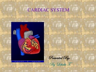
- 1. CARDIAC SYSTEM Presented By: By Lehulu T.
- 2. Normal anatomy & physiology CVS • Consists: heart, arteries, veins, capillaries • Functions: 1. circulation of blood 2. delivery of oxygen and other nutrients to tissues of the body 3. removal of carbon dioxide and other products of cellular metabolism
- 3. HEART Anatomy and physiology: A. Heart wall/ Layers 1. pericardium a. fibrous b. serous 2. epicardium 3. myocardium 4. endocardium
- 4. B. Chambers 1. Atria a. right b. left 2. Ventricles a. right b. left C. Valves 1. Atrioventricular valves a. Mitral valve/bicuspid b. Tricuspid valve
- 5. AV valve Function - permit unidirectional flow of blood from specific atrium to specific ventricle during ventricular diastole - prevent reflux of blood during ventricular systole - valve leaflets open during ventricular diastole and close during ventricular systole; valve closure produces the first heart sounds (S1)
- 6. 2. Semilunar valves a. Pulmonic valve b. Aortic valve Functions: - permit unidirectional flow of blood from specific ventricle to arterial vessel during ventricular systole -prevent reflux during ventricular diastole - open when ventricles contract and close during ventricular diastole; closure produces the second heart sound (S2)
- 7. D. Conduction System 1. Sino atrial (SA) node 2. Intermodal Tracts – at upper right atrium 3. Atrioventricular (AV) node 4. Bundle of His – group of muscle fibers - right bundle branch - left bundle branch 5. Purkinje fibers – through out ventricles * Electrical activity of heart can be visualized by ECG
- 8. E. Coronary Circulation 1. Arteries a. right coronary artery b. left coronary artery 2. Veins a. coronary sinus veins – group of small veins
- 9. VASCULAR SYSTEM Functions: a. supply tissues with blood b. remove wastes c. carry deoxygenated blood back to the heart
- 10. TYPES OF BLOOD VESSELS A. Arteries B. Arterioles C. Capillaries: the following exchanges occur: - oxygen and carbon dioxide - solutes between the blood and tissues - fluid volume transfer between the plasma and interstitial spaces D. Veins E. Venules
- 11. ASSESSMENT HISTORY TAKING A. Presenting problem 1. Nonspecific symptoms include:- - fatigue - cough - Headache - weight loss/gain - syncope (dizziness) - difficulty of sleeping - anorexia
- 12. 2. Specific signs and symptoms a. chest pain b. dyspnea (shortness/difficulty of breath) c. orthopnea / paroxysmal nocturnal dyspnea d. palpitations e. edema f. cyanosis B. Lifestyle: occupation, hobbies, financial status, stressors, exercise, smoking, living conditions
- 13. C. Use of medications: OTC drugs, contraceptives, cardiac drugs D. Personality: Type A, manic-depressive, anxieties E. Nutrition: dietary habits, cholesterol, salt intake, alcohol consumption F. Past Medical History G. Family history: heart disease (congenital, acute, chronic); risk factors (DM, hypertension, obesity)
- 14. PHYSICAL EXAMINATION A. Skin and mucous membranes: - color/texture, temperature, hair distribution on extremities, atrophy or edema, petechial B. Peripheral pulses: - palpate and rate all arterial pulses (temporal, carotid, brachial, radial, femoral, popliteal, dorsalis pedis and posterior tibia) on scale of: 0=absent 1=weak 2=normal 3=full /bounding
- 15. C. Measure and record blood pressure D. Inspect and palpate the neck vessels: a. jugular veins: note; location, characteristics, jugular venous pressure b. carotid arteries: location and characteristics E. Auscultate heart sounds - normal - (S1, S2) - abnormal - gallop (S3, S4) - murmur - pericardial friction rub
- 16. LABORATORY / DIAGNOSTIC TESTS A. Chemistry and electrolyte analysis 1. Cardiac enzymes: in MI a. Troponin T: detected 3-12 hours after chest pain b. Troponin I: detected 3-12 hrs. c. creatinine phosphokinase (CPK ): 6-12Hrs d. Aspartate aminotransferase (AST) (SGOT): with in 24 Hrs. after chest pain e. Lactic dehydrogenase (LDH): with in 36 Hrs.
- 17. 2. Electrolytes a. Sodium (Na): 135-145meq/L - hyponatremia: may cause fluid excess - hypernatremia: fluid deficit b. Potassium (K): 3.5-5 meq/L - Inc. or Dec. levels can cause dysrhythmias c. Magnesium (Mg): 1.3-2.1 meq/L - decrease levels can cause dysrhythmias
- 18. d. Calcium (Ca): 4.5-5.3 meq/L: -Normal values for blood clotting and neuromuscular activity - decrease levels cause tetany, - Inc. levels causes muscle atony - Dec. and Inc. levels causes dysrhythmias 3. Serum Lipids a. Total Cholesterol : 150-200mg/dl: - high levels predispose to atherosclerotic Heart disease
- 19. b. High density lipids (HDL): 30-85 mg/dl - low levels predispose to CVD c. Low density lipids (LDL): 50-140 mg/dl: - high levels predispose to atherosclerotic plaque formation d. Triglycerides :10-150 mg/dl: - high levels increase risk of atherosclerotic heart disease
- 20. B. Hematologic Studies 1. CBC – their number,size,infections 2. Coagulation time: 5-15mins; Inc. levels indicate bleeding tendency, used to monitor heparin Rx. 3. Prothrombin time (PT): 9.5-12sec.; used to monitor warfarin Rx. 4. Activated partial thromboplastin time (APTT): 20-45sec; used to monitor heparin therapy 5. Erythrocyte sedimentation rate(ESR) : <20mm/hr; Inc. level indicate inflammatory process
- 21. C. Urine Studies (U/A) D. Electrocardiogram (ECG/EKG) 1. Noninvasive ECG – a graphic record of the electrical activity of the heart 2. Portable recorder (Holter monitor) – provides continuous recording of ECG for up to 24 hrs. 3. Exercise ECG (stress test): the ECG is recorded during prescribed exercise; may show heart disease when resting ECG does not. E. Echocardiogram: noninvasive recording of the cardiac structures using ultrasound
- 22. F. Cardiac catheterization: invasive, but often definitive test for diagnosis of cardiac disease. 1. A catheter is inserted into the right or left side of the heart to obtain information 2. Purpose: to measure intracardiac pressures and oxygen levels in various parts of the heart; with injection of a dye, • allows visualization of the heart chambers, blood vessels and blood flow (angiography)
- 23. 3. Nursing care for C.catheterization prior to the test - informed consent - any allergies especially to iodine - keep client on NPO for 8-12 hrs. - record V/S, height, weight - inform client that a feeling of warmth and fluttering sensation as catheter is inserted
- 24. post test - assess circulation to the extremity used for catheter insertion - check peripheral pulses, color, sensation of affected extremity - if protocol requires, keep affected extremity straight for approximately 8 hrs. - observe catheter insertion site for swelling, bleeding - assess V/S and report for significant changes
- 25. G. Coronary arteriography 1. visualization of coronary arteries by injection of radiopaque dye/ contrast medium and recording on a movie film. 2. Purpose: evaluation of heart disease and angina, location of areas of infarction and extent of lesions, ruling out coronary artery disease in clients with MI. 3. Nursing care: same as cardiac catheterization
- 26. ANALYSIS Nursing diagnosis for the client with CVD include A. Fluid volume excess r/t…as evidenced by.. B. Decreased cardiac output C. Altered peripheral tissue perfusion D. Impaired skin integrity E. Risk for activity intolerance F. Pain G. Ineffective coping H. Fear I. Anxiety
- 27. PLANNING GOALS A. Fluid imbalance will be resolved, edema will be minimized B. Cardiac output will be improved. C. peripheral tissue perfusion will be improved D. Adequate skin integrity will be maintained E. Activity intolerance will progressively resolved F. Pain in the chest will be diminished G. Client’s level of fear and anxiety will be decreased
- 28. INTERVENTIONS CARDIAC MONITORING A. ECG 1. P wave: produced by atrial depolarization; indicates SA node function 2. P-R interval a. indicates AV conduction time or the time it takes an impulse to travel from the atria down and through the AV node b. measured from beginning of P wave to beginning of QRS complex
- 29. 3. QRS complex a. indicates ventricular depolarization b. measured from onset of Q wave to end of S wave 4. ST segment a. indicates time interval between complete depolarization of ventricles and repolarization of ventricles b. measured after QRS complex to beginning of T wave 5. T wave a. represents ventricular repolarization b. follows ST segment
- 30. HEMODYNAMIC MONITORING (Swan Ganz Catheter) A. A multilumen catheter with a balloon tip that is advanced through the superior vena cava into the RA, RV, and Pulmonary Artery/PA. When it is wedged it is in the distal arterial branch of the pulmonary artery. B. Purpose: 1. Proximal port: measures RA pressure 2. Distal port: a. measures PA pressure
- 31. b. normal values: PA pressure (systolic and diastolic) less than 20mmHg C. Nursing care 1. a sterile dry dressing should be applied to site and changed Q 24 hours; inspect site daily and report signs of infection 2. if catheter is inserted via an extremity, immobilize extremity to prevent catheter dislodgment or trauma.
- 32. 3. Observe catheter site for leakage 4. Continuously monitor PA systolic and diastolic pressures and report significant variations 5. Maintain client in same position for each reading 6. Record PA systolic and diastolic readings at least every hour
- 33. Central venous pressure (cvp) A. Obtained by inserting a catheter into the external jugular, ante cubital, or femoral vein and threading it into the vena cava. The catheter is attached to an IV infusion and H2O manometer by a three way stopcock B. Purposes: 1. Reveals RA pressure, reflecting alterations in the RV pressure
- 34. 2. Provides information concerning blood volume and adequacy of central venous return 3. Provides an IV route for drawing blood samples, administering fluids or medication, and possibly inserting a pacing catheter C. Normal range is 4-10 cmH20; - elevation indicates hypervolemia, - decreased level indicates hypovolemia D. Nursing care 1. Ensure client is relaxed
- 35. 2. Maintain zero point of manometer always at level of right atrium (miaxillary line) 3. Determine patency of catheter by opening IV infusion line 4. Turn stopcock to allow IV solution to run into manometer to a level of 10-20cm above expected pressure reading 5. Turn stopcock to allow IV solution to flow from manometer into catheter; fluid level in manometer fluctuates with respiration
- 36. 6. Stop ventilatory assistance during measurement of CVP 7. After CVP reading, return stopcock to IV infusion position 8. Record CVP reading and position of client EVALUATION • Act of examining improvements previously stated problems • May lead to re assessment
- 37. • Thanks