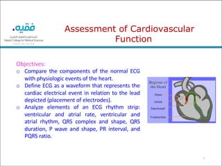
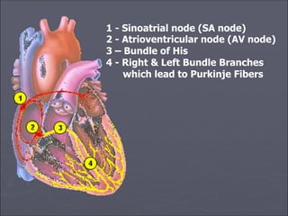
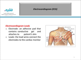
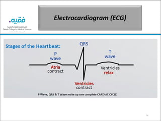
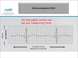
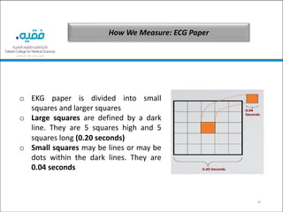
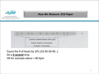
This document provides an overview of cardiac anatomy, physiology, and assessment. It discusses the components of the cardiac conduction system including the sinoatrial node, atrioventricular node, bundle of His, and Purkinje fibers. It also describes how electrocardiograms work and the parts of an ECG strip. Key aspects of cardiac function like contractility, preload, afterload, and stroke volume are defined. The document outlines steps for assessing a patient's cardiac status including vital signs, risk factors, and diagnostic tests.
This document provides an overview of electrocardiograms (ECGs) including what they are used for, how they work, and how to interpret the different parts of an ECG tracing. An ECG is a test that records the heart's electrical activity through electrodes placed on the skin. It can be used to diagnose heart conditions by examining features such as intervals, waves, rates, and voltages. A normal ECG shows distinct P, QRS, and T waves along with standardized intervals between them. Abnormalities in the tracing can indicate issues like arrhythmias, enlarged heart chambers, or poor blood flow.
This document provides an overview of electrocardiograms (ECGs) including what they are used for, how they work, and how to interpret the different parts of an ECG tracing. An ECG records the electrical signals of the heart and can be used to diagnose heart conditions. It describes the normal anatomy and function of the heart as well as defining the typical waves, intervals and rates that make up a normal ECG tracing such as the P, QRS, T waves and PR interval. Abnormalities that can be detected from an irregular heartbeat like atrial fibrillation are also discussed.
The document discusses cardiovascular anatomy and physiology. It describes the structures of the heart including the layers (pericardium, epicardial fat, myocardium, endocardium), chambers (left and right atria and ventricles), and valves. It also discusses the cardiac conduction system including the sinoatrial node, atrioventricular node, bundle of His, and Purkinje fibers. Hemodynamic monitoring techniques are covered such as intra-arterial blood pressure monitoring, central venous pressure monitoring, pulmonary artery pressure monitoring, and mixed venous oxygen saturation monitoring. Physical exam skills related to cardiovascular assessment are also outlined.
The document provides an overview of a course on cardiovascular diseases (CVD). The 16-hour course will cover the etiology, pathophysiology, manifestations, risk factors, and management of various CVDs. It will also address diagnostic tests, medications, treatments, and rehabilitation services for CVD patients. The main objective is for students to acquire knowledge and skills to promote health, prevent illness, diagnose, manage and coordinate rehabilitation of CVD patients. Specific topics to be covered include anatomy and physiology of the heart and vessels, assessment of the cardiovascular system, and common CVDs and their management.
This document provides an overview of cardiovascular disorders and ischemic heart disease (IHD). It defines IHD as heart weakening caused by reduced blood flow to the heart, typically due to coronary artery disease where the coronary arteries narrow. It discusses the anatomy of the heart including the four chambers, great vessels, valves, and cardiac muscle cells. It also covers ECGs, cardiac conduction, circulatory system functions, common cardiovascular diseases like IHD, strokes, peripheral artery disease, aortic disease, and high blood pressure, as well as types of angina.
The document discusses electrocardiography (ECG), which involves using electrodes to detect and record the electrical activity of the heart over time. It provides a brief history of ECG development and describes the modern ECG machine. It then explains the electrical conduction system of the heart and the waves that make up a normal ECG, including the P, QRS, and T waves. The cardiac cycle and heart sounds are also summarized.
The document discusses the mechanisms that regulate blood pressure in the short term, including the nervous system and chemicals. It explains that the nervous system, including the baroreceptor reflex and chemoreceptors, controls blood pressure by changing peripheral resistance within seconds or minutes in response to changes in blood pressure. The document also outlines the roles of the vasomotor center, sympathetic and parasympathetic activity, and adrenal glands in short term blood pressure regulation.
The document discusses various diagnostic measures used in cardiology to diagnose and treat cardiovascular abnormalities. It describes stress tests, echocardiography, radiographic tests like chest x-rays and CT angiography, electrocardiographic tests including electrocardiograms and Holter monitoring, invasive tests like cardiac catheterization and electrophysiologic studies, and laboratory tests like measuring central venous pressure and pulmonary capillary wedge pressure. These diagnostic tests evaluate the structure and function of the heart and blood vessels.
The document provides information about the cardiovascular system including:
1. It describes the main functions of the cardiovascular system which includes transporting oxygen, carbon dioxide, nutrients, hormones, and removing waste from the body.
2. It explains the double circulatory pathway where blood travels through the heart twice - first to the lungs then to the body, before returning to the heart.
3. It outlines the key structures of the circulatory system including the heart, blood vessels (arteries, veins, capillaries), blood, and provides a diagram of heart chambers and circulation.
The electrocardiogram (ECG or EKG) measures and records the electrical activity of the heart. It was developed in 1893 by Willem Einthoven, who received the Nobel Prize for his work. An ECG works by detecting the tiny electrical changes on the skin that occur with each heartbeat. It shows the heart's rate and rhythm, as well as any damage to heart muscle. A standard 12-lead ECG provides multiple views of the heart and can help diagnose conditions like heart attacks.
This document provides an overview of basic ECG interpretation and nursing management. It begins with the anatomy and physiology of the cardiovascular system, including the heart chambers, valves, vessels and conduction system. It then covers electrophysiology, describing the cardiac cycle, waveforms and intervals on an ECG. The document provides steps for analyzing rhythm strips and discusses various sinus arrhythmias like sinus tachycardia, bradycardia, arrhythmia and arrest.
The document provides an overview of the cardiovascular system, including the anatomy and physiology of the heart and blood vessels. It discusses the chambers of the heart, valves, coronary circulation, conduction system, and nerve supply. It also covers measurements of various pressures like mean arterial pressure, central venous pressure, and pulmonary artery pressure. Other topics include cardiac output, stroke volume, contractility, and the determinants of cardiac performance. Graphs of the cardiac cycle and Wigger's diagram are presented showing the mechanical events in systole and diastole.
Cardiac diagnostics and laboratory tests Dr. Rima Das
The document provides information on various cardiac diagnostic and laboratory tests including:
1. ECG - Analyzes electrical events of the cardiac cycle and can provide insight into pathophysiology. Important aspects to analyze include rhythm, intervals, axis, and waves.
2. Treadmill test - Progressive exercise test to evaluate cardiovascular response. Used to detect ischemia by analyzing ECG changes, symptoms, and hemodynamics.
3. Echocardiography - Ultrasound of the heart to assess structure, function, valves, and flow. Provides different views of the heart chambers and valves.
4. Holter monitoring - Mobile ECG recording over 24-72 hours to detect arrhythmias and ST-T changes
crème de la crème basics to understand electrocardiographic analysis in an easy & simple way with some specifications to its use in Emergency medicine/clinical toxicology practice.
This document provides an overview of electrocardiography (ECG) and how to interpret ECG strips in the context of toxicology. It begins by defining the objectives of learning ECG and its importance in toxicology. It then describes the basic components of an ECG including the waves, intervals, complexes, leads, and normal values. The document explains how to assess heart rate and rhythm on an ECG strip. Finally, it demonstrates some common ECG changes that can occur due to toxic exposures, such as sinus tachycardia, sinus bradycardia, and heart block.
Cardiovascular assessment and diagnostic proceduresANILKUMAR BR
The document provides an overview of cardiovascular anatomy and physiology, including the structure of the heart, chambers of the heart, heart valves, cardiac cycle, and coronary arteries. It also discusses the cardiac conduction system, including the sinoatrial node, atrioventricular node, bundle of His, and Purkinje fibers. Common diagnostic tests and manifestations of cardiovascular disease are also mentioned.
The document discusses the anatomy, physiology, and assessment of the cardiovascular system. It describes the heart's location, chambers, valves, blood supply, conduction system, cardiac cycle, and normal functioning. Assessment methods covered include vital signs, heart sounds, ECG, echocardiography, nuclear imaging scans, and diagnostic tests like chest x-rays. Nurses' responsibilities in preparing patients and monitoring them during tests are also outlined.
1. The document provides an overview of cardiovascular anatomy and physiology, including the structure and function of the heart, blood vessels, conduction system, cardiac cycle, and heart sounds.
2. Key concepts covered include cardiac output, preload and afterload, the 8 phases of the cardiac cycle, heart sounds such as S1, S2, S3, S4, and murmurs.
3. Assessment techniques for the cardiovascular system such as auscultation locations and heart sounds are demonstrated.
This document provides information on various cardiovascular tests used to assess heart function and identify heart disorders. It discusses the following tests: medical history and physical examination, laboratory tests of cardiac enzymes and biomarkers, chest radiography, electrocardiography, echocardiography, exercise stress testing, pharmacologic stress testing, and cardiac catheterization. For each test, the document outlines the procedure, what is evaluated, and normal versus abnormal findings.
Diagnostic tests are used in cardiology to confirm data from a patient's history and physical assessment. Common tests include blood studies to detect cardiac biomarkers released during injury, imaging studies like echocardiograms and CT scans to visualize the heart structures, and stress tests to evaluate the heart's response to physical or pharmacological stress. Electrocardiograms are also routinely performed to analyze the heart's electrical activity. More invasive procedures like cardiac catheterization can further evaluate conditions and guide treatment. The results of diagnostic tests along with the clinical picture are important for cardiologists to diagnose and manage cardiovascular conditions.
Cardiovascular tests are used to assess the function of the heart and to identify the disorders associated with the pathological heart function. Following are the tests used to assess cardiovascular function
The heart functions to pump blood throughout the body via two circulatory systems - pulmonary and systemic. It generates blood pressure and ensures one-way blood flow. Cardiac output, the amount of blood pumped, is determined by heart rate and stroke volume. Intrinsic factors like the Frank-Starling mechanism and extrinsic neural and hormonal controls regulate cardiac output in response to the body's changing needs.
Dr. Awadhesh Kumar Sharma is an interventional cardiologist who has had an excellent academic career. The goal of this session is to provide a basic understanding of ECG waves and intervals, how to interpret ECGs, and describe key aspects of using ECGs clinically. An ECG represents the heart's electrical activity and can be used to identify arrhythmias, ischemia, chamber abnormalities, and other conditions. It is important to carefully analyze standardized ECGs by examining features like rhythm, intervals, voltages and assessing for any abnormalities.
Cardiovascular disruptions can be caused by a lack of blood supply to the heart, infections of the heart, or immune-mediated inflammatory conditions. A lack of blood supply can result in ischemia, angina, heart attacks, and heart failure. Infections can directly damage the heart valves and decrease cardiac output. Common infections include infective endocarditis from bacteria entering the bloodstream, and rheumatic heart disease from an untreated streptococcal infection which causes inflammation of the heart valves.
The ECG records the electrical activity of the heart over time and is the gold standard for diagnosing cardiac arrhythmias and conduction abnormalities. It detects three main waves - the P wave from atrial depolarization, the QRS complex from ventricular depolarization, and the T wave from ventricular repolarization. Abnormalities in conduction through the AV node can cause first-, second-, or third-degree heart block visible on the ECG. Higher degrees of block impair conduction more severely and require treatment such as pacemaker implantation.
Jill Pizzola's Tenure as Senior Talent Acquisition Partner at THOMSON REUTERS...dsnow9802
Jill Pizzola's tenure as Senior Talent Acquisition Partner at THOMSON REUTERS in Marlton, New Jersey, from 2018 to 2023, was marked by innovation and excellence.
How to Prepare for Fortinet FCP_FAC_AD-6.5 Certification?NWEXAM
Begin Your Preparation Here: https://bit.ly/3VfYStG — Access comprehensive details on the FCP_FAC_AD-6.5 exam guide and excel in the Fortinet Certified Professional - Network Security certification. Gather all essential information including tutorials, practice tests, books, study materials, exam questions, and the syllabus. Solidify your knowledge of Fortinet FCP_FAC_AD-6.5 certification. Discover everything about the FCP_FAC_AD-6.5 exam, including the number of questions, passing percentage, and the time allotted to complete the test.
Leadership Ambassador club Adventist modulekakomaeric00
Aims to equip people who aspire to become leaders with good qualities,and with Christian values and morals as per Biblical teachings.The you who aspire to be leaders should first read and understand what the ambassador module for leadership says about leadership and marry that to what the bible says.Christians sh
Learnings from Successful Jobs SearchersBruce Bennett
Are you interested to know what actions help in a job search? This webinar is the summary of several individuals who discussed their job search journey for others to follow. You will learn there are common actions that helped them succeed in their quest for gainful employment.
IT Career Hacks Navigate the Tech Jungle with a RoadmapBase Camp
Feeling overwhelmed by IT options? This presentation unlocks your personalized roadmap! Learn key skills, explore career paths & build your IT dream job strategy. Visit now & navigate the tech world with confidence! Visit https://www.basecamp.com.sg for more details.
A Guide to a Winning Interview June 2024Bruce Bennett
This webinar is an in-depth review of the interview process. Preparation is a key element to acing an interview. Learn the best approaches from the initial phone screen to the face-to-face meeting with the hiring manager. You will hear great answers to several standard questions, including the dreaded “Tell Me About Yourself”.
In the intricate tapestry of life, connections serve as the vibrant threads that weave together opportunities, experiences, and growth. Whether in personal or professional spheres, the ability to forge meaningful connections opens doors to a multitude of possibilities, propelling individuals toward success and fulfillment.
Eirini is an HR professional with strong passion for technology and semiconductors industry in particular. She started her career as a software recruiter in 2012, and developed an interest for business development, talent enablement and innovation which later got her setting up the concept of Software Community Management in ASML, and to Developer Relations today. She holds a bachelor degree in Lifelong Learning and an MBA specialised in Strategic Human Resources Management. She is a world citizen, having grown up in Greece, she studied and kickstarted her career in The Netherlands and can currently be found in Santa Clara, CA.
Joyce M Sullivan, Founder & CEO of SocMediaFin, Inc. shares her "Five Questions - The Story of You", "Reflections - What Matters to You?" and "The Three Circle Exercise" to guide those evaluating what their next move may be in their careers.
1. Adult Health Nursing 1 :
Cardiac disorders 1
Fakeeh College for Medical Sciences
Fourth Year – Level 4
2020-2021, Semester II
Dr Imad AL-Jarrah, PhD,
RN
2. 2
Objectives:
o Compare the components of the normal ECG
with physiologic events of the heart.
o Define ECG as a waveform that represents the
cardiac electrical event in relation to the lead
depicted (placement of electrodes).
o Analyze elements of an ECG rhythm strip:
ventricular and atrial rate, ventricular and
atrial rhythm, QRS complex and shape, QRS
duration, P wave and shape, PR interval, and
PQRS ratio.
Assessment of Cardiovascular
Function
3. 3
Anatomy and Physiology
• The heart compose of three layers
– Endocardium: the inner layer of endothelial tissue
– Myocardium: the middle layer of the muscle fiber
responsible for pumping action
– Epicardium: the outer layer
4. 4
Anatomy and Physiology
• Right heart consists of Rt atrium, Rt ventricle,
distribute deoxygenated blood.
• Left heart consists of Lt atrium, Lt ventricle ,
distribute oxygenated blood.
• Varying thickness of a trial and ventricular wall
and left and right ventricles related to workload.
7. 7
Anatomy and Physiology
• The heart has large metabolic requirement, 70-
80% of delivered oxygen
• Coronary arteries are perfused during diastole
• Left coronary artery branches (LAD & LCX)
• Right coronary artery branch (RCA)
• Posterior wall received blood by (PDCA)
• Blood return to heart through the coronary sinus
Located in the Right atrium
9. 9
Cardiac Conduction
• Cardiac electrical cell is characterized by
– Atomicity: ability to initiate electrical
impulses
– Excitability: ability to respond to impulses
– Conductivity: ability to transmit
impulses
– Conductive System:(SA node – AV node –
bundle of His–Purkinje Fibers).
10. Components of the Conduction System
• Sinoatrial Node (Part I):
• located in back wall of the right atrium near the
entrance of vena cava
• initiates impulses 60-100 times per minute
without any nerve stimulation from brain
• establishes basic rhythm of the heartbeat
• called the pacemaker of the heart
10
11. Components of the Conduction System
• Atrioventricular Node (Part II):
• located in the bottom of the right atrium near the
septum
• cells in the AV node conduct impulses more
slowly, so there is a delay as impulses travel
through the node
• initiates impulses 40-60 times per minute without
any nerve stimulation from brain
11
12. Components of the Conduction System
• Atrioventricular Bundle (Part III):
• These branch a lot to form the Purkinje fibers that transmit
the impulses to the myocardium
• The bundle of His, bundle branches and Purkinje fibers
transmit quickly and cause both ventricles to contract at
the same time (Like a “phone tree”)
12
13. 1 - Sinoatrial node (SA node)
2 - Atrioventricular node (AV node)
3 – Bundle of His
4 - Right & Left Bundle Branches
which lead to Purkinje Fibers
14. ELECTROCARDIOGRAM (ECG)
14
The Electrical Basis of the EKG/ECG
o Electrical impulses are present on the skin surface at a very low
voltage; The EKG machine picks up these impulses and amplifies
them.
o Electrical activity is sensed by Electrodes are placed on the skin
surface to pick up these impulses and give us a picture of how they
are traveling in the form of an Electrocardiogram. This is printed on
EKG paper and is called a Rhythm strip or an EKG strip
15. Electrocardiogram (ECG)
15
Electrocardiogram Leads
o Electrode: an adhesive pad that
contains conductive gel and
attaches to patient’s skin
o Leads: the lead wires connect the
electrodes to the cardiac monitor
18. 18
How We Measure: ECG Paper
o EKG paper is divided into small
squares and larger squares
o Large squares are defined by a dark
line. They are 5 squares high and 5
squares long (0.20 seconds)
o Small squares may be lines or may be
dots within the dark lines. They are
0.04 seconds
19. 19
How We Measure: ECG Paper
Count the # of beats by 10’s (10-20-30-40…)
On a 6 second strip
HR for example above = 80 bpm
20. 20
Cardiac homodynamic
– Cardiac Out put: the amount of blood your heart
pumps each minute.
– cardiac output in terms of the following equation:
Cardiac output = stroke volume × heart rate.
– Values for cardiac output are usually denoted as L/min. For a
healthy person weighing 70 kg, the cardiac output at rest
averages about 5 L/min; assuming a heart rate of 70 beats/min,
the stroke volume would be approximately 70 ml.
21. 21
Cardiac homodynamic
– STROKE VOLUME : the amount of blood
ejected by Lt ventricle in each beat which is 70
ml determined by
•Preload
•After load
•Contractility
– EJECTION FRACTION: 42% for right heart and
50% for the left heart
22. Four determinants of cardiac output
22
Four determinants of cardiac output
23. Four determinants of cardiac output
• Heart rate: the faster the heart beats, the more blood can be pumped over a
particular period of time.
• Contractility: This can be equated to an increased contractility of the heart
muscle, resulting in increased cardiac output. Too little pedal power, or
impaired contractility, will reduce cardiac output.
• Preload: Is the stretch of myocardium or end-diastolic volume of the
ventricles and most frequently refers to the volume in a ventricle just before
the Contraction (start of systole)
• Afterload: Is the force against which the ventricles must act in order
to eject blood, and is largely dependent on the arterial blood
pressure and vascular tone.
23
24. 24
ASSESSMENT
• Health history and clinical
manifestations
–Acute symptoms: current medication,
allergies, general appearance, hemodynamic status
–Stable patients: complete history, spouse or
partner, demographic information
25. 25
ASSESSMENT
• Cardiac S&S
–Chest pain or pressure
–Shortness of breath
–Edema and weight gain
–Palpitations
–Fatigue
–Dizziness or loss of consciousness
26. 26
PHYSICAL ASSESSMENT
• Blood pressure
– Normal is 120/80 mmhg ( 100/60 – 140/90 ) invasive
and non invasive
• Pulse pressure:
– Difference between systolic and diastolic, 30-40 mmhg,
reflects strock volume & ejection velocity, vascular
resistance,
– Less than 30 mmhg = serious reduction in output
28. 28
RISK FACTORS
– Nonmodifiable: age, positive family history,
race & gender
– Modifiable: hyperglycemia, hyperlipidemia,
hypertension, inactivity, smoking, obesity &
type A personality
29. 29
Laboratory tests
– Cardiac enzymes
• Iso enzymes are more specific, createnine
kinase ( CK ) and its iso enzyme ( CK-MB ),
lactic dehydrogenase (LDH) ,troponin I
30. 30
Laboratory tests
– Blood chemistry
•Lipid profile:
A- Cholesterol (less than 200 mg/dl),
B- Triglycerides ( 40 – 150 mg / dl ) source of
energy, cell wall, store in a depose tissue.
C- LDL ( less than 130 mg / dl )
D- HDL (35 – 65 mg/dl M, 35 – 85 mg/dl F)
31. 31
Laboratory tests
• Serum electrolytes
– K, Na, Ca and other electrolytes can reflect the heart
function as well as fluid & electrolyte disturbances
• BUN
– May indicates impaired renal function and impaired
cardiac output
32. 32
Laboratory tests
• Coagulation studies
– Partial Thromboplastine Time (PTT): 25 – 40
sec, used to regulate heparin dosage,1.5–2.5 is
the therapeutic range
– Prothrombin Time (PT): less than13 sec, used
to regulate warfarin, 1.5 – 2.5 times of PT is the
therapeutic range
– International Normalized Ratio (INR):
standardized method for reporting PT level,
used for regulating warfarin dosage
33. 33
Diagnostic Evaluation
• Chest X-Ray ( CXR ) fluoroscopy
– Assess size, position of the heart, cardiothoracic ratio (
CTR ), position of central lines
• Electrocardiography (ECG)
– Can be either on bed side or from a distance, 12 leads
ECG, continuous monitoring, telemetry monitoring 2 or
3 leads monitoring)
34. 34
Diagnostic Evaluation
• Cardiac Stress Test
During time of increased demand, abnormalities
in cardiovascular functions are more likely to
be detected, used to evaluate the heart
function, coronary arteries as well as the cause
of chest pain
35. 35
Diagnostic Evaluation
–Exercise stress test: pt walk on a treadmill
or pedals (stationary bicycle), the goal is to increase HR
and monitored for ECH changes, arrhythmias,
hypotension, pain, dyspnea and dizziness. Pt fast 4
hours before test, nurse needs to instruct pt about the
test
36. 36
• Cardiac catheterization
– Invasive diagnostic procedure involves
introduction of specific catheter into Rt & Lt side
blood vessels under fluoroscopy. Its used to
evaluate coronary arteries potency, heart
function as a pump, vascular system and heart
structure