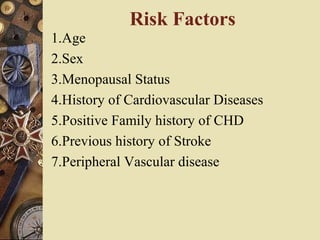
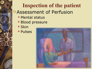
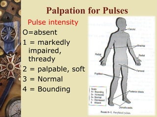
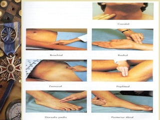
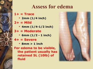
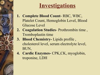
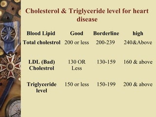
4. īˇ Chest pain
īˇ Shortness of breath or dyspnea
īˇ Edema and weight gain
īˇ Weakness and fatigue
īˇ Palpitation
īˇ Dizziness and syncope or loss of conscious
Manifestation of heart disease
5. Cardiac assessment includes
īĄ History (patientâs story)
īĄ Assessment of perfusion
īŦ Mental status
īŦ Blood pressure
īŦ Pulses
īĄ Inspection of the patient
īŦ Evaluation for dependent edema
īŦ Auscultation of the heart
īĄ Chemistries(cardiac enzyme)
6. Health History
īˇ The nurse should obtain the history by using
the specific question about the onset &
severity of the chest discomfort, associated
symptoms, current medication& allergy.
īˇ History include the following history
1.History of present illness
2.Past illness
3.Allergic history
4.Medication history
5.Family history
6.Personal history
7. Physical Assessment
īˇ General appearance
īˇ Cognition
īˇ Inspection of the skin
īˇ Vital sign
īˇ Arterial pulse
īˇ Jugular venous pulsation
īˇ Heart inspection, palpation ,
percussion and auscultation
īˇ Inspection of extremities
īˇ Respiratory
assessment
Physical assessment of the cardiac patient
include :
8. History (patientâs story)
īĄ Patientâs presenting sign
or symptoms
īŦ Pain:
īĄ Where is it?
īĄ Does it move or stay
in one place?
īĄ What is it like?
īĄ How severe is it?
īĄ How frequent?
īĄ What are you doing
when it occurs?
īĄ What can you do to
make it go away?
īŦ Other symptoms
9. Evaluation of Pain
īĄ The reddened area is the most common area
of discomfort in ACS
īĄ The pain is usually not a sharp, jabbing pain,
but a sensation of pressure, fullness,
squeezing or aching
10. Evaluation of Pain
īą Distress may extend from
the chest into one or both
arms or may appear in the
arms alone
īą It may be mistaken for
arthritis, bursitis, or
muscle strain
īą To tell the difference ask
the patient to lift arms
above head, heart pain
will not be aggravated by
this while the other
causes will
11. Evaluation of Pain
īĸ Discomfort may radiate into
the neck and jaws or one or
both sides and in front or
back
īĸ It may be mistaken for a
tooth ache, arthritis or stiff
neck
īĸ To test: turn your head or
bend your neck, heart pain
will not be aggravated,
whereas most pain originating
in the neck will be
īĸ Differentiation of toothache
from heart attack may be
more difficult
12. Evaluation of Pain
īĸ Pain â usually pressure,
fullness, squeezing or
aching may appear in the
upper abdomen where it
is often mistaken for
indigestion
īĸ Nausea or vomiting may
occur with this pain
13. Evaluation of Pain
īĸ Back pain may be the only
sign of heart attack
īĸ Usually this is located
between the shoulder
blades and is similar to the
tired hurt experienced
after tedious work
involving protracted use of
the arms and hands and
stooping of the shoulders
14. Evaluation of Pain
īą Heart pain often occurs in a combination of
patterns in all these areas
īą Not infrequently pain the neck, jaws, abdomen,
arms, and back may be even more severe than
that in the chest
īą Nausea or vomiting, sweating, also should be
considered a sign
15. Evaluation of Pain
īą Pain in the left chest wall centering on the
left nipple is almost never a sign of heart
attack
īą This pain may be a jabbing sensation
lasting a second or two, a dull soreness
lasting for minutes or hours, or a
combination of the two
16. Pain Assessment
R/O Pain Symptoms
Esophageal
Reflex
īĄSub sternal chest pain
īĄChest tightness
īĄIndigestion
Pleural Pain īĄSharp
īĄPositional
īĄIncreased with deep breath
Pericardial
Pain
īĄLeft chest radiating into clavicle area
or back
īĄSharp and positional
īĄIncreased with deep breath
īĄFever
17. Pain Assessment
R/O Pain Symptoms
Gallbladder
Pain
īĄChest pain â abdominal pain
īĄIndigestion
Muscular /
Skeletal
īĄPositional
īĄTender to palpation
Pleural Pain īĄSharp
īĄPositional
īĄIncreased with deep breath
īĄCatching respirations
Pancreatitis īĄSevere sharp pain
īĄPositional ( pt. Tends to want to sit and
lean forward
18. Inspection of the patient
īĄ Assessment of Perfusion
īŦMental status
īŦBlood pressure
īŦSkin
īŦPulses
19. Skin & Nails
īĸ Color
īĸ Moisture/
texture
īĸ Temperature
īĸ Turger
īĸ Capillary refill
īŦnormal <3 seconds
īĸ Clubbing
-associated with
cyanotic heart disease
as well as lung disorders
22. Assess for edema
1+ = Trace
īŦ 2mm (1/4 inch)
2+ = Mild
īŦ 4mm (1/4-1/2 inch)
3+ = Moderate
īŦ 6mm (1/2 - 1 inch)
4+ = Severe
īŦ 8mm > 1 inch
For edema to be visible,
the patient usually has
retained 5L (10lb) of
fluid
23. Blood Pressure Assessments
īĄ Left and right BP should always be compared to
detect any differences
īŦ Normally the BP varies 5-10 mm Hg
īŦ A difference of 20 mm Hg or more suggests arterial
compression or obstruction.
īŦ A difference between arm pressures and/or leg BP
could mean a dissecting aortic aneurysm
īĄ BP should always be taken at heart level
īŦ Falsely elevated readings are obtained if the arm is
lower than the heart
īŦ Falsely lower readings are obtained if the arm is
higher than the heart
īĄ Cuffs size: cover the upper arm by 40%
īŦ Too small can give falsely high readings
īŦ Too large can give falsely low readings
24. Homenâs Sign
īą It is present when brisk flexion of
the foot causes pain in the calf of
the leg
īą May be assessed when the patient
is examined for edema
īą In some patients it may indicate
deep-vein thrombosis
25. Neck
īĸ No jugular vein
distension should be
present at 30 °
elevation position
Procedure for evaluation
īĸ elevate pt. head of bed
to 30°
īĸ locate external jugular
vein ( may briefly
occlude to identify it
īĸ release pressure and
observe for distension
26. Neck - jugular vein distension
īą Head supported and relaxed at 30° - 40 °
īą Highest point of pulsation observed during
exhalation
īą Vertical distance between pulsation and sternal
angles is estimated in cm
īą Normal value is 4cm or less
28. Carotid Auscultation
īą Using bell of
stethoscope on carotid
artery
īą Auscultate for bruits
īą blood flow through
tortuous or partially
occluded vessel
īą high flow through a
normal vessel
29. First heart sound - S1
īą âLUBâ
īą Caused by rapid deceleration of blood flow
when the AV (mitral and tricuspid) close at
the beginning of systole
īą Dull, low-pitched, louder than S2 at the
mitral area
īą Best heard and 4th
intercostal space left of
the sternal border and apex
30. Second heart sound â S2
īĸ âDUBâ
īĸ Closing of the aortic and pulmonic valves at
the end of systole
īĸ Higher pitched, snappier and shorter in
duration, louder than S1 at the Aortic area
īĸ Heard best at the base of the heart (2nd
intercostal space to the right and left of the
sternum)
31. Third Heart Sound-S3
īĸ Ventricular diastolic gallop
( Ventricular Disorder rhythm)
īĸ âKen-tuck-yâ
īĸ Due to rapid filling of the left ventricle
īĸ Can be normal in young adults and children
īĸ After age 40 S3 is likely to be
associated with heart diseases
(congestive heart failure)
Best heart with the bell
over apex
32. Fourth Heart Sound - S4
īą Atrial diastolic gallop ( Artrial Disorder rhythm)
īą âTen-ne-seeâ
īą Occurs during diastole with atrial contraction (atrial kick)
īą Note: Cannot be heard with patients in atrial-fib
īą Associated with a noncompliant ventricle â AMI,
hypertension, hypertrophy
īą Not commonly heard in adults
īą Athletes may have a benign S4
Best heart with the bell at
the apex with the pt. lying in
the left lateral position
33. Causes of Murmurs
īĸ Flow through damaged valves
īĸ Increased flow through normal valves and
vessels
īŦ fever, exercise
īĸ Backflow across an incompetent valve
īĸ Blood flow across a partial obstruction or
irregularity
īĸ Shunting of blood from an abnormal passage
from high to low pressure (patent foramen
ovale)
īĸ Decreased strength of myocardial
contraction
36. Blood Lipid Good Borderline high
Total cholestrol 200 or less 200-239 240&Above
LDL (Bad)
Cholestrol
130 OR
Less
130-159 160 & above
Triglyceride
level
150 or less 150-199 200 & above
Cholesterol & Triglyceride level for heart
disease
37. Diagnostic Tests
1. X- ray- to evaluate internal structure.
2. Electrocardiogram- to evaluate the
interpretation of the electrical activity of the
heart.
3. Cardiac stress test- to determine the heart
response with the increased oxygen need.
4. Echocardiogram-it help in record the blood flow
through heart, valvular abnormality, congenital
cardiac defect & cardiac function
5. Holter Monitoring- Monitor ECG rhythm 24-48
& correlating the rhythm changes with symptoms
recorded in the diary
38. Diagnostic Tests
6. Stress echocardiogram-The combination
of exercise test with echocardiogram are
done in the resting image as well as after
exercise.
7. Nuclear Cardiology- it involve the I V
injection of radioisotopes & uptake is
counted over the heart by scintillation
camera to identify myocardial contractility
8. Magnetic resonance angiograph- In the
MRA, with the use of gadolinium as I V
contrast to evaluate the arterial disease
39. 9. PET scan- In this two radionuclide
Nitrogen-13- ammonia is injected I V first
& scan evaluated for myocardial
perfusion.
10. Coronary Angiogram- The injection of
the radiopaque contrast medium directly
into coronary artery same as cardiac
catheterization to evaluate the patency of
coronary artery.
11.Cardiac catheterization- to identify the
oxygen saturation and the pressure
reading as well as heart chamber
Diagnostic Tests
40. 12. Electrophysiology study- the test used to
record intra-cardiac electrical activity using
catheters inserted via the femoral vein into
the right side of the heart. It recorded the
electrical activity of the cardiac structure.
13.Peripheral arteriography & venography
â the injection of the radiopaque medium is
inserted in the artery and vein after that
serial X- rays taken to detect & visualize
the atherosclerotic plaque, occlusion,
aneurysm & traumatic injury
Diagnostic Tests
41. 14. Haemodynamic monitoring- the
haemodynamic monitering of the arterial
blood pressure, pulmonary artery
pressure, pulmonary wedge pressure &
cardiac out put is done to evaluate the
cardiovascular status & response of the
patient towards the treatment.
Diagnostic Tests