INTRODUCTION
Some interesting
facts aboutCVS
A brief introduction to
Cardiovascular system
structure and the way its
components are organized.
Some fun Exercises.
Functions of
Cardiovascular system
conditions that affect the
heart's structure and
function.
01
04
02
05
03
06
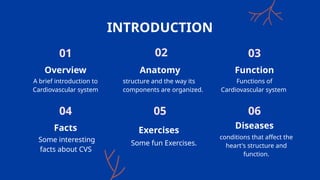
Overview Anatomy Function
Facts Exercises
Diseases
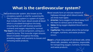
What is thecardiovascular system?
Blood vessels form an intricate network of
highways through which blood travels. There
are three main types:
1. Arteries: Carry oxygen-rich blood away from
the heart to various parts of the body.
2. Veins: Bring oxygen-depleted blood back to
the heart.
3. Capillaries: Tiny vessels where the exchange
of oxygen, nutrients, and waste products
occurs.
Blood is a fluid composed of red blood cells, white
blood cells, platelets, and plasma, responsible
for transporting oxygen, nutrients, hormones,
and waste products.
The cardiovascular system, also known as the
circulatory system, is vital for sustaining life.
The Circulatory system is a system of organs
that includes the heart, blood vessels & blood
which is circulated throughout the body,
ensuring that each cell receives essential
nutrients and oxygen required for survival.
The heart is the central component, acting as a
powerful pump. This muscular organ ensures
blood circulates throughout the body,
providing oxygen and nutrients to tissues and
removing waste products.
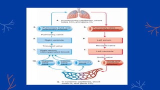
6.
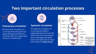
Two important circulationprocesses
The systemic circulation is a
circuit loop that delivers
oxygenated blood from the left
heart to the rest of the body &
returns deoxygenated blood
back to the right heart via large
veins known as Venae Cavae.
The Pulmonary circulation is a
circuit loop from the right heart
taking deoxygenated blood to
the lungs where it is oxygenated
& return to the left heart.
Pulmonary circulation Systemic circulation
7.
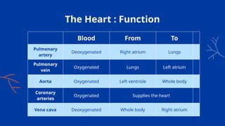
The Heart :Function
Blood From To
Pulmonary
artery
Deoxygenated Right atrium Lungs
Pulmonary
vein
Oxygenated Lungs Left atrium
Aorta Oxygenated Left ventricle Whole body
Coronary
arteries
Oxygenated Supplies the heart
Vena cava Deoxygenated Whole body Right atrium
8.
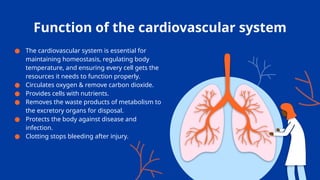
Function of thecardiovascular system
● The cardiovascular system is essential for
maintaining homeostasis, regulating body
temperature, and ensuring every cell gets the
resources it needs to function properly.
● Circulates oxygen & remove carbon dioxide.
● Provides cells with nutrients.
● Removes the waste products of metabolism to
the excretory organs for disposal.
● Protects the body against disease and
infection.
● Clotting stops bleeding after injury.
9.
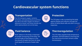
Cardiovascular system functions
TheCVS transports oxygen, nutrients,
hormones, and waste products to and from
the cells in the body. Oxygen-rich blood is
delivered to tissues, and carbon dioxide and
other waste products are carried away.
The CVS plays a role in protecting the body
against disease and infection through the
action of white blood cells and other immune
responses.
Fluid balance in the body is important
for proper functioning of the body. The
core principle of fluid balance is that
the amount of water lost from thr body
must equal the amount of water take
in.
The CVS helps regulate body temperature,
pH levels, and fluid balance, ensuring that
the internal environment remains stable
(homeostasis)
Transportation
Fluid balance
Protection
Thermoregulation
10.
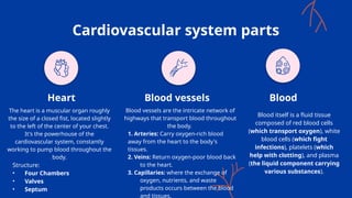
Cardiovascular system parts
Theheart is a muscular organ roughly
the size of a closed fist, located slightly
to the left of the center of your chest.
It's the powerhouse of the
cardiovascular system, constantly
working to pump blood throughout the
body.
Structure:
• Four Chambers
• Valves
• Septum
Blood vessels are the intricate network of
highways that transport blood throughout
the body.
1. Arteries: Carry oxygen-rich blood
away from the heart to the body's
tissues.
2. Veins: Return oxygen-poor blood back
to the heart.
3. Capillaries: where the exchange of
oxygen, nutrients, and waste
products occurs between the blood
and tissues.
Blood itself is a fluid tissue
composed of red blood cells
(which transport oxygen), white
blood cells (which fight
infections), platelets (which
help with clotting), and plasma
(the liquid component carrying
various substances).
Heart Blood vessels Blood
11.
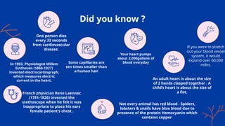
Did you know?
In 1903, Physiologist Willem
Einthoven (1860-1927)
invented electrocardiograph,
which measures electric
current in the heart.
French physician Rene Laennec
(1781-1826) invented the
stethoscope when he felt it was
inappropriate to place his ears
female patient's chest .
Some capillaries are
ten times smaller than
a human hair
If you were to stretch
out your blood vessel
system, it would
expand over 60,000
miles.
Your heart pumps
about 2,000gallons of
blood everyday
Not every animal has red blood . Spiders,
lobsters & snails have blue blood due to
presence of the protein Hemocyanin which
contains copper
An adult heart is about the size
of 2 hands clasped together . A
child’s heart is about the size of
a fist.
One person dies
every 33 seconds
from cardiovascular
disease.
12.
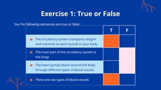
Exercise 1: Trueor False
Say the following sentences are true or false:
T F
● The circulatory system transports oxygen
and nutrients to each muscle in your body
● The main part of the circulatory system is
the lungs
● The heart pumps blood around the body
through different types of blood vessels
● There are two types of blood vessels
13.
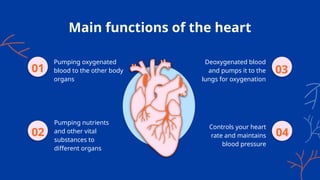
Main functions ofthe heart
Pumping oxygenated
blood to the other body
organs
01
Pumping nutrients
and other vital
substances to
different organs
Deoxygenated blood
and pumps it to the
lungs for oxygenation
Controls your heart
rate and maintains
blood pressure
02
03
04
14.
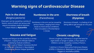
Warning signs ofcardiovascular Disease
Chest pain can be caused by a variety of
factors, some of which are serious and
require immediate medical attention.
1.Heart Attack 2. Myocarditis
Numbness in the arm can be caused by
various factors, ranging from minor
issues to more serious conditions.
1 .Poor Circulation 2.Pinched Nerve
3. Stroke
Nausea and fatigue can be caused by a variety
of factors, ranging from lifestyle habits to
underlying medical conditions.
1.Poor Diet 2. Lack of Sleep 3. Excessive Alcohol
or Caffeine 4. Infections 5. Neurological
Conditions 6. Chronic Fatigue Syndrome
Chronic coughing is defined as a cough that lasts for
eight weeks or longer in adults, or four weeks in
children.t can be caused by a variety of factors,
including:
1.Asthma 2. Chronic Obstructive Pulmonary Disease
(COPD) 3. Infections 4. Medications 5. Environmental
Triggers 6. Acid Reflux (GERD)
Shortness of breath, is a feeling of not
being able to get enough air into your
lungs
Pain in the chest
(Angina pectoris)
Numbness in the arm
(Paresthesia)
Shortness of breath
(Dyspnea)
Nausea and fatigue Chronic coughing
15.
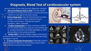
Diagnosis, Blood Testof cardiovascular system
Here are some of the most common diagnostic methods:
1. Electrocardiogram (ECG or EKG): This test records
the electrical activity of the heart to identify abnormal
rhythms or damage to the heart muscle.
2. Echocardiography: Uses ultrasound waves to create
images of the heart. It helps assess the heart's
structure and function, including the heart's
chambers, valves, and walls.
3. Cardiac Angiography: Involves inserting a catheter
into the blood vessels and using an X-ray contrast dye
to visualize blood flow and detect blockages or other
abnormalities in the coronary arteries.
4. Cardiac MRI: Provides detailed images of the heart's
structure and can identify areas of the heart that have been
damaged or scarred.
5. CT Scan (Computed Tomography): Especially useful in
detecting calcium deposits in the coronary arteries which
might indicate coronary artery disease.
16.
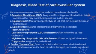
Diagnosis, Blood Testof cardiovascular system
Here are some common blood tests related to cardiovascular health:
1.Complete Blood Count (CBC): Analyzes different types of blood cells to detect
conditions that may mimic heart problems, such as anemia.
2. Lipoprotein (a): Measures a specific type of LDL that can increase the risk of
heart disease.
3. Lipid Profile: Measures cholesterol and triglycerides. It includes:
• Total Cholesterol
• Low-Density Lipoprotein (LDL) Cholesterol: Often referred to as "bad"
cholesterol.
• High-Density Lipoprotein (HDL) Cholesterol: Known as "good" cholesterol.
• Triglycerides: A type of fat in the blood.
4. Cardiac Troponin Test: Detects a protein called troponin, which is released
into the bloodstream when the heart muscle is damaged, such as during a heart
attack.
17.
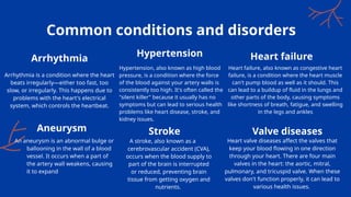
Hypertension, also knownas high blood
pressure, is a condition where the force
of the blood against your artery walls is
consistently too high. It's often called the
"silent killer" because it usually has no
symptoms but can lead to serious health
problems like heart disease, stroke, and
kidney issues.
Heart failure, also known as congestive heart
failure, is a condition where the heart muscle
can't pump blood as well as it should. This
can lead to a buildup of fluid in the lungs and
other parts of the body, causing symptoms
like shortness of breath, fatigue, and swelling
in the legs and ankles
Common conditions and disorders
Arrhythmia is a condition where the heart
beats irregularly—either too fast, too
slow, or irregularly. This happens due to
problems with the heart's electrical
system, which controls the heartbeat.
An aneurysm is an abnormal bulge or
ballooning in the wall of a blood
vessel. It occurs when a part of
the artery wall weakens, causing
it to expand
A stroke, also known as a
cerebrovascular accident (CVA),
occurs when the blood supply to
part of the brain is interrupted
or reduced, preventing brain
tissue from getting oxygen and
nutrients.
Heart valve diseases affect the valves that
keep your blood flowing in one direction
through your heart. There are four main
valves in the heart: the aortic, mitral,
pulmonary, and tricuspid valve. When these
valves don't function properly, it can lead to
various health issues.
Arrhythmia
Hypertension Heart failure
Aneurysm Stroke Valve diseases
18.
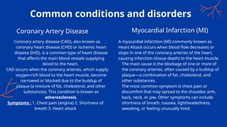
Common conditions anddisorders
Coronary artery disease (CAD), also known as
coronary heart disease (CHD) or ischemic heart
disease (IHD), is a common type of heart disease
that affects the main blood vessels supplying
blood to the heart.
CAD occurs when the coronary arteries, which supply
oxygen-rich blood to the heart muscle, become
narrowed or blocked due to the buildup of
plaque (a mixture of fat, cholesterol, and other
substances). This condition is known as
atherosclerosis.
Symptoms : 1. Chest pain (angina) 2. Shortness of
breath 3. Heart attack
Coronary Artery Disease Myocardial Infarction (MI)
A myocardial infarction (MI) commonly known as
Heart Attack occurs when blood flow decreases or
stops in one of the coronary arteries of the heart,
causing infarction (tissue death) to the heart muscle.
The main cause is the blockage of one or more of
the coronary arteries, often caused by a buildup of
plaque—a combination of fat, cholesterol, and
other substances.
The most common symptom is chest pain or
discomfort that may spread to the shoulder, arm,
back, neck, or jaw. Other symptoms can include
shortness of breath, nausea, lightheadedness,
sweating, or feeling unusually tired.
19.
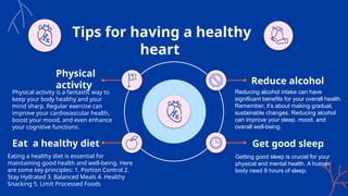
Tips for havinga healthy
heart
Physical activity is a fantastic way to
keep your body healthy and your
mind sharp. Regular exercise can
improve your cardiovascular health,
boost your mood, and even enhance
your cognitive functions.
Physical
activity
Eat a healthy diet
Eating a healthy diet is essential for
maintaining good health and well-being. Here
are some key principles: 1. Portion Control 2.
Stay Hydrated 3. Balanced Meals 4. Healthy
Snacking 5. Limit Processed Foods
Get good sleep
Getting good sleep is crucial for your
physical and mental health. A human
body need 8 hours of sleep.
Reduce alcohol
Reducing alcohol intake can have
significant benefits for your overall health.
Remember, it’s about making gradual,
sustainable changes. Reducing alcohol
can improve your sleep, mood, and
overall well-being.
20.
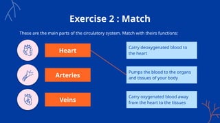
Exercise 2 :Match
These are the main parts of the circulatory system. Match with theirs functions:
Carry deoxygenated blood to
the heart
Pumps the blood to the organs
and tissues of your body
Carry oxygenated blood away
from the heart to the tissues
Heart
Arteries
Veins
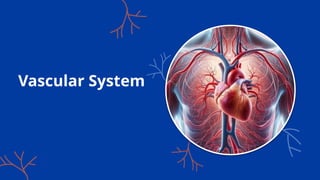
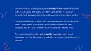
• The cardiovascularsystem contributes to homeostasis of other body systems
by transporting and distributing blood throughout the body to deliver
materials (such as oxygen, nutrients, and hormones) and carry away wastes.
• The structures involved in these important tasks are the blood vessels, which
form a closed system of tubes that carries blood away from the heart,
transports it to the tissues of the body, and then returns it to the heart.
• Three major types of vessels—artery, capillary, and vein—carry blood
throughout the body. Each type of vessel differs in structure, depending on its
function.
23.
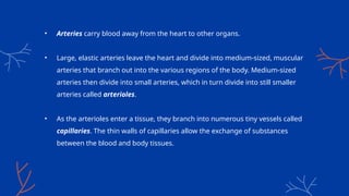
• Arteries carryblood away from the heart to other organs.
• Large, elastic arteries leave the heart and divide into medium-sized, muscular
arteries that branch out into the various regions of the body. Medium-sized
arteries then divide into small arteries, which in turn divide into still smaller
arteries called arterioles.
• As the arterioles enter a tissue, they branch into numerous tiny vessels called
capillaries. The thin walls of capillaries allow the exchange of substances
between the blood and body tissues.
24.
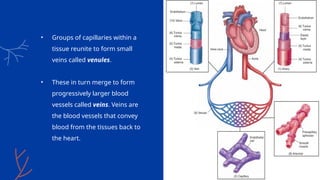
• Groups ofcapillaries within a
tissue reunite to form small
veins called venules.
• These in turn merge to form
progressively larger blood
vessels called veins. Veins are
the blood vessels that convey
blood from the tissues back to
the heart.
25.
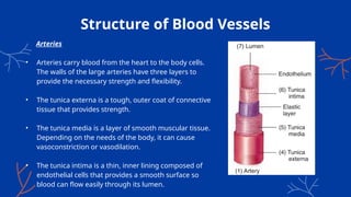
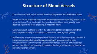
Structure of BloodVessels
Arteries
• Arteries carry blood from the heart to the body cells.
The walls of the large arteries have three layers to
provide the necessary strength and flexibility.
• The tunica externa is a tough, outer coat of connective
tissue that provides strength.
• The tunica media is a layer of smooth muscular tissue.
Depending on the needs of the body, it can cause
vasoconstriction or vasodilation.
• The tunica intima is a thin, inner lining composed of
endothelial cells that provides a smooth surface so
blood can flow easily through its lumen.
26.
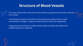
Structure of BloodVessels
• The surge of blood felt in the arteries when blood is pumped from the heart is referred
to as a pulse.
• Arterial blood (except for that found in the pulmonary artery) contains a high
concentration of oxygen. It appears bright red and is said to be oxygenated.
• Oxygenated blood travels to smaller arteries called arterioles and finally to the
smallest vessels, the capillaries.
27.
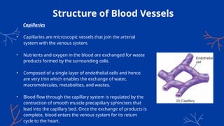
Structure of BloodVessels
Capillaries
• Capillaries are microscopic vessels that join the arterial
system with the venous system.
• Nutrients and oxygen in the blood are exchanged for waste
products formed by the surrounding cells.
• Composed of a single layer of endothelial cells and hence
are very thin which enables the exchange of water,
macromolecules, metabolites, and wastes.
• Blood flow through the capillary system is regulated by the
contraction of smooth muscle precapillary sphincters that
lead into the capillary bed. Once the exchange of products is
complete, blood enters the venous system for its return
cycle to the heart.
28.
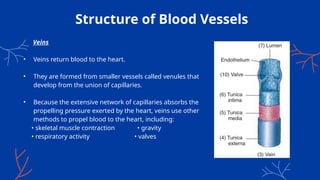
Structure of BloodVessels
Veins
• Veins return blood to the heart.
• They are formed from smaller vessels called venules that
develop from the union of capillaries.
• Because the extensive network of capillaries absorbs the
propelling pressure exerted by the heart, veins use other
methods to propel blood to the heart, including:
• skeletal muscle contraction • gravity
• respiratory activity • valves
29.
Structure of BloodVessels
• The valves are small structures within veins that prevent the backflow of blood.
• Valves are found predominantly in the extremities and are especially important for
returning blood from the legs to the heart because blood must travel a long
distance against the force of gravity to reach the heart.
• Large veins such as those found in the abdomen contain smooth muscle that
contract peristaltically to propel blood toward the heart against gravity.
• Blood carried in the veins (except for the blood in the pulmonary veins) contains a
low concentration of oxygen (deoxygenated) with a corresponding high
concentration of carbon dioxide. Deoxygenated blood takes on a characteristic
purple color. Blood continuously circulates to the lungs so that carbon dioxide can
be exchanged for oxygen.
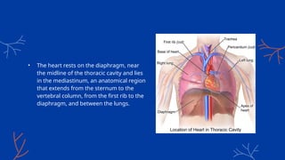
• The heartrests on the diaphragm, near
the midline of the thoracic cavity and lies
in the mediastinum, an anatomical region
that extends from the sternum to the
vertebral column, from the first rib to the
diaphragm, and between the lungs.
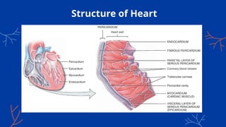
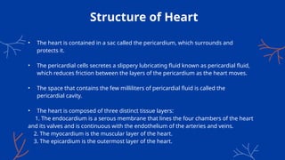
Structure of Heart
•The heart is contained in a sac called the pericardium, which surrounds and
protects it.
• The pericardial cells secretes a slippery lubricating fluid known as pericardial fluid,
which reduces friction between the layers of the pericardium as the heart moves.
• The space that contains the few milliliters of pericardial fluid is called the
pericardial cavity.
• The heart is composed of three distinct tissue layers:
1. The endocardium is a serous membrane that lines the four chambers of the heart
and its valves and is continuous with the endothelium of the arteries and veins.
2. The myocardium is the muscular layer of the heart.
3. The epicardium is the outermost layer of the heart.
34.
Structure of Heart
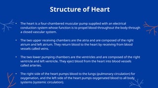
•The heart is a four-chambered muscular pump supplied with an electrical
conduction system whose function is to propel blood throughout the body through
a closed vascular system.
• The two upper receiving chambers are the atria and are composed of the right
atrium and left atrium. They return blood to the heart by receiving from blood
vessels called veins.
• The two lower pumping chambers are the ventricles and are composed of the right
ventricle and left ventricle. They eject blood from the heart into blood vessels
called arteries.
• The right side of the heart pumps blood to the lungs (pulmonary circulation) for
oxygenation, and the left side of the heart pumps oxygenated blood to all body
systems (systemic circulation).
35.
Structure of Heart
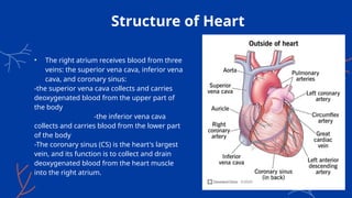
•The right atrium receives blood from three
veins: the superior vena cava, inferior vena
cava, and coronary sinus:
-the superior vena cava collects and carries
deoxygenated blood from the upper part of
the body
-the inferior vena cava
collects and carries blood from the lower part
of the body
-The coronary sinus (CS) is the heart's largest
vein, and its function is to collect and drain
deoxygenated blood from the heart muscle
into the right atrium.
36.
Structure of Heart
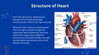
•From the right atrium, blood passes
through the tricuspid valve (right
atrioventricular valve) to the right ventricle.
• When the heart contracts, blood passes
from the right ventricle through the
pulmonary valve (pulmonary semilunar
valve) into a large artery called the
pulmonary trunk, which divides into right
and left pulmonary arteries and carries
blood to the lungs.
37.
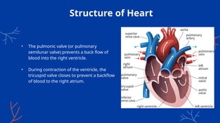
Structure of Heart
•The pulmonic valve (or pulmonary
semilunar valve) prevents a back flow of
blood into the right ventricle.
• During contraction of the ventricle, the
tricuspid valve closes to prevent a backflow
of blood to the right atrium.
38.
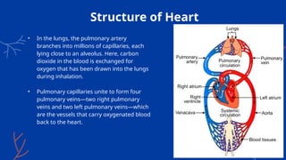
Structure of Heart
•In the lungs, the pulmonary artery
branches into millions of capillaries, each
lying close to an alveolus. Here, carbon
dioxide in the blood is exchanged for
oxygen that has been drawn into the lungs
during inhalation.
• Pulmonary capillaries unite to form four
pulmonary veins—two right pulmonary
veins and two left pulmonary veins—which
are the vessels that carry oxygenated blood
back to the heart.
39.
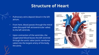
Structure of Heart
•Pulmonary veins deposit blood in the left
atrium.
• From here, blood passes through the mitral
valve (bicuspid/ left atrioventricular valve)
to the left ventricle.
• Upon contraction of the ventricles, the
oxygenated blood leaves the left ventricle
through the aortic valve (aortic semilunar
valve) into the largest artery of the body,
the aorta.
40.
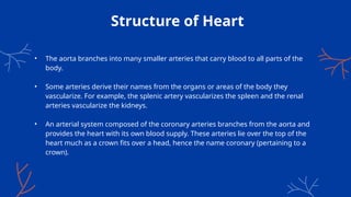
Structure of Heart
•The aorta branches into many smaller arteries that carry blood to all parts of the
body.
• Some arteries derive their names from the organs or areas of the body they
vascularize. For example, the splenic artery vascularizes the spleen and the renal
arteries vascularize the kidneys.
• An arterial system composed of the coronary arteries branches from the aorta and
provides the heart with its own blood supply. These arteries lie over the top of the
heart much as a crown fits over a head, hence the name coronary (pertaining to a
crown).
41.
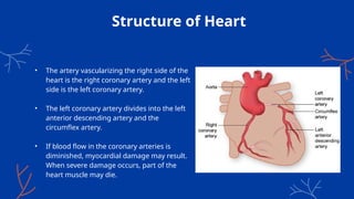
Structure of Heart
•The artery vascularizing the right side of the
heart is the right coronary artery and the left
side is the left coronary artery.
• The left coronary artery divides into the left
anterior descending artery and the
circumflex artery.
• If blood flow in the coronary arteries is
diminished, myocardial damage may result.
When severe damage occurs, part of the
heart muscle may die.
43.
QUIZ
1. Which ofthe following structures is responsible for transporting oxygenated
blood from the lungs to the heart?
A) Pulmonary veins B) Pulmonary arteries
C) Aorta D) Vena cava
2. Which chamber of the heart pumps deoxygenated blood to the lungs?
A) Left ventricle B) Right atrium
C) Right ventricle D) Left atrium
3. Which blood vessel carries oxygenated blood away from the heart to the rest
of
the body?
A) Pulmonary artery B) Vena cava
C) Aorta D) Jugular vein
44.
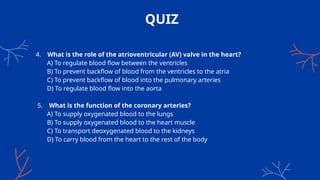
QUIZ
4. What isthe role of the atrioventricular (AV) valve in the heart?
A) To regulate blood flow between the ventricles
B) To prevent backflow of blood from the ventricles to the atria
C) To prevent backflow of blood into the pulmonary arteries
D) To regulate blood flow into the aorta
5. What is the function of the coronary arteries?
A) To supply oxygenated blood to the lungs
B) To supply oxygenated blood to the heart muscle
C) To transport deoxygenated blood to the kidneys
D) To carry blood from the heart to the rest of the body
45.
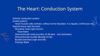
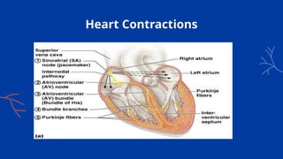
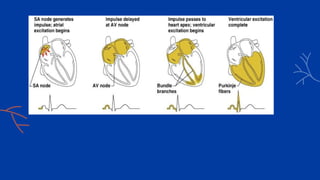
The Heart: ConductionSystem
• Intrinsic conduction system
(nodal system)
• Heart muscle cells contract, without nerve impulses, in a regular, continuous way
• Special tissue sets the pace
• Sinoatrial node (right atrium)
• Pacemaker
• Atrioventricular node (junction of r&l atria and ventricles)
• Atrioventricular bundle (Bundle of His)
• Bundle branches (right and left)
• Purkinje fibers
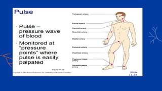
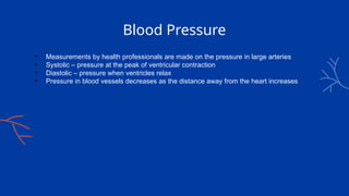
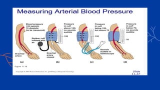
Blood Pressure
• Measurementsby health professionals are made on the pressure in large arteries
• Systolic – pressure at the peak of ventricular contraction
• Diastolic – pressure when ventricles relax
• Pressure in blood vessels decreases as the distance away from the heart increases
51.
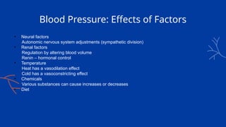
Blood Pressure: Effectsof Factors
• Neural factors
Autonomic nervous system adjustments (sympathetic division)
• Renal factors
Regulation by altering blood volume
Renin – hormonal control
• Temperature
Heat has a vasodilation effect
Cold has a vasoconstricting effect
• Chemicals
Various substances can cause increases or decreases
• Diet
52.
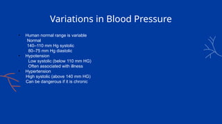
Variations in BloodPressure
• Human normal range is variable
Normal
140–110 mm Hg systolic
80–75 mm Hg diastolic
• Hypotension
Low systolic (below 110 mm HG)
Often associated with illness
• Hypertension
High systolic (above 140 mm HG)
Can be dangerous if it is chronic
53.
Reference
1. Gylys BA,Mary Ellen Wedding. Medical terminology systems : a body systems approach.
Philadelphia: F.A. Davis; 2005.
2. Tortora GJ, Derrickson B. Principles of anatomy and physiology. 12th ed. Hoboken, N.J.: Wiley;
2009.
3. https://en.wikipedia.org/wiki/Circulatory_system#Structure
4. https://en.wikipedia.org/wiki/Myocardial_infarction?form=MG0AV3
5. Elaine N. Marieb.Essentials of Human Anatomy & Physiology.Seventh Edition. 11
Chapter The Cardiovascular System
#25 Because blood is propelled thorough the arteries by the pumping action of the heart, the walls of the arteries must be able to withstand the surge of blood that results from each contraction of the heart.
vasoconstriction (reduction of the lumen diameter caused by smooth muscle contraction) or vasodilation (widening of the lumen caused by smooth muscle relax ation)


![anatomy presentation [Autosaved]thesis.pptx](https://cdn.slidesharecdn.com/ss_thumbnails/anatomypresentationautosaved-250224130828-3fb23976-thumbnail.jpg?width=640&height=640&fit=bounds)


![CTEV [ clubfoot] DR ARUN LAL ,DR MOHAMED ASHRAF travancore medical college k...](https://cdn.slidesharecdn.com/ss_thumbnails/ctevclubfootdrarunlaldrmohamedashraftravancoremedicalcollegekollamkeralaindia-260208063247-18fc466c-thumbnail.jpg?width=640&height=640&fit=bounds)


