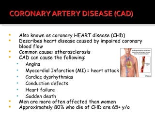
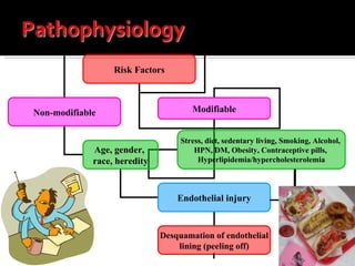
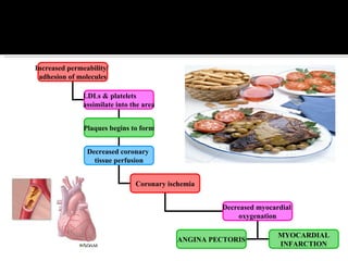
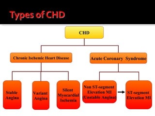
Management of Clients  with Functional Cardiac Disorders
Also known as coronary HEART disease (CHD) Describes heart disease caused by impaired coronary blood flow Common cause: atherosclerosis CAD can cause the following: Angina Myocardial Infarction (MI) = heart attack Cardiac dysrhythmias Conduction defects Heart failure Sudden death Men are more often affected than women Approximately 80% who die of CHD are 65+ y/o
Risk Factors Non-modifiable Modifiable Age, gender,  race, heredity Endothelial injury Stress, diet, sedentary living, Smoking, Alcohol,  HPN,   DM, Obesity, Contraceptive pills,  Hyperlipidemia/hypercholesterolemia Desquamation of endothelial lining (peeling off)
Increased permeability/  adhesion of molecules LDLs & platelets assimilate into the area Plaques begins to form Decreased coronary  tissue perfusion Coronary ischemia Decreased myocardial  oxygenation ANGINA PECTORIS MYOCARDIAL INFARCTION
Inspection: Skin color Neck vein distention (jugular vein) Respiration Peripheral edema Palpation: Peripheral pulses
Auscultation: Heart sounds (presence of S 3  in adults & S 4 ) Murmurs – audible vibrations of the heart & great vessels produced by turbulent blood flow Pericardial friction rub – extra heart sound originating from the pericardial sac  - may be a sign of inflammation, infection, or infiltration - described as a short, high-pitched scratchy sound
Dyspnea  Dyspnea on exertion – may indicate decreased cardiac reserve Orthopnea – a symptom of more advanced heart failure Paroxysmal nocturnal dyspnea – severe SOB that usually occurs 2-5hrs after onset of sleep Chest Pain – may be due to decreased coronary tissue perfusion or compression & irritation of nerve endings Edema – increased hydrostatic pressure in venous system causes shifting of plasma resulting to interstitial fluid accumulation Syncope – due to decreased cerebral tissue perfusion Palpitations Fatigue
ECG (Electrocardiography) – graphical recording of the heart’s electrical activities; 1 st  diagnostic test done when cardiovascular disorder is suspected Waves: P wave – atrial depolarization (contraction/stimulation)  QRS complex – ventricular depolarization (changes are irreversible) ST segment – ventricular repolarization (changes are reversible) U wave – hypokalemia  PR interval (time for impulse to travel) = 0.12-0.20s (3-5 squares) √ for AV block QRS = 0.10s or (<2squares) √ for electrolyte &/or ventricular imbalance
Abnormalities:  absent P wave = atrial fibrillation saw-tooth pattern = atrial flutter elevated ST segment = MI 3rd degree heart block = prolonged PR then progressively prolonged
 
 
Cardiac Enzymes (Cardiac Markers): 1 st :  Myoglobin a. urine  = 0 – 2mg/dL  (↑within 30mins – 2hrs after MI) b. blood = <70mg/dL 2 nd : Troponin* - regulates calcium-mediated contractile process released during MI (Troponin T & I)    - blood = <0.6mg/dL - ↑ within 3-6hrs after MI & remains elevated for 21 days upon onset of attack 3 rd : Creatinine kinase (CK) – intracellular enzymes found in muscles converting ATP to ADP   CK-MB – specific to myocardial tissue  (↑within 4-6hrs & decreases to normal within 2-3days)  male = 12-70 mg/dL female = 10-55 mg/dL  4 th : LDH (specifically LDH 1 - most sensitive indicator of myocardial damage) = 45-90mg/dL - ↑within 3-4 days & remains elevated for 14 days
Stress Test / Treadmill Test (Treadmill Stress Test) – ECG monitoring during a series of activities of patient on a treadmill Purposes: identify ischemic heart disease evaluate patients with chest pain evaluate effectiveness of therapy develop appropriate fitness program Instructions to patient: get adequate sleep prio r   to test - avoid: caffeinated beverages, tea, alcohol, on the day before until the test day - wear comfortable, loose-fitting clothes & rubber-soled shoes on the test day - light breakfast on the day of the test - inform physician of any unusual sensations during the test - rest after the test
Pharmacologic Stress Test – use of intravenous injection of pharmacologic vasodilator (dipyridamole, adenosine, or dobutamine) in combination of radionuclide myocardial imaging To evaluate presence of significant CHD for patients contraindicated in TST Dipyradamole blocks cellular re-absorption of adenosine (endogenous vasodilator) & increases coronary blood flow 3-5x above baseline levels If with CHD, the resistance vessels distal to the stenosis already are maximally dilated to maintain normal resting flow, thus, further vasodilatation does not produce increased blood flow Dobutamine – used in patients with bronchospastic pulmonary disease   - increases myocardial O 2  demand by increasing cardiac contractility, HR, & BP
Cardiac Catheterization – involves passage of  flexible catheters  into great vessels & heart chambers under local  anesthesia - lab is equipped for viewing & recording fluoroscopic images & for measuring pressures in the heart & great vessels, cardiac output studies, & for obtaining ABG samples - Epinephrine – to counteract possible allergic reactions Right heart Catheterization – catheter inserted into peripheral veins (basilic or femoral) then advanced into the right heart Left heart Catheterization – catheter inserted retrograde through peripheral artery (brachial or femoral) into the aorta & left heart Coronary Angiogram – injection of radiographic contrast medium into the heart so that an outline of moving structures are visualized & filmed Coronary Arteriography - injection of radiographic contrast medium into the coronary arteries permits visualization of lesions in these vessels
 
Before Procedure: Check consent  form √  for allergies to  seafood &  iodine  NPO post midnight Baseline V/S Explain that warm or flushing sensation  may be  felt upon administr ation   of  the dye; “fluttering” sensation may be felt as catheter enters the heart Administer sedatives as ordered Have the client void prior to transport to cath lab After Procedure: Bed rest – upper extremity catheter = until stable v/s, HOB not more than 30 °   - lower extremity = 24hrs, flat on bed for 6hrs Apply pressure (5lb-sand bag) over puncture site & monitor for bleeding Monitor v/s q15 for 1 st  2hrs then q1 until stable v/s, esp. peripheral pulses Immobilize affected extremity in extension for adequate circulation Monitor for color & temperature changes of extremities Instruct client to report tingling sensations
Swan-Ganz Catheterization – to determine & monitor cardiovascular status; inserted via antecubital vein into the right side of the heart & is floated into the pulmonary artery 4 lumens:   1. CVP – specific to right heart  RA = 0-12  RV = 5-12  Indications: increased CVP = heart failure -decreased CVP = hypovolemia 2. Pulmonary pressures: PAP (pulmonary artery pressure) = 20-30mmHg PCWP (pulmonary capillary wedge pressure) = 8-13mmHg (√ for pulmonary edema) 3. Specimen collection tube – also used for administering meds 4. Balloon
Echocardiography – uses ultrasound to assess cardiac structure & mobility Doppler U/S – to detect blood flow of artery & vein specifically of lower extremities (No smoking 1hr before the test)  Holter Monitoring – portable 24hr ECG monitoring which attempts to assess activities which precipitate dysrhythmias & its time of the day MRI – magnetic fields & radiowaves are used to detect & define abnormalities in tissues (aorta, tumors, cardiomyopathy, pericardiac disease) - shows actual beating & blood flow; image over 3 spatial dimensions Secure consent Assess for claustrophobia Remove metal items (jewelries, eyeglasses) Instruct client to remain still during the entire procedure Inform client of the duration (45-60mins) CI: clients with pacemakers, prosthetic valves, recently implanted clips or wires
CHD Chronic Ischemic Heart Disease Acute Coronary  Syndrome Stable  Angina Variant  Angina Silent  Myocardial Ischemia Non ST-segment  Elevation MI (Unstable Angina) ST-segment  Elevation MI
Ischemia – suppressed blood flow Angina – to choke Occurs when blood supply is inadequate to meet the heart’s metabolic demands Symptomatic paroxysmal chest pain or pressure sensation associated with transient ischemia
Causes: Atherosclerosis, HPN, DM, Buerger’s Disease, Polycythemia Vera, Aortic regurgitation Reduced coronary tissue perfusion Decreased myocardial oxygenation Anaerobic metabolism Increased lactic acid production (lactic acidosis) Chest pain
Stable angina – the common initial manifestation of  a heart disease Common cause: atherosclerosis (although those with advance atherosclerosis do not develop angina) Pain is precipitated by increased work demands of the heart (i.e.. physical exertion, exposure to cold, & emotional stress) Pain location: precordial or substernal chest area Pain characteristics:  con stricting, squeezing, or suffocating sensation Usua lly steady, increasing in intensity only at the onset &  end  of attack May  radiate to left shoulder, arm, jaw, or other chest areas Dura tion: < 15mins Relie ved by rest (preferably sitting or standing with support) or by use of  NTG
Variant/Vasospastic Angina (Prinzmetal Angina) 1 st  described by Prinzmetal & Associates in 1659 Cause: spasm of coronary arteries (vasospasm) due to coronary artery stenosis Mechanism is uncertain (may be from hyperactive sympathetic responses, mishandling defects of calcium in smooth vascular muscles, reduced prostaglandin I 2  production) Pain Characteristics: occurs during rest or with minimal exercise - commonly follows a cyclic or regular pattern of occurrence (i.e.. Same time each day usually at early hours) If client is for cardiac cath, Ergonovine (nonspecific vasoconstrictor) may be administered to evoke anginal attack & demonstrate the presence & location of spasm
Nocturnal Angina -  frequently occurs  nocturnally (may be  associated with  REM stage of sleep) Angina Decubitus – paroxysmal chest pain occurs when client sits or stands up Post-infarction Angina – occurs after MI when residual ischemia may cause episodes of angina
Dx: detailed pain history, ECG, TST,  angiogram  may be used to confirm & describe type of angina Tx: directed towards MI prevention\ Lifestyle modification (individualized regular exercise program, smoking cess a tion) Stress reduction Diet changes Avoidance of cold PTCA (percutaneous transluminal coronary angioplasty) may be indicated if with severe artery  occlusion
Nitroglycerin (NTGs) – vasodilators:  patch (Deponit, Transderm-NTG) sublingual (Nitrostat) oral (Nitroglyn) IV (Nitro-Bid) Β -adrenergic blockers:  Propanolol (Inderal) Atenolol (Tenormin) Metoprolol (Lopressor) Calcium channel blockers:  Nifedipine (Calcibloc, Adalat) Diltiazem (Cardizem) Lipid lowering agents –statins:  Simvastatin Anti-coagulants:  ASA (Aspirin) Heparin sodium Warfarin (Coumadin)
Class I – angina  occurs with strenuous, rapid, or  prolonged  exertion at work or recreation Class II – angina  occurs on walking or going up the stairs rapidly or after meals, walking uphill, walking more than 2 blocks on the level or going more than 1 flight of ordinary stairs at normal pace, under emotional stress, or in cold Class III – angina occurs on walking 1-2 blocks on the level or going 1 flight of ordinary stairs at normal pace Class IV – angina occurs even at rest
Diet instructions  (low salt, low fat,  low cholesterol , high fiber); avoid animal fats E.g.. White meat – chicken w/o skin, fish Stop smoking & avoid alcohol Activity restrictions are placed within client’s limitations NTGs – max of 3doses at 5-min intervals Stinging sensation under the tongue for SL is normal Advise clients to always carry 3 tablets Store meds in cool, dry place, air-tight amber bottles & change stocks every 6months Inform clients that headache, dizziness, flushed face are common side effects.  Do not discontinue the drug. For patches, rotate skin sites usually on chest wall Instruct on evaluation of effectiveness based on pain relief Propanolols causes bronchospasm & hypoglycemia, do not administer to asthmatic & diabetic clients Heparin – monitor bleeding tendencies (avoid punctu res , use of soft-bristled toot hbrush ); monitor PTT levels; use d   for  2wks max; do not  massage if via  SC; have protamine  sulfate  available Coumadin – monitor for bleeding & PT; always  have  vit K readily available (avoid green leafy  veggies)
Unstab le Angina/Non ST-Segment Elevation MI –  a clinical   syndro me of myocardial ischemia Causes: atherosclerotic plaque disruption or significant CHD, cocaine use (risk factor) Defining guidelines: (3 presentations) Symptoms at rest (usually prolonged, i.e.. >20mins) New onset exertional angina (increased in severity of at least 1 class – to at least class III) in <2months Recent acceleration of angina to at least class III in <2months Dx: based on pain severity & presenting sympto ms , ECG findings & serum cardiac markers When chest pain has been unremitting for >20mins, possibility of ST-Segment Elevation MI is usually considered
ST-Segment Elevation MI (Heart Attack) Characterized by ischemic death of myocardial  tis sue associated with atherosclerotic disease of coro nar y arteries Area of infarction is determined by the affected coronary artery & its distribution of blood flow (right coronary artery, left anterior descending artery, left circumflex artery) Dx: based on presenting S/Sx, serum markers, & ECG (changes may not be present immediately after symptoms except dysrhythmias; PVCs/premature ventricular contractions are common after MI) Typical ECG changes: ST-segment elevation, Q wave prolongation, T wave inversion
Manifestations:  chest pain – severe crushing, constricting, “someone sitting on my chest” - substernal radiating to left arm, neck or jaw - prolonged (>35mins) & not relieved by rest Shortness of breath, profuse perspiration Feeling of impending doom Complications: death (usually within 1 hr of onset) Heart fail ure & cardiogenic shock – profound LV failure from  massive MI  resulting to low cardiac output Thromboe mboli – leads to immobility & impaired cardiac function  contributi ng to blood stasis in veins Rupture  of myocardium Ventricul ar aneurysms – decreases pumping efficiency of heart &  increase s  work of LV
Causes: atherosclerotic heart disease, thrombosis/embolism,  shock &/or hemorrhage, direct trauma Myocardial ischemia ↑ cellular  hypoxia ↓ myocardial  O 2  supply ↓  myocardial contractility ↓ cardiac output ↓ arterial pressure Stimulation of  sympathetic receptors ↑ peripheral  vasoconstriction ↑  myocardial  contractility ↑  afterload ↑ myocardial  O 2  demand   ↑  HR ↑ diastolic filling  ↓ myocardial  tissue perfusion
Time after Onset Type of Injury & Gross Tissue Changes 0-0.5hrs Reversible injury 1-2hrs Onset of irreversible injury 4-12hrs Beginning of coagulation necrosis 18-24hrs Continued necrosis; gross pallor of infected tissue 1-3days Total necrosis; onset of acute inflammatory process 3-7days Infarcted area becomes soft with a yellow-brown center & hyperemic edges 7-10days Minimally soft & yellow with vascularized edges; scar tissue generation begins (fibroplastic activity) 8 th  week Complete scar tissue replacement
Initial  Management:  OMEN   -  O 2  therapy via nasal prongs  - adequate analgesia ( M orphine via IV – also has vasodilator property) -  E CG monitoring -sublingual  N TG (unless contraindicated; IV may be given to limit infarction size & most effective if given within 4hrs of onset) Thrombolytic Therapy – best results occur if initiated within 60-90mins of onset (Streptokinase & Urokinase – promote conversion of plasminogen to plasmin) Anti-arrhythmics: lidocaine, atropine, propano lol Anticoagulants & antiplatelets: ASA, heparin Stool softeners
Surgery : Revascularization PTCA Coronary stent implantation Coronary Artery Bypass Graft (CABG) – no response to medical treatment & PTCA Resection – aneurysm
 
Promote oxygenation & tissue perfusion (place client on semi-fowler’s, O 2  via nasal cannula, monitor v/s changes, remind client on his activity limitations & restrictions) Promote comfort & rest Monitor the ff perimeters: v/s, ECG, rate & rhythm of pulse, effects of ADLs on cardiac status Diet: low salt, low cholesterol, low calories, avoid alcohol & smoking Take prescribe meds at regular basis Stress management  Resume sexual activity after 4-6wks from discharge or when client can go up 2 flights of stairs without difficulty Assume less tiring position (non-MI partner takes active role). Perform sexual activity in a cool, familiar place. Take prescribed NTG before sexual activity Refrain from sexual activity after a large meal or during a tiring day. Moderation should be observed if palpitations, dizziness or dyspnea is observed
 
Also known as Thromboangiitis obliterans Usually a disease of heavy cigarette smoker/tobacco user men, 25-40y/o Inflammatory arterial disorder that causes thrombus formation often extends to adjacent veins & nerves Affects medium-sized arteries (usually plantar & digital vessels in the foot or lower legs) unknown pathogenesis but it had been suggested that: tobacco may trigger an immune response or  unmask a clotting defect;  ->  these 2 can incite an inflammatory reaction of the vessel wall
Pain –  predominant symptom; R/T distal arterial i schemia Intermittent claudication in the arch of foot & digits Increased sensitivity to cold (due to impaired circulation Absent/diminished peripheral pulses Color changes in extremity (cyanotic on dependent position; digits may turn reddish blue) Thick malformed nails (chronic ischemia) Disease progression ulcerate tissues & gangrenous changes may arise; may necessitate amputation
Diagnostic methods – those that assess blood flow (Doppler ultrasound & MRI) Tx: mandatory to stop smoking or using tobacco Meds to increase blood flow to extremities Surgery (surgical sympathectomy) amputation
Mechanism: intensive  vasospasm of arteries &  arterioles in the   fi ngers Cause: unknown Usually affects young women Precipitated by exposure to cold &  strong  emotions Raynaud’s phenomenon – associated with previous injury (i.e.. Frostbite, occupation al  trauma associated with use of heavy vibr ating  tools, collagen diseases, neuro d/o, chro nic  arterial occlusive d/o)
Period of ischemia (ischemia due to vasospasm)  change in skin color = pallor to cyanotic 1 st  noticed at the fingertips later moving to distal phalanges Cold sensation Sensory perception changes (numbness & tingling) Period of hyperemia – intense redness Throbbing Paresthesia  Return to normal color Note: although all of the fingers are affected symmetrically, only 1-2digits may be involved Severe cases: arthritis may arise (due to nutritional impairment) Brittle nails Thickening of the skin of fingertips Ulceration & superficial gangrene of fingers (rare occasions)
Dx: initial = based on Hx of vasospastic attacks Immersion of hand in cold water to initiate attack aids in the Dx Doppler flow velocimetry – used to quantify blood flow during temperature changes Serial Computed thermography (finger skin temp) – for diagnosing the extent of disease Tx: directed towards eliminating factors causing vasospasm & protecting fingers from injury during ischemic attacks PRIORITIES: Abstinence in smoking & protection from cold  Avoidance of emotional stress (anxiety & stress  may  precipitate vascular spasm) Meds: avoid vasoconstrictors (i.e..  Decongestants) -Calcium channel blockers (Diltiazem, Nifedip ine , Nicardipine) – decrease episodes of attacks
Assessment: Hx of symptoms (pain, esp. chest pain; palpitations; dyspnea) v/s Nursing Dx:  ineffective tissue perfusion  (cardio pulmonary) Impaired  gas exchange Anxiety  due to fear of  death  (clients with MI  or  An gina) Goals: Relief of pain & symptoms Prevention of further cardiac damage Nursing Interventions: Pain control Proper medications Decrease client’s anxiety Health teachings (meds, activities, diet, exercise, etc)
Thank You for Listening!

Cardio2

  • 1.
    Management of Clients with Functional Cardiac Disorders
  • 2.
    Also known ascoronary HEART disease (CHD) Describes heart disease caused by impaired coronary blood flow Common cause: atherosclerosis CAD can cause the following: Angina Myocardial Infarction (MI) = heart attack Cardiac dysrhythmias Conduction defects Heart failure Sudden death Men are more often affected than women Approximately 80% who die of CHD are 65+ y/o
  • 3.
    Risk Factors Non-modifiableModifiable Age, gender, race, heredity Endothelial injury Stress, diet, sedentary living, Smoking, Alcohol, HPN, DM, Obesity, Contraceptive pills, Hyperlipidemia/hypercholesterolemia Desquamation of endothelial lining (peeling off)
  • 4.
    Increased permeability/ adhesion of molecules LDLs & platelets assimilate into the area Plaques begins to form Decreased coronary tissue perfusion Coronary ischemia Decreased myocardial oxygenation ANGINA PECTORIS MYOCARDIAL INFARCTION
  • 5.
    Inspection: Skin colorNeck vein distention (jugular vein) Respiration Peripheral edema Palpation: Peripheral pulses
  • 6.
    Auscultation: Heart sounds(presence of S 3 in adults & S 4 ) Murmurs – audible vibrations of the heart & great vessels produced by turbulent blood flow Pericardial friction rub – extra heart sound originating from the pericardial sac - may be a sign of inflammation, infection, or infiltration - described as a short, high-pitched scratchy sound
  • 7.
    Dyspnea Dyspneaon exertion – may indicate decreased cardiac reserve Orthopnea – a symptom of more advanced heart failure Paroxysmal nocturnal dyspnea – severe SOB that usually occurs 2-5hrs after onset of sleep Chest Pain – may be due to decreased coronary tissue perfusion or compression & irritation of nerve endings Edema – increased hydrostatic pressure in venous system causes shifting of plasma resulting to interstitial fluid accumulation Syncope – due to decreased cerebral tissue perfusion Palpitations Fatigue
  • 8.
    ECG (Electrocardiography) –graphical recording of the heart’s electrical activities; 1 st diagnostic test done when cardiovascular disorder is suspected Waves: P wave – atrial depolarization (contraction/stimulation) QRS complex – ventricular depolarization (changes are irreversible) ST segment – ventricular repolarization (changes are reversible) U wave – hypokalemia PR interval (time for impulse to travel) = 0.12-0.20s (3-5 squares) √ for AV block QRS = 0.10s or (<2squares) √ for electrolyte &/or ventricular imbalance
  • 9.
    Abnormalities: absentP wave = atrial fibrillation saw-tooth pattern = atrial flutter elevated ST segment = MI 3rd degree heart block = prolonged PR then progressively prolonged
  • 10.
  • 11.
  • 12.
    Cardiac Enzymes (CardiacMarkers): 1 st : Myoglobin a. urine = 0 – 2mg/dL (↑within 30mins – 2hrs after MI) b. blood = <70mg/dL 2 nd : Troponin* - regulates calcium-mediated contractile process released during MI (Troponin T & I) - blood = <0.6mg/dL - ↑ within 3-6hrs after MI & remains elevated for 21 days upon onset of attack 3 rd : Creatinine kinase (CK) – intracellular enzymes found in muscles converting ATP to ADP CK-MB – specific to myocardial tissue (↑within 4-6hrs & decreases to normal within 2-3days) male = 12-70 mg/dL female = 10-55 mg/dL 4 th : LDH (specifically LDH 1 - most sensitive indicator of myocardial damage) = 45-90mg/dL - ↑within 3-4 days & remains elevated for 14 days
  • 13.
    Stress Test /Treadmill Test (Treadmill Stress Test) – ECG monitoring during a series of activities of patient on a treadmill Purposes: identify ischemic heart disease evaluate patients with chest pain evaluate effectiveness of therapy develop appropriate fitness program Instructions to patient: get adequate sleep prio r to test - avoid: caffeinated beverages, tea, alcohol, on the day before until the test day - wear comfortable, loose-fitting clothes & rubber-soled shoes on the test day - light breakfast on the day of the test - inform physician of any unusual sensations during the test - rest after the test
  • 14.
    Pharmacologic Stress Test– use of intravenous injection of pharmacologic vasodilator (dipyridamole, adenosine, or dobutamine) in combination of radionuclide myocardial imaging To evaluate presence of significant CHD for patients contraindicated in TST Dipyradamole blocks cellular re-absorption of adenosine (endogenous vasodilator) & increases coronary blood flow 3-5x above baseline levels If with CHD, the resistance vessels distal to the stenosis already are maximally dilated to maintain normal resting flow, thus, further vasodilatation does not produce increased blood flow Dobutamine – used in patients with bronchospastic pulmonary disease - increases myocardial O 2 demand by increasing cardiac contractility, HR, & BP
  • 15.
    Cardiac Catheterization –involves passage of flexible catheters into great vessels & heart chambers under local anesthesia - lab is equipped for viewing & recording fluoroscopic images & for measuring pressures in the heart & great vessels, cardiac output studies, & for obtaining ABG samples - Epinephrine – to counteract possible allergic reactions Right heart Catheterization – catheter inserted into peripheral veins (basilic or femoral) then advanced into the right heart Left heart Catheterization – catheter inserted retrograde through peripheral artery (brachial or femoral) into the aorta & left heart Coronary Angiogram – injection of radiographic contrast medium into the heart so that an outline of moving structures are visualized & filmed Coronary Arteriography - injection of radiographic contrast medium into the coronary arteries permits visualization of lesions in these vessels
  • 16.
  • 17.
    Before Procedure: Checkconsent form √ for allergies to seafood & iodine NPO post midnight Baseline V/S Explain that warm or flushing sensation may be felt upon administr ation of the dye; “fluttering” sensation may be felt as catheter enters the heart Administer sedatives as ordered Have the client void prior to transport to cath lab After Procedure: Bed rest – upper extremity catheter = until stable v/s, HOB not more than 30 ° - lower extremity = 24hrs, flat on bed for 6hrs Apply pressure (5lb-sand bag) over puncture site & monitor for bleeding Monitor v/s q15 for 1 st 2hrs then q1 until stable v/s, esp. peripheral pulses Immobilize affected extremity in extension for adequate circulation Monitor for color & temperature changes of extremities Instruct client to report tingling sensations
  • 18.
    Swan-Ganz Catheterization –to determine & monitor cardiovascular status; inserted via antecubital vein into the right side of the heart & is floated into the pulmonary artery 4 lumens: 1. CVP – specific to right heart RA = 0-12 RV = 5-12 Indications: increased CVP = heart failure -decreased CVP = hypovolemia 2. Pulmonary pressures: PAP (pulmonary artery pressure) = 20-30mmHg PCWP (pulmonary capillary wedge pressure) = 8-13mmHg (√ for pulmonary edema) 3. Specimen collection tube – also used for administering meds 4. Balloon
  • 19.
    Echocardiography – usesultrasound to assess cardiac structure & mobility Doppler U/S – to detect blood flow of artery & vein specifically of lower extremities (No smoking 1hr before the test) Holter Monitoring – portable 24hr ECG monitoring which attempts to assess activities which precipitate dysrhythmias & its time of the day MRI – magnetic fields & radiowaves are used to detect & define abnormalities in tissues (aorta, tumors, cardiomyopathy, pericardiac disease) - shows actual beating & blood flow; image over 3 spatial dimensions Secure consent Assess for claustrophobia Remove metal items (jewelries, eyeglasses) Instruct client to remain still during the entire procedure Inform client of the duration (45-60mins) CI: clients with pacemakers, prosthetic valves, recently implanted clips or wires
  • 20.
    CHD Chronic IschemicHeart Disease Acute Coronary Syndrome Stable Angina Variant Angina Silent Myocardial Ischemia Non ST-segment Elevation MI (Unstable Angina) ST-segment Elevation MI
  • 21.
    Ischemia – suppressedblood flow Angina – to choke Occurs when blood supply is inadequate to meet the heart’s metabolic demands Symptomatic paroxysmal chest pain or pressure sensation associated with transient ischemia
  • 22.
    Causes: Atherosclerosis, HPN,DM, Buerger’s Disease, Polycythemia Vera, Aortic regurgitation Reduced coronary tissue perfusion Decreased myocardial oxygenation Anaerobic metabolism Increased lactic acid production (lactic acidosis) Chest pain
  • 23.
    Stable angina –the common initial manifestation of a heart disease Common cause: atherosclerosis (although those with advance atherosclerosis do not develop angina) Pain is precipitated by increased work demands of the heart (i.e.. physical exertion, exposure to cold, & emotional stress) Pain location: precordial or substernal chest area Pain characteristics: con stricting, squeezing, or suffocating sensation Usua lly steady, increasing in intensity only at the onset & end of attack May radiate to left shoulder, arm, jaw, or other chest areas Dura tion: < 15mins Relie ved by rest (preferably sitting or standing with support) or by use of NTG
  • 24.
    Variant/Vasospastic Angina (PrinzmetalAngina) 1 st described by Prinzmetal & Associates in 1659 Cause: spasm of coronary arteries (vasospasm) due to coronary artery stenosis Mechanism is uncertain (may be from hyperactive sympathetic responses, mishandling defects of calcium in smooth vascular muscles, reduced prostaglandin I 2 production) Pain Characteristics: occurs during rest or with minimal exercise - commonly follows a cyclic or regular pattern of occurrence (i.e.. Same time each day usually at early hours) If client is for cardiac cath, Ergonovine (nonspecific vasoconstrictor) may be administered to evoke anginal attack & demonstrate the presence & location of spasm
  • 25.
    Nocturnal Angina - frequently occurs nocturnally (may be associated with REM stage of sleep) Angina Decubitus – paroxysmal chest pain occurs when client sits or stands up Post-infarction Angina – occurs after MI when residual ischemia may cause episodes of angina
  • 26.
    Dx: detailed painhistory, ECG, TST, angiogram may be used to confirm & describe type of angina Tx: directed towards MI prevention\ Lifestyle modification (individualized regular exercise program, smoking cess a tion) Stress reduction Diet changes Avoidance of cold PTCA (percutaneous transluminal coronary angioplasty) may be indicated if with severe artery occlusion
  • 27.
    Nitroglycerin (NTGs) –vasodilators: patch (Deponit, Transderm-NTG) sublingual (Nitrostat) oral (Nitroglyn) IV (Nitro-Bid) Β -adrenergic blockers: Propanolol (Inderal) Atenolol (Tenormin) Metoprolol (Lopressor) Calcium channel blockers: Nifedipine (Calcibloc, Adalat) Diltiazem (Cardizem) Lipid lowering agents –statins: Simvastatin Anti-coagulants: ASA (Aspirin) Heparin sodium Warfarin (Coumadin)
  • 28.
    Class I –angina occurs with strenuous, rapid, or prolonged exertion at work or recreation Class II – angina occurs on walking or going up the stairs rapidly or after meals, walking uphill, walking more than 2 blocks on the level or going more than 1 flight of ordinary stairs at normal pace, under emotional stress, or in cold Class III – angina occurs on walking 1-2 blocks on the level or going 1 flight of ordinary stairs at normal pace Class IV – angina occurs even at rest
  • 29.
    Diet instructions (low salt, low fat, low cholesterol , high fiber); avoid animal fats E.g.. White meat – chicken w/o skin, fish Stop smoking & avoid alcohol Activity restrictions are placed within client’s limitations NTGs – max of 3doses at 5-min intervals Stinging sensation under the tongue for SL is normal Advise clients to always carry 3 tablets Store meds in cool, dry place, air-tight amber bottles & change stocks every 6months Inform clients that headache, dizziness, flushed face are common side effects. Do not discontinue the drug. For patches, rotate skin sites usually on chest wall Instruct on evaluation of effectiveness based on pain relief Propanolols causes bronchospasm & hypoglycemia, do not administer to asthmatic & diabetic clients Heparin – monitor bleeding tendencies (avoid punctu res , use of soft-bristled toot hbrush ); monitor PTT levels; use d for 2wks max; do not massage if via SC; have protamine sulfate available Coumadin – monitor for bleeding & PT; always have vit K readily available (avoid green leafy veggies)
  • 30.
    Unstab le Angina/NonST-Segment Elevation MI – a clinical syndro me of myocardial ischemia Causes: atherosclerotic plaque disruption or significant CHD, cocaine use (risk factor) Defining guidelines: (3 presentations) Symptoms at rest (usually prolonged, i.e.. >20mins) New onset exertional angina (increased in severity of at least 1 class – to at least class III) in <2months Recent acceleration of angina to at least class III in <2months Dx: based on pain severity & presenting sympto ms , ECG findings & serum cardiac markers When chest pain has been unremitting for >20mins, possibility of ST-Segment Elevation MI is usually considered
  • 31.
    ST-Segment Elevation MI(Heart Attack) Characterized by ischemic death of myocardial tis sue associated with atherosclerotic disease of coro nar y arteries Area of infarction is determined by the affected coronary artery & its distribution of blood flow (right coronary artery, left anterior descending artery, left circumflex artery) Dx: based on presenting S/Sx, serum markers, & ECG (changes may not be present immediately after symptoms except dysrhythmias; PVCs/premature ventricular contractions are common after MI) Typical ECG changes: ST-segment elevation, Q wave prolongation, T wave inversion
  • 32.
    Manifestations: chestpain – severe crushing, constricting, “someone sitting on my chest” - substernal radiating to left arm, neck or jaw - prolonged (>35mins) & not relieved by rest Shortness of breath, profuse perspiration Feeling of impending doom Complications: death (usually within 1 hr of onset) Heart fail ure & cardiogenic shock – profound LV failure from massive MI resulting to low cardiac output Thromboe mboli – leads to immobility & impaired cardiac function contributi ng to blood stasis in veins Rupture of myocardium Ventricul ar aneurysms – decreases pumping efficiency of heart & increase s work of LV
  • 33.
    Causes: atherosclerotic heartdisease, thrombosis/embolism, shock &/or hemorrhage, direct trauma Myocardial ischemia ↑ cellular hypoxia ↓ myocardial O 2 supply ↓ myocardial contractility ↓ cardiac output ↓ arterial pressure Stimulation of sympathetic receptors ↑ peripheral vasoconstriction ↑ myocardial contractility ↑ afterload ↑ myocardial O 2 demand ↑ HR ↑ diastolic filling ↓ myocardial tissue perfusion
  • 34.
    Time after OnsetType of Injury & Gross Tissue Changes 0-0.5hrs Reversible injury 1-2hrs Onset of irreversible injury 4-12hrs Beginning of coagulation necrosis 18-24hrs Continued necrosis; gross pallor of infected tissue 1-3days Total necrosis; onset of acute inflammatory process 3-7days Infarcted area becomes soft with a yellow-brown center & hyperemic edges 7-10days Minimally soft & yellow with vascularized edges; scar tissue generation begins (fibroplastic activity) 8 th week Complete scar tissue replacement
  • 35.
    Initial Management: OMEN - O 2 therapy via nasal prongs - adequate analgesia ( M orphine via IV – also has vasodilator property) - E CG monitoring -sublingual N TG (unless contraindicated; IV may be given to limit infarction size & most effective if given within 4hrs of onset) Thrombolytic Therapy – best results occur if initiated within 60-90mins of onset (Streptokinase & Urokinase – promote conversion of plasminogen to plasmin) Anti-arrhythmics: lidocaine, atropine, propano lol Anticoagulants & antiplatelets: ASA, heparin Stool softeners
  • 36.
    Surgery : RevascularizationPTCA Coronary stent implantation Coronary Artery Bypass Graft (CABG) – no response to medical treatment & PTCA Resection – aneurysm
  • 37.
  • 38.
    Promote oxygenation &tissue perfusion (place client on semi-fowler’s, O 2 via nasal cannula, monitor v/s changes, remind client on his activity limitations & restrictions) Promote comfort & rest Monitor the ff perimeters: v/s, ECG, rate & rhythm of pulse, effects of ADLs on cardiac status Diet: low salt, low cholesterol, low calories, avoid alcohol & smoking Take prescribe meds at regular basis Stress management Resume sexual activity after 4-6wks from discharge or when client can go up 2 flights of stairs without difficulty Assume less tiring position (non-MI partner takes active role). Perform sexual activity in a cool, familiar place. Take prescribed NTG before sexual activity Refrain from sexual activity after a large meal or during a tiring day. Moderation should be observed if palpitations, dizziness or dyspnea is observed
  • 39.
  • 40.
    Also known asThromboangiitis obliterans Usually a disease of heavy cigarette smoker/tobacco user men, 25-40y/o Inflammatory arterial disorder that causes thrombus formation often extends to adjacent veins & nerves Affects medium-sized arteries (usually plantar & digital vessels in the foot or lower legs) unknown pathogenesis but it had been suggested that: tobacco may trigger an immune response or unmask a clotting defect; -> these 2 can incite an inflammatory reaction of the vessel wall
  • 41.
    Pain – predominant symptom; R/T distal arterial i schemia Intermittent claudication in the arch of foot & digits Increased sensitivity to cold (due to impaired circulation Absent/diminished peripheral pulses Color changes in extremity (cyanotic on dependent position; digits may turn reddish blue) Thick malformed nails (chronic ischemia) Disease progression ulcerate tissues & gangrenous changes may arise; may necessitate amputation
  • 42.
    Diagnostic methods –those that assess blood flow (Doppler ultrasound & MRI) Tx: mandatory to stop smoking or using tobacco Meds to increase blood flow to extremities Surgery (surgical sympathectomy) amputation
  • 43.
    Mechanism: intensive vasospasm of arteries & arterioles in the fi ngers Cause: unknown Usually affects young women Precipitated by exposure to cold & strong emotions Raynaud’s phenomenon – associated with previous injury (i.e.. Frostbite, occupation al trauma associated with use of heavy vibr ating tools, collagen diseases, neuro d/o, chro nic arterial occlusive d/o)
  • 44.
    Period of ischemia(ischemia due to vasospasm) change in skin color = pallor to cyanotic 1 st noticed at the fingertips later moving to distal phalanges Cold sensation Sensory perception changes (numbness & tingling) Period of hyperemia – intense redness Throbbing Paresthesia Return to normal color Note: although all of the fingers are affected symmetrically, only 1-2digits may be involved Severe cases: arthritis may arise (due to nutritional impairment) Brittle nails Thickening of the skin of fingertips Ulceration & superficial gangrene of fingers (rare occasions)
  • 45.
    Dx: initial =based on Hx of vasospastic attacks Immersion of hand in cold water to initiate attack aids in the Dx Doppler flow velocimetry – used to quantify blood flow during temperature changes Serial Computed thermography (finger skin temp) – for diagnosing the extent of disease Tx: directed towards eliminating factors causing vasospasm & protecting fingers from injury during ischemic attacks PRIORITIES: Abstinence in smoking & protection from cold Avoidance of emotional stress (anxiety & stress may precipitate vascular spasm) Meds: avoid vasoconstrictors (i.e.. Decongestants) -Calcium channel blockers (Diltiazem, Nifedip ine , Nicardipine) – decrease episodes of attacks
  • 46.
    Assessment: Hx ofsymptoms (pain, esp. chest pain; palpitations; dyspnea) v/s Nursing Dx: ineffective tissue perfusion (cardio pulmonary) Impaired gas exchange Anxiety due to fear of death (clients with MI or An gina) Goals: Relief of pain & symptoms Prevention of further cardiac damage Nursing Interventions: Pain control Proper medications Decrease client’s anxiety Health teachings (meds, activities, diet, exercise, etc)
  • 47.
    Thank You forListening!