Downloaded 246 times


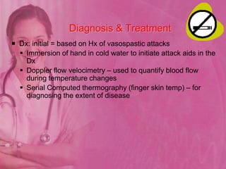


This document discusses cardiovascular diseases and provides information on their assessment and management. It covers topics like angina pectoris, myocardial infarction, congestive heart failure, and hyperlipidemia. For angina, it describes the different types, symptoms, pathophysiology, diagnosis, and treatment including medications and lifestyle modifications. It also discusses assessing and managing acute coronary syndromes and MI, including complications. Nursing care plans are provided for MI focusing on pain management, circulation, oxygenation, and emotional support. Congestive heart failure and its pathophysiology, signs, diagnoses, and a nursing care plan are also outlined. The document concludes with a brief overview of hyperlipidemia causes.