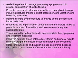
Nelia B. Perez RN, MSN Northeastern College Santiago City Common Respiratory Diseases
C O P D hronic bstructive ulmonary isease
Chronic Obstructive Pulmonary Disease Includes diseases that cause airflow obstruction  Chronic Bronchitis Emphysema Risk Factors include environmental exposures and host factors Primary symptoms are cough, sputum production and dyspnea
 
EMPHYSEMA
Definition is a chronic obstructive  pulmonary disease . It is often caused by exposure to  toxic   chemicals , including long-term exposure to  tobacco smoke .
Smoking an pollution can lead to  emphysema  and  lung cancer .
 
Infectious Allergens  Smoking  Hereditary  Air pollution agents  Predisposing Factors
Dyspnea with insidious onset progressing to Chronic cough, sputum production severe dyspnea with slight exertion  On inspection, hyper- inflated “barrel chest” wheezing, fatigue, and tachypnea  due to air trapping, muscle wasting, and pursed- lip breathing ( pink- puffers)  On auscultation, diminished breath sounds with crackles, wheezes, rhonchi, and Hyperresonance with percussion and a decrease in fremitus prolonged expiration  Hypoxemia and hypercarpnia, Anorexia, weight loss, weakness, and inactivity  Inflammatory reactions and infections from morning headaches in advance stages  pooled secretions  Clinical Manifestations
NURSING INTERVENTIONS Encourage the client to be in a smoking cessation program  Encourage the client to do pursed- lip breathing which can help to slow and control the rate and depth of expiration, prevents lung collapse and airways Instruct the client about having a proper activity pacing Instruct client to have inspiratory muscle training  Encourage lifestyle modifications Administer oxygen therapy at low volumes as prescribed
Nursing Management Patient education Breathing exercises Inspiratory muscle training Activity pacing Self-care activities Physical conditioning Oxygen and nutritional therapy Coping measures
MEDICATIONS Bronchodilators;  Albuterol (Ventolin), levalbuterol (Xopenax)  Corticosteriods;  Budesonide ( Pulmicort), beclomethasone (Beclovent)
Surgical Management Bullectomy Lung Volume Reduction Surgery Lung Transplantation
CHRONIC BRONCHITIS
DEFINITION A disease of the airways The presence of cough and sputum production for at least three months in each of two consecutive years. Chronic exposure to smoke or other pollutant irritates the airways, resulting in hypersecretion of mucus and inflammation, thickened bronchial walls and narrow bronchial lumen.
PREDISPOSING FACTORS Hereditary Exposure to irritants History of cigarette smoking Frequent respiratory infections
Clinical Manifestation Chronic, productive cough in winter months (earliest sign) -  cough is exacerbated by cold weather, dampness, and pulmonary irritants Shortness of breath
Nursing Interventions Monitor dyspnea and hypoxia.  Encourage high fluid intake to liquefy secretions.  Instruct client in directed or controlled coughing.  Eliminate pulmonary irritants.  Instruct client in effective breathing techniques.  Recommend the client to adopt a lifestyle of moderate activity, ideally in a climate with minimal shifts in temperature and humidity.  Encourage client to avoid emotional disturbances and stressful situations.  Instruct client and family about signs and symptoms of infection or other complications and to report changes in physical and cognitive status.
DRUGS Antibiotics  amoxicillin (Amoxil) doxycycline (Vibramycin)  Bronchodilators ephedrine (Primatene ) salmeterol (Serevent )  salbutamol (Ventolin)  Corticosteroids prednisone (Deltasone) ipratropium (Atrovent)
Asthma
definition Asthma is a disease in which inflammation of the airways causes airflow into and out of the lungs to be restricted. When an asthma attack occurs, the muscles of the bronchial tree become tight and the lining of the air passages swells, reducing airflow and producing the characteristic wheezing sound. Mucus production is increased.  The word asthma is derived from the Greek aazein, meaning \"sharp breath\".
Predisposing factors Allergens – it is one of the predisposing factors because asthma attacks are mostly caused by allergens. It is the most common predisposing factor.  Too much activity (play) – too much activity may lead to shortness of breath that causes compensation that results to hyperventilation or increased RR.  Air pollution – it can be a predisposing factor because pollution in the air may lead to irritation of the airway.
Clinical Manifestations shortness of breath (dyspnea)  Wheezing  cough  rapid breathing (tachypnea)  prolonged expiration  a rapid heart rate (tachycardia)  rhonchous lung sounds (audible through a stethoscope)  over-inflation of the chest.  in-drawing of tissues between the ribs and above the sternum and clavicles  Presence of paradoxical pulse
Nursing Interventions Put patient in a comfortable position (semi-fowler’s to promote lung expansion)  Encourage the client’s mother to lessen the client’s activity.  Teach about the importance of deep breathing exercises.  Provide comfort measures.  Health Teaching on ways to avoid asthma attack like preventing excessive exposure to allergens  Provide good nutrition for the client for the immune system  If possible, encourage the client to wear a mask.
DRUGS Albuterol Sulfate  Ventolin  Salbutamol
Pulmonary Embolism
DEFINITION Pulmonary embolism refers to the obstruction of the base or one or more branches of the pulmonary arteries by a thrombus (or thrombus) that originates somewhere in the venous system or in the right side of the heart. Gas exchange is impaired in the lung mass supplied by the obstructed vessel. Massive pulmonary embolism is life-threatening and can cause death within the first 1 to 2 hours after the embolic event.
 
It is a common disorder associated with trauma, surgery (orthopedic, major abdominal, pelvic, gynecologic), pregnancy, oral contraceptive use, congestive heart failure, age older than 50 years, hypercoagulable states, and prolonged immobility. Most thrombi originate in the deep veins of the legs.
PREDISPOSING FACTORS Inactivity  Prolonged bed rest  Certain surgical procedures  Some medical conditions  Being overweight  Pacemakers or venous catheters  Pregnancy and childbirth  Supplemental estrogen  Family history Smoking
CLINICAL MANIFESTATIONS Symptoms depend on the size of the thrombus and the area of the pulmonary artery occlusion.  Dyspnea is the most common symptom.  Tachypnea is the most frequent sign.  Chest pain is common, usually sudden in onset and pleuritic in nature; it can be substernal and may mimic angina pectoris.  Fever  Tachycardia
Others… Apprehension Cough  Diaphoresis  Hemoptysis  Syncope  Shock  Sudden death may occur  Multiple small emboli in the terminal pulmonary arterioles simulate symptoms of bronchopneumonia or heart failure.
Nursing Management 1. Providing General Care Ensure  Understanding of need for continuous oxygen therapy.  Provide nebulizers, incentive spirometry, or percussion and postural drainage.  Encourage deep-breathing exercises.
2. Preventing Thrombus Formation    Encourage early ambulation and active and passive leg exercises.  Instruct patient to move legs in a \"pumping\" exercise.  Advise patient to avoid prolonged sitting, immobility, and constrictive clothing.  Do not permit dangling of legs and feet in a dependent position.  Instruct patient to place feet on floor or chair and to avoid crossing legs.  Do not leave intravenous catheters in veins for prolonged periods.
3. Monitoring Anticoagulant and Thrombolytic Therapy    Advise bed rest, monitor vital signs every 2 hours, limit invasive procedures.    Measure PT or activated PTT every 3 to 4 hours after thrombolytic infusion is started to confirm activation of fibrinolytic systems.    Perform only essential ABG studies on upper extremities, with manual compression of puncture site for at least 30 minutes.
4. Minimizing Chest Pain, Pleuritic   Place patient in semi-Fowler's position; turn and reposition frequently.   Administer analgesics as prescribed for severe pain.
5. Alleviating Anxiety   Encourage patient to express feelings and concerns.   Answer questions concisely and accurately.   Explain therapy, and describe how to recognize untoward effects early.
6. Managing Oxygen Therapy   Assess for hypoxia (pulse oximetry), deep breathing, incentive spirometry, nebulizer therapy, percussion, and postnasal drainage.
7. Providing Postoperative Nursing Care     Measure pulmonary arterial pressure and urinary output.    Assess insertion site of arterial catheter for hematoma formation and infection.    Maintain blood pressure to ensure perfusion of vital organs.    Encourage isometric exercises, antiembolism stockings, and walking when permitted out of bed; elevate foot of bed when patient is resting.    Discourage sitting; hip flexion compresses large veins in the legs.
8. Promoting Home and Community-Based Care Teaching Patients Self-Care    Before discharge and at follow-up clinic or home visits, teach patient how to prevent recurrence and which signs and symptoms should alert patient to seek medical attention.    Teach patient to look for bruising and bleeding when taking anticoagulants and to avoid bumping into objects.    Advise patient to use a toothbrush with soft bristles to prevent gingival bleeding.
  Instruct patient not to take aspirin (an anticoagulant) or antihistamine drugs while taking warfarin sodium (Coumadin).    Advise patient to check with physician before taking any medication, including over-the-counter drugs.    Advise patient to continue wearing antiembolism stockings as long as directed.    Instruct patient to avoid laxatives, which affect vitamin K absorption (vitamin K promotes coagulation). Teach patient to avoid sitting with legs crossedor for prolonged periods.
  Recommend that patient change position regularly when traveling, walk occasionally, and do active exercises of legs and ankles.    Advise patient to drink plenty of liquids.    Teach patient to report dark, tarry stools immediately.    Recommend that patient wear identification stating that he or she is taking anticoagulants.
DRUGS •  Anti-coagulant: – Heparin – Warfarin    •  Thrombolytics: – Reteplase (Retavase) – Tenecteplase (Tnkase) – Streptokinase (Streptase)
Cystic Fibrosis
This is an autosomal recessive inheritance disease caused by a defective gene which causes the body to produce thick, sticky mucus that clogs the lungs, making it difficult to breathe and causing life-threatening lung infections.
PREDISPOSING FACTORS Caucasian people  Cystic fibrosis is an autosomal recessive condition - that means that a person needs two defective genes (one from each parent) to develop the condition. The gene that has been thought to cause cystic fibrosis is called cystic fibrosis transmembrane conductance regulator or CFTR. This gene has been located on chromosome 7
Clinical Manifestations Productive cough  Wheezing  Gastrointestinal problems (eg. abdominal pain, weight loss)  Hyperinflation of the lung fields on chest x-ray  Nasal polyps  Sinusitis  Male and female infertility  Clubbing of the extremities
Assist the patient to manage pulmonary symptoms and to prevent complication of cystic fibrosis  Promote removal of pulmonary secretions; chest physiotherapy, including postural drainage, chest percussion, and vibration, and breathing exercises  Remind client to avoid exposure to crowds and to persons with known infection  Emphasize the importance of adequate fluid and dietary intake to promote removal of secretions and to ensure an adequate nutritional status  Teach to modify daily activities to accommodate their symptoms and treatment modalities  Improved nutrition ( high calorie diet, vitamin and mineral rich) to maintain weight and ensure essential nutrients are consumed  Refer for counselling and support groups as chronic diseases can cause a great amount of stress for the patient and family
  Bronchodilators (such as albuterol or salmeterol), which are used to make breathing easier. Bronchodilators may also make it easier to cough up mucus.    DNase (such as Pulmozyme), which is used to thin mucus in the lungs.    Mucolytics (such as Mucomyst), to thin mucus in the lungs and also in the intestines. These are not used very much, because they can irritate the lungs.    An inhaled saltwater solution (hypertonic saline), sometimes used to help clear mucus from the lungs. It is low-cost, and it may help reduce inflammation in the airways.    Antibiotics are medicines that kill bacteria that cause infections.
Respiratory Arrest
Definition Previously known as the adult respiratory distress syndrome  Severe form of lung injury  Characterized by a sudden and progressive pulmonary edema, increasing bilateral infiltrates, on chest x-ray, hypoxemia, refractory to oxygenation, and reduced lung compliance
Acute Respiratory Failure Decreased respiratory drive Dysfunction of the chest wall Dysfunction of lung parenchyma Inadequate ventilation Treat underlying cause
Acute Respiratory Distress Syndrome Sudden and progressive pulmonary edema, increasing bilateral infiltrates, hypoxemia refractory to oxygen supplementation and reduced lung compliance Result of inflammatory trigger Treat underlying condition Ventilator considerations
Predisposing Factors Aspiration  Drug ingestion and overdose  Hematologic disorders such as DIC, massive transfusions, cardiopulmonary bypass  Prolonged inhalation of high concentrations of oxygen, smoke and corrosive substances  Localized infection such as viral pneumonia  Metabolic diseases such as pancreatitis and uremia  Shock  Trauma such as pulmonary contusion, multiple fractures, and head injury  Major surgery  Fat or air embolism  Systemic Sepsis
Clinical Manifestation Dyspnea  Increased respiratory rate  Increased heart rate  Bilateral edema  Shortness of breathe  Hypoxia  Signs and symptoms of the incumbent respiratory disease
Nursing Management Close monitoring in ICU  Nebulizer therapy  Tracheostomy care  Suctioning  Assisting on ET intubation and bronchoscopy  Enteral feeding of 35-45 kg/cal per day  Turn the patient frequently to improve ventilation and perfusion of the lungs and secretion drainage  Oxygenation on prone position  Mechanical ventilation with PEEP  Fluid Management
Drugs METHYLPREDNISOLONE(glucocorticoids)  Oral: Medrol, Meprolone (CAN) methylprednisolone sodium succinate  IV, IM injection: A-Methapred, Solu-Medrol Pregnancy Category C
Bronchiectasis Chronic, irreversible dilation of bronchi and bronchioles Chronic cough and purulent sputum production Postural drainage promotes clearing of secretions Antibiotics may be prescribed
Atelectasis Atelectasis - Closure or collapse of alveoli Pleural effusion Pneumothorax Hemothorax
 
 
Respiratory Infections Acute tracheobronchitis Pneumonia Shock and respiratory failure
 
Nursing Management Improving airway patency Promoting rest Promoting fluid intake and nutrition Monitoring and managing complications
Pulmonary Tuberculosis Mycobacterium tuberculosis Airborne transmission Tuberculin skin testing Pharmacologic therapy- multi-drug regimens and prophylaxis
 
Lung Abscess Complication of bacterial pneumonia or caused by aspiration or oral anaerobes Mild productive cough may lead to acute illness Pleural friction rub IV antibiotics
Pleural Conditions Pleurisy Pleural effusion Empyema
Pulmonary Edema Most often occurs as result of abnormal cardiac function Crackles Orthopnea Treat underlying disease
Pulmonary Hypertension Systolic pulmonary artery pressure > 30 mm Hg. or mean pulmonary artery pressure >25 mm Hg. Primary is idiopathic Secondary results from existing cardiac or pulmonary disease Manage underlying disease
Pulmonary Heart Disease  (Cor Pulmonale) Right ventricle enlarges with or without right-sided heart failure Caused by severe COPD Improve ventilation with supplemental oxygen, chest physical therapy, and bronchial hygiene
Sarcoidosis Multisystem granulomatous disease of unknown etiology Involves lungs, lymph nodes, liver, spleen, CNS, skin, eyes, fingers, and parotid glands Hypersensitivity response Corticosteroid therapy or other cytotoxic and immunosuppressive agents may be used
Chest Trauma Blunt trauma  Flail chest Penetrating trauma Pneumothorax
Management of Patients with Upper Respiratory Tract Disorders
Upper Airway Infections Rhinitis vs. Viral Rhinitis Acute Sinusitis vs. Chronic Sinusitis Chronic Pharyngitis Tonsillitis and Adenoiditis Peritonsillar Abscess Laryngitis
 
Nursing Management Maintain a patent airway Promote comfort Promote communication Encourage fluid intake Teaching self-care
Obstruction and Trauma of the Upper Respiratory Airway Obstruction during sleep Epistaxis  Nasal obstruction Fractures Laryngeal obstruction
 
Cancer of Larynx Hoarseness of more than 2 weeks duration Cough and/or sore throat Pain and/or burning  Laryngectomy
Thank you…  Not only for listening and for coming today… But also for the wonderful 2 nd  semester…  I hope to see all of you on Summer…. GOD BLESS…

Waterlily

  • 1.
    Nelia B. PerezRN, MSN Northeastern College Santiago City Common Respiratory Diseases
  • 2.
    C O PD hronic bstructive ulmonary isease
  • 3.
    Chronic Obstructive PulmonaryDisease Includes diseases that cause airflow obstruction Chronic Bronchitis Emphysema Risk Factors include environmental exposures and host factors Primary symptoms are cough, sputum production and dyspnea
  • 4.
  • 5.
  • 6.
    Definition is achronic obstructive pulmonary disease . It is often caused by exposure to toxic chemicals , including long-term exposure to tobacco smoke .
  • 7.
    Smoking an pollutioncan lead to emphysema and lung cancer .
  • 8.
  • 9.
    Infectious Allergens Smoking Hereditary Air pollution agents Predisposing Factors
  • 10.
    Dyspnea with insidiousonset progressing to Chronic cough, sputum production severe dyspnea with slight exertion On inspection, hyper- inflated “barrel chest” wheezing, fatigue, and tachypnea due to air trapping, muscle wasting, and pursed- lip breathing ( pink- puffers) On auscultation, diminished breath sounds with crackles, wheezes, rhonchi, and Hyperresonance with percussion and a decrease in fremitus prolonged expiration Hypoxemia and hypercarpnia, Anorexia, weight loss, weakness, and inactivity Inflammatory reactions and infections from morning headaches in advance stages pooled secretions Clinical Manifestations
  • 11.
    NURSING INTERVENTIONS Encouragethe client to be in a smoking cessation program Encourage the client to do pursed- lip breathing which can help to slow and control the rate and depth of expiration, prevents lung collapse and airways Instruct the client about having a proper activity pacing Instruct client to have inspiratory muscle training Encourage lifestyle modifications Administer oxygen therapy at low volumes as prescribed
  • 12.
    Nursing Management Patienteducation Breathing exercises Inspiratory muscle training Activity pacing Self-care activities Physical conditioning Oxygen and nutritional therapy Coping measures
  • 13.
    MEDICATIONS Bronchodilators; Albuterol (Ventolin), levalbuterol (Xopenax) Corticosteriods; Budesonide ( Pulmicort), beclomethasone (Beclovent)
  • 14.
    Surgical Management BullectomyLung Volume Reduction Surgery Lung Transplantation
  • 15.
  • 16.
    DEFINITION A diseaseof the airways The presence of cough and sputum production for at least three months in each of two consecutive years. Chronic exposure to smoke or other pollutant irritates the airways, resulting in hypersecretion of mucus and inflammation, thickened bronchial walls and narrow bronchial lumen.
  • 17.
    PREDISPOSING FACTORS HereditaryExposure to irritants History of cigarette smoking Frequent respiratory infections
  • 18.
    Clinical Manifestation Chronic,productive cough in winter months (earliest sign) - cough is exacerbated by cold weather, dampness, and pulmonary irritants Shortness of breath
  • 19.
    Nursing Interventions Monitordyspnea and hypoxia. Encourage high fluid intake to liquefy secretions. Instruct client in directed or controlled coughing. Eliminate pulmonary irritants. Instruct client in effective breathing techniques. Recommend the client to adopt a lifestyle of moderate activity, ideally in a climate with minimal shifts in temperature and humidity. Encourage client to avoid emotional disturbances and stressful situations. Instruct client and family about signs and symptoms of infection or other complications and to report changes in physical and cognitive status.
  • 20.
    DRUGS Antibiotics amoxicillin (Amoxil) doxycycline (Vibramycin) Bronchodilators ephedrine (Primatene ) salmeterol (Serevent ) salbutamol (Ventolin) Corticosteroids prednisone (Deltasone) ipratropium (Atrovent)
  • 21.
  • 22.
    definition Asthma isa disease in which inflammation of the airways causes airflow into and out of the lungs to be restricted. When an asthma attack occurs, the muscles of the bronchial tree become tight and the lining of the air passages swells, reducing airflow and producing the characteristic wheezing sound. Mucus production is increased. The word asthma is derived from the Greek aazein, meaning \"sharp breath\".
  • 23.
    Predisposing factors Allergens– it is one of the predisposing factors because asthma attacks are mostly caused by allergens. It is the most common predisposing factor. Too much activity (play) – too much activity may lead to shortness of breath that causes compensation that results to hyperventilation or increased RR. Air pollution – it can be a predisposing factor because pollution in the air may lead to irritation of the airway.
  • 24.
    Clinical Manifestations shortnessof breath (dyspnea) Wheezing cough rapid breathing (tachypnea) prolonged expiration a rapid heart rate (tachycardia) rhonchous lung sounds (audible through a stethoscope) over-inflation of the chest. in-drawing of tissues between the ribs and above the sternum and clavicles Presence of paradoxical pulse
  • 25.
    Nursing Interventions Putpatient in a comfortable position (semi-fowler’s to promote lung expansion) Encourage the client’s mother to lessen the client’s activity. Teach about the importance of deep breathing exercises. Provide comfort measures. Health Teaching on ways to avoid asthma attack like preventing excessive exposure to allergens Provide good nutrition for the client for the immune system If possible, encourage the client to wear a mask.
  • 26.
    DRUGS Albuterol Sulfate Ventolin Salbutamol
  • 27.
  • 28.
    DEFINITION Pulmonary embolismrefers to the obstruction of the base or one or more branches of the pulmonary arteries by a thrombus (or thrombus) that originates somewhere in the venous system or in the right side of the heart. Gas exchange is impaired in the lung mass supplied by the obstructed vessel. Massive pulmonary embolism is life-threatening and can cause death within the first 1 to 2 hours after the embolic event.
  • 29.
  • 30.
    It is acommon disorder associated with trauma, surgery (orthopedic, major abdominal, pelvic, gynecologic), pregnancy, oral contraceptive use, congestive heart failure, age older than 50 years, hypercoagulable states, and prolonged immobility. Most thrombi originate in the deep veins of the legs.
  • 31.
    PREDISPOSING FACTORS Inactivity Prolonged bed rest Certain surgical procedures Some medical conditions Being overweight Pacemakers or venous catheters Pregnancy and childbirth Supplemental estrogen Family history Smoking
  • 32.
    CLINICAL MANIFESTATIONS Symptomsdepend on the size of the thrombus and the area of the pulmonary artery occlusion. Dyspnea is the most common symptom. Tachypnea is the most frequent sign. Chest pain is common, usually sudden in onset and pleuritic in nature; it can be substernal and may mimic angina pectoris. Fever Tachycardia
  • 33.
    Others… Apprehension Cough Diaphoresis Hemoptysis Syncope Shock Sudden death may occur Multiple small emboli in the terminal pulmonary arterioles simulate symptoms of bronchopneumonia or heart failure.
  • 34.
    Nursing Management 1.Providing General Care Ensure Understanding of need for continuous oxygen therapy. Provide nebulizers, incentive spirometry, or percussion and postural drainage. Encourage deep-breathing exercises.
  • 35.
    2. Preventing ThrombusFormation Encourage early ambulation and active and passive leg exercises. Instruct patient to move legs in a \"pumping\" exercise. Advise patient to avoid prolonged sitting, immobility, and constrictive clothing. Do not permit dangling of legs and feet in a dependent position. Instruct patient to place feet on floor or chair and to avoid crossing legs. Do not leave intravenous catheters in veins for prolonged periods.
  • 36.
    3. Monitoring Anticoagulantand Thrombolytic Therapy  Advise bed rest, monitor vital signs every 2 hours, limit invasive procedures.  Measure PT or activated PTT every 3 to 4 hours after thrombolytic infusion is started to confirm activation of fibrinolytic systems.  Perform only essential ABG studies on upper extremities, with manual compression of puncture site for at least 30 minutes.
  • 37.
    4. Minimizing ChestPain, Pleuritic  Place patient in semi-Fowler's position; turn and reposition frequently.  Administer analgesics as prescribed for severe pain.
  • 38.
    5. Alleviating Anxiety  Encourage patient to express feelings and concerns.  Answer questions concisely and accurately.  Explain therapy, and describe how to recognize untoward effects early.
  • 39.
    6. Managing OxygenTherapy  Assess for hypoxia (pulse oximetry), deep breathing, incentive spirometry, nebulizer therapy, percussion, and postnasal drainage.
  • 40.
    7. Providing PostoperativeNursing Care  Measure pulmonary arterial pressure and urinary output.  Assess insertion site of arterial catheter for hematoma formation and infection.  Maintain blood pressure to ensure perfusion of vital organs.  Encourage isometric exercises, antiembolism stockings, and walking when permitted out of bed; elevate foot of bed when patient is resting.  Discourage sitting; hip flexion compresses large veins in the legs.
  • 41.
    8. Promoting Homeand Community-Based Care Teaching Patients Self-Care  Before discharge and at follow-up clinic or home visits, teach patient how to prevent recurrence and which signs and symptoms should alert patient to seek medical attention.  Teach patient to look for bruising and bleeding when taking anticoagulants and to avoid bumping into objects.  Advise patient to use a toothbrush with soft bristles to prevent gingival bleeding.
  • 42.
     Instructpatient not to take aspirin (an anticoagulant) or antihistamine drugs while taking warfarin sodium (Coumadin).  Advise patient to check with physician before taking any medication, including over-the-counter drugs.  Advise patient to continue wearing antiembolism stockings as long as directed.  Instruct patient to avoid laxatives, which affect vitamin K absorption (vitamin K promotes coagulation). Teach patient to avoid sitting with legs crossedor for prolonged periods.
  • 43.
     Recommendthat patient change position regularly when traveling, walk occasionally, and do active exercises of legs and ankles.  Advise patient to drink plenty of liquids.  Teach patient to report dark, tarry stools immediately.  Recommend that patient wear identification stating that he or she is taking anticoagulants.
  • 44.
    DRUGS • Anti-coagulant: – Heparin – Warfarin   • Thrombolytics: – Reteplase (Retavase) – Tenecteplase (Tnkase) – Streptokinase (Streptase)
  • 45.
  • 46.
    This is anautosomal recessive inheritance disease caused by a defective gene which causes the body to produce thick, sticky mucus that clogs the lungs, making it difficult to breathe and causing life-threatening lung infections.
  • 47.
    PREDISPOSING FACTORS Caucasianpeople Cystic fibrosis is an autosomal recessive condition - that means that a person needs two defective genes (one from each parent) to develop the condition. The gene that has been thought to cause cystic fibrosis is called cystic fibrosis transmembrane conductance regulator or CFTR. This gene has been located on chromosome 7
  • 48.
    Clinical Manifestations Productivecough Wheezing Gastrointestinal problems (eg. abdominal pain, weight loss) Hyperinflation of the lung fields on chest x-ray Nasal polyps Sinusitis Male and female infertility Clubbing of the extremities
  • 49.
    Assist the patientto manage pulmonary symptoms and to prevent complication of cystic fibrosis Promote removal of pulmonary secretions; chest physiotherapy, including postural drainage, chest percussion, and vibration, and breathing exercises Remind client to avoid exposure to crowds and to persons with known infection Emphasize the importance of adequate fluid and dietary intake to promote removal of secretions and to ensure an adequate nutritional status Teach to modify daily activities to accommodate their symptoms and treatment modalities Improved nutrition ( high calorie diet, vitamin and mineral rich) to maintain weight and ensure essential nutrients are consumed Refer for counselling and support groups as chronic diseases can cause a great amount of stress for the patient and family
  • 50.
     Bronchodilators(such as albuterol or salmeterol), which are used to make breathing easier. Bronchodilators may also make it easier to cough up mucus.  DNase (such as Pulmozyme), which is used to thin mucus in the lungs.  Mucolytics (such as Mucomyst), to thin mucus in the lungs and also in the intestines. These are not used very much, because they can irritate the lungs.  An inhaled saltwater solution (hypertonic saline), sometimes used to help clear mucus from the lungs. It is low-cost, and it may help reduce inflammation in the airways.  Antibiotics are medicines that kill bacteria that cause infections.
  • 51.
  • 52.
    Definition Previously knownas the adult respiratory distress syndrome Severe form of lung injury Characterized by a sudden and progressive pulmonary edema, increasing bilateral infiltrates, on chest x-ray, hypoxemia, refractory to oxygenation, and reduced lung compliance
  • 53.
    Acute Respiratory FailureDecreased respiratory drive Dysfunction of the chest wall Dysfunction of lung parenchyma Inadequate ventilation Treat underlying cause
  • 54.
    Acute Respiratory DistressSyndrome Sudden and progressive pulmonary edema, increasing bilateral infiltrates, hypoxemia refractory to oxygen supplementation and reduced lung compliance Result of inflammatory trigger Treat underlying condition Ventilator considerations
  • 55.
    Predisposing Factors Aspiration Drug ingestion and overdose Hematologic disorders such as DIC, massive transfusions, cardiopulmonary bypass Prolonged inhalation of high concentrations of oxygen, smoke and corrosive substances  Localized infection such as viral pneumonia  Metabolic diseases such as pancreatitis and uremia Shock Trauma such as pulmonary contusion, multiple fractures, and head injury Major surgery Fat or air embolism Systemic Sepsis
  • 56.
    Clinical Manifestation Dyspnea Increased respiratory rate Increased heart rate Bilateral edema Shortness of breathe Hypoxia Signs and symptoms of the incumbent respiratory disease
  • 57.
    Nursing Management Closemonitoring in ICU Nebulizer therapy Tracheostomy care Suctioning Assisting on ET intubation and bronchoscopy Enteral feeding of 35-45 kg/cal per day Turn the patient frequently to improve ventilation and perfusion of the lungs and secretion drainage Oxygenation on prone position Mechanical ventilation with PEEP Fluid Management
  • 58.
    Drugs METHYLPREDNISOLONE(glucocorticoids) Oral: Medrol, Meprolone (CAN) methylprednisolone sodium succinate IV, IM injection: A-Methapred, Solu-Medrol Pregnancy Category C
  • 59.
    Bronchiectasis Chronic, irreversibledilation of bronchi and bronchioles Chronic cough and purulent sputum production Postural drainage promotes clearing of secretions Antibiotics may be prescribed
  • 60.
    Atelectasis Atelectasis -Closure or collapse of alveoli Pleural effusion Pneumothorax Hemothorax
  • 61.
  • 62.
  • 63.
    Respiratory Infections Acutetracheobronchitis Pneumonia Shock and respiratory failure
  • 64.
  • 65.
    Nursing Management Improvingairway patency Promoting rest Promoting fluid intake and nutrition Monitoring and managing complications
  • 66.
    Pulmonary Tuberculosis Mycobacteriumtuberculosis Airborne transmission Tuberculin skin testing Pharmacologic therapy- multi-drug regimens and prophylaxis
  • 67.
  • 68.
    Lung Abscess Complicationof bacterial pneumonia or caused by aspiration or oral anaerobes Mild productive cough may lead to acute illness Pleural friction rub IV antibiotics
  • 69.
    Pleural Conditions PleurisyPleural effusion Empyema
  • 70.
    Pulmonary Edema Mostoften occurs as result of abnormal cardiac function Crackles Orthopnea Treat underlying disease
  • 71.
    Pulmonary Hypertension Systolicpulmonary artery pressure > 30 mm Hg. or mean pulmonary artery pressure >25 mm Hg. Primary is idiopathic Secondary results from existing cardiac or pulmonary disease Manage underlying disease
  • 72.
    Pulmonary Heart Disease (Cor Pulmonale) Right ventricle enlarges with or without right-sided heart failure Caused by severe COPD Improve ventilation with supplemental oxygen, chest physical therapy, and bronchial hygiene
  • 73.
    Sarcoidosis Multisystem granulomatousdisease of unknown etiology Involves lungs, lymph nodes, liver, spleen, CNS, skin, eyes, fingers, and parotid glands Hypersensitivity response Corticosteroid therapy or other cytotoxic and immunosuppressive agents may be used
  • 74.
    Chest Trauma Blunttrauma Flail chest Penetrating trauma Pneumothorax
  • 75.
    Management of Patientswith Upper Respiratory Tract Disorders
  • 76.
    Upper Airway InfectionsRhinitis vs. Viral Rhinitis Acute Sinusitis vs. Chronic Sinusitis Chronic Pharyngitis Tonsillitis and Adenoiditis Peritonsillar Abscess Laryngitis
  • 77.
  • 78.
    Nursing Management Maintaina patent airway Promote comfort Promote communication Encourage fluid intake Teaching self-care
  • 79.
    Obstruction and Traumaof the Upper Respiratory Airway Obstruction during sleep Epistaxis Nasal obstruction Fractures Laryngeal obstruction
  • 80.
  • 81.
    Cancer of LarynxHoarseness of more than 2 weeks duration Cough and/or sore throat Pain and/or burning Laryngectomy
  • 82.
    Thank you… Not only for listening and for coming today… But also for the wonderful 2 nd semester… I hope to see all of you on Summer…. GOD BLESS…