The document provides an overview of cardiovascular conditions including ischemia, infarction, coronary artery disease, myocardial infarction, and cardiac arrhythmias. It discusses diagnostic testing and evaluation, management, treatment options, and medications for these conditions. Key points include diagnostic markers for infarction like cardiac enzymes, symptoms of angina and myocardial infarction, management of conditions through diet, medication, procedures like angioplasty and bypass surgery, and treatment of arrhythmias through medications, cardioversion, defibrillation, and pacing.
![MEDICAL-SURGICAL NURSING
UNIVERSITY*OF*SANTO*TOMAS*–*COLLEGE*OF*NURSING*|*FJCP*2017*|
α
α
α
α
α*NOTES*
1.7.4 Beta Blockers (propranol)
1.8 TREATMENT
1.8.1 Percutaneous Transluminal Corornary
Angioplasty (PTCA)
1.8.2 Intravascular Stenting: Done to prevent
restenosis after PTCA; Given
Coumadin, ASA (prevent clotting;
lifetime medication for stents)
2. MI
*Loose circulation to whole body: Brain and Kidneys
(U/O monitor)
2.1 CAUSES
2.1.1 Total occlusion of one of the coronary
arteries causing ischemia, injury and
NECROSIS
2.1.2 Infarction most common to the LV
2.1.3 RF: same risk of Angina
2.1.4 Not relieved by rest or NTG
2.2 LEVINE’S SIGN: clutching at the chest due to
Pain; Most likely having an MI
2.3 ASSESSMENT
2.3.1 Crushing substernal pain (radiate to jaw,
back and arms; unrelieved by rest and
NTG)
2.3.2 Dyspnea, Diaphoresis
2.3.3 Hypoxia causes Arrhythmias (V
Fibrillation: MC cause of Death)
*PULSELESS V- -
TACH and V FIB: Shockable rhythm
2.4 DX
2.4.1 Diagnostic evaluation: ECG shows
deep, wide Q wave (All layers are
infarcted: epi, myo and endocardium),
Elevated or depressed ST-segment, T-
wave inversion
2.4.2 Coupled with Cardiac Enzymes, you
will be able to determine the presence
of MI; Increased Cardiac Enzymes
2.5 MANAGEMENT
2.5.1 Bedrest with Bedside Commode (used
as a chair and makes it easier to
defecate and urinate; just beside the
bed)
2.5.2 CABG: Saphenous Vein
(Contraindicated with varicose Veins);
Internal Mammary Artery (may be
used); The number CBG x5 meaning 5
vessels were bypassed
2.5.3 Angioplasty
2.5.4 Low calorie, low cholesterol, low fat
diet
2.5.5 Monitor labs (ABG, CK, electrolyte and
Troponin)
2.5.6 02
2.5.7 CPR (if patient becomes unconscious
and defibrillate when ECG shows V
Fibrillation)
*CPR within um of 15 minutes
5-10 minutes; Brain maxim
anoxic; Longer, becomes stroked-out (vegetable)
2.6 CPR
2.6.1 Assess consciousness: hey hey are you
ok: Unresponsive
2.6.2 Check the Pulse: Carotid Pulse for at
least 10 seconds
2.6.3 Assess Respiration: rise and fall [New:
MONITOR for absence of breathing or
abnormal breathing {GASPING for
breath}]
2.6.4 Call for help: CODE
2.6.5 Start Chest Compression: 30:2
Compression to Breathe; ;
2 INCHES
HIGH QUALITY CPR Adequate rate
and Adequate Depth: 100-120
compressions per minute;
2.6.6 Advance Airway (Ambubag): 1 breathe
every 6 seconds; Avoid excessive
ventilation; REMEMBER TO COUNT
2.6.7 ABC: Compression, Airway, Breathing
2.6.8 New: 2 minutes of Full compression not
unless someone brings the Defibrillator
2.6.9 Defibrillator: Apex and Sternum (for
the paddles and lubricate); Once placed
on the chest, APPLY 10 lbs of pressure;
Setting must be on MODE:
ASYNCHRONIZED (It does not
matter where it will hit; the rhythm is
chaotic; No need to be careful; Shock
will hopefully go back to any rhythm
good for perfusion); JOULES: It
depends on the defibrillator being used;
2.6.9.1 MONOPHASIC: One way and
does not travel back to the heart;
360 Joules
2.6.9.2 BIPHASIC: Goes back and forth
with an AC Current with lesser
joules or damage to the muscles;
200 Joules
*CARDIOVERSION: SYNCHRONIZED
2.6.9 if does not wake up: KEEP CPR and Inject
needed meds: EPINEPHRINE; Then Continue
Defibrillation with same joules
2.7 MEDICATION
2.7.1 MONA
2.7.2 MORPHINE: vasodilator and
analgesics (reduces Myocardial 02
demand)
2.7.3 O2
2.7.4 NTG
0 0](https://image.slidesharecdn.com/1-medical-surgicalcompress-230601155911-5ddb901f/85/1-medical-surgical_compress-pdf-2-320.jpg)
![MEDICAL-SURGICAL NURSING
UNIVERSITY*OF*SANTO*TOMAS*–*COLLEGE*OF*NURSING*|*FJCP*2017*|
α
α
α
α
α*NOTES*
2.7.5 ASA: Anti-Platelet
2.7.6 Beta Blocker and CA Channel
2.7.7 Thrombolytics: Given 3 4 hrs
- after
S/SX
2.7.8 Anticoagulant
2.8 VENTRICULAR FIBRILLATION: Chaotic
Discharge; rate >300/min, may result to clinical
death; IMMEIDIATE DEFIBRILATION and
EPINEPHRINE
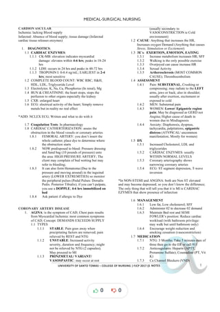
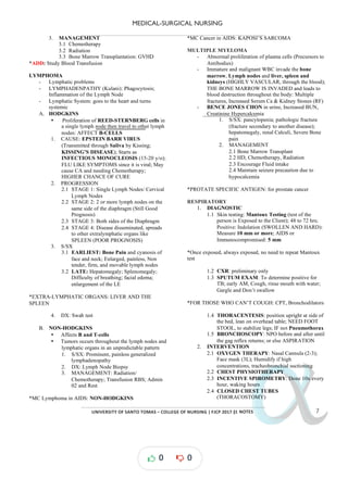
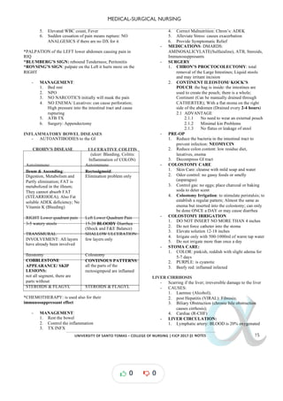
CARDIAC ARRYTHMIAS
- Abnormal electrical conduction or automaticity that
changes cardiac rhythm rate
- PROPERTIES: [1] Automaticity (initiating its own
heart beats); [2] Conductivity (Traveling from one
point to another); [3] Contractility (the ability to
empty the content of the heart)
- SA NODE: the intrinsic pacemaker of the heart
IMPULSE WAVE ACTIVITY
SA NODE
HR: 60-100
bpm
P-wave Atrial
Depolarization:
Contraction
AV NODE
HR: 40-60
bpm
Time travel &
Impulse from SA to
AV:
PR interval: 0.12-
20 seconds
BUNDLE
OF HIS
PURKINJE
FIBERS:
20 40bpm
-
QRS: 0.4-0.12
Seconds
Ventricular
Depolarization
T-Wave Ventricular
Repolarization:
Resting Heart
CARDIAC DYRRYTHMIAS
TYPES LABEL BPM MGMT
SINUS
Normal Sinus
Rhythm
All the
available
waves
(PQRST)
Sinus
Bradycardia
Still from SA
Node; But
slower;
Sleeping
Asymtomatic:
No tx
Symptomatic:
Atropine S04
Sinus
Tachycardia
Exertion
ATRIAL
Atrial Flutter P-wave Jagged
Saw teeth
250-
350
Quinidine (Na
channel
Blocker)
Atrial
Fibrillation
Jiggling;
Beating to fast,
shaking
atrium;
Presence of
Blood clot
>350 Anti-
coagulants
PAC Ectopic Heart
beat
VENTRICULAR
Ventricular
Tachycardia
All QRS 101-
250
Lidocaine;
VTach Pulse:
Cardioversion
VTach w/o
Pulse:
Defibrillation
Ventricular
Fibrillation
Chaotic
HEART BLOCK
First Degree
Heart Block
Prolonged PR
interval
HR at
40 50
-
No treatment
Second
Degree
Type 1:
Wincklebach
PR interval:
Long, longer,
longest drop
No treatment
Type 2 PR Interval:
Norma,
normal,
normal, drop
If requires tx if
VR falls too
low to
maintain
adequate CO
Third Degree No impulse
form SA node
reaches the
ventricles; no
association;
cant determine
which wave is
which
Pace makers
*IMPAIRED TISSUE PERFUSION: all Cardiac
Dysrhythmias
1. CAUSES
1.1 Congenital, Drug toxicity, Electrolyte
imbalances (HYPOKALEMIA: most dangerous;
Sister is Magnesium so also low); Heart Disease
(MI, CHF)
2. TREATMENT
2.1 Anti arrhythmic card
, Synchronized ioversion,
CPR, Defibrillation
2.2 ICD (Implanted Cardiac Defibrillator; detects
fibrillation and shocks; for post VFIb/MI
patients), Transcutaneous pacing
3. CARDIOVERSION: 50-200 Joules only
3.1 Synchronized mode: you wait for a particular or
certain wave; WAIT FOR THE R-WAVE
3.2 DON’T HIT the T-WAVE (the heart is resting)
0 0](https://image.slidesharecdn.com/1-medical-surgicalcompress-230601155911-5ddb901f/85/1-medical-surgical_compress-pdf-3-320.jpg)
![MEDICAL-SURGICAL NURSING
UNIVERSITY*OF*SANTO*TOMAS*–*COLLEGE*OF*NURSING*|*FJCP*2017*|
α
α
α
α
α*NOTES*
VALVULAR HEART DISEASE
- Can result from endocarditis and inflammation then
HF
1. TYPES
1.1 STENOSIS or NARROWING: does not open
due to or hardening (CHF)
stiffening
1.2 INSUFFICIENCY: Incomplete closure of the
valve (CHF)
1.3 PROLAPSE OF THE VALVE: protrudes into
the La (MC in Mitral/ Bicuspid valve)
2. FORMS
2.1 Aortic insufficiency: Left Sided CHF
2.2 Mitral insufficiency: Left Sided CHF
2.3 Mitral Stenosis
2.4 Mitral Valve Prolapse
2.5 Tricuspid Insufficiency
3. MANAGEMENT
3.1 PORCINE: Pork grafting for valves; Temporary
3.2 Prosthetic: Permanent
3.2.1 HANCOC: Tricuspid
HTN
1. CLASSIFICATION OF BP FOR ADULTS
SYSTOLIC DIASTOLIC
Optimal 120 80
Normal <130 <85
High 130 139
- 85 89
-
STAGE 1 140 159
- 90 99
-
STAGE 2
(+20)
160 179
- 100 109
-
STAGE 3
(+10)
>180 >110
*To diagnose with HTN; 2 Consecutive reading over a 2-week
period
2. ASSESSMENT
2.1 Essential: no particular reason
2.2 Asymptomatic
2.3 Elevated BP
2.4 Dizziness
2.5 H/A
2.6 L Ventricular Hypertrophy
2.7 HF
2.8 Cerebral Ischemia
2.9 RENAL FAILURE
*Renal ischemia: low blood supply; It will release RENIN à
Angiotensin I ACE
à à ANGIOTENSIN II: High BP
2.10Visual Disturbances inc. Blindness: in the
FRONTAL LOBE
2.11Epistaxis : Pressure in the BV
3. DX
3.1 Increased BUN, creatinine, Na and cholesterol
4. ANEURYSM: GOAL: maintain NORMAL BP;
Don’t make it rupture
4.1 TYPES
4.1.1 Saccular: one side pouch
4.1.2 Fusiform: Both sides
4.1.3 Dissecting
4.1.4 False Aneurysm
4.2 SURGERY: Clipping of Aneurysm; Coiling (did
not treat just block not really stop the aneurysm)
5. MANAGEMENT
5.1 Regular exercise to reduce weight
5.2 Low sodium diet and limitation of alcohol
5.3 Assess CV status and VS; Take an average of 2
or more readings to establish HTN
5.4 Assess neurologic Disorders and observe for
changes that may indicate alteration in cerebral
perfusion (CVA)
5.5 Monitor I&O and weight
6. MEDICATION
6.1 Maintain quiet environment to reduce stress:
AWAY FROM NX station and elevator
ARTERIAL DISEASES
1. REYNAUD’S DISEASE: Vasospasms of the
arterioles and arteries of the extremities
(Oxygenated Blood); Ischemic à NECROSIS
1.1 ETIOLOGY
1.1.1 Cold [ICE]; Stress; Smoking
1.2 UPPER EXTREMITIES
1.3 ASSESSMENT
1.3.1 Blanching of ext. followed by cyanosis:
WHITE à à
BLUE RED
1.3.2 Reddened tissue
1.3.3 Numbness, tingling sensation
(Paresthesia: Impaired circulation)
1.4 MANAGEMENT
1.4.1 Stop Smoking
1.4.2 VASODILATORS
1.4.3 Avoid Precipitating factors
1.4.4 Warm Clothing
1.4.5 Avoid injuries to hands and fingers
2. BUERGER’S DISEASE/ THROMBO
ANGINITIS OBLITERANS: occlusive disease of
the median and small arteries and veins by clot
formation
2.1 RF: 30-35 y/o; Males; Men who SMOKE
2.2 ETIOLOGY:
2.2.1 Unknown, smoking, males
2.3 LOWER EXTREMITIES
2.4 ASSESSMENT
2.4.1 Intermittent Claudication
2.4.2 Ischemic Pain occurring in the digits
while at rest
2.4.3 Cool, Numb, tingling sensation
2.4.4 Diminished pulse at distal extremities
2.4.5 Ulceration
2.5 Amputation
2.6 MANAGEMENT
2.6.1 Instruct to stop smoking
2.6.2 Monitor Pulses
0 0](https://image.slidesharecdn.com/1-medical-surgicalcompress-230601155911-5ddb901f/85/1-medical-surgical_compress-pdf-5-320.jpg)
![MEDICAL-SURGICAL NURSING
UNIVERSITY*OF*SANTO*TOMAS*–*COLLEGE*OF*NURSING*|*FJCP*2017*|
α
α
α
α
α*NOTES*
and lung will still continue being in mediastinal
shifting
- ASSESSMENT: Dyspnea, decreased 02 at;
Diminished or absent breath sound; Sharp chest pain
that increases with exertion; tracheal shift to
unaffected side (tension); SUCKING SOUNDS
WITH OPEN CHEST WOUND
- MGMT: apply vented dressing; Position in High
Fowlers; Prepare for chest tube placement until the
lungs has fully expanded; Monitor hypotension,
tachycardia and tachypnea (mediastinal shifting);
Administer 02 and Monitor chest tube drainage
system
CHEST TUBE
- Returns negative pressure to intrapleural space
- Used to remove abnormal accumulation of air and
fluid from pleural space
- CHAMBERS
1. COLELLCTION CHAMBER: Drainage
2. WATER AND SEAL CHAMBER: Tip of the
tube under water (2cm) allowing fluid and air
from entering the pleural space; Immersed so
that the air will not go back; Water will show
INTERMITTENT BUBBLING
3. SUCTION CONTROL CHAMBER: gentle
bubbling normal high
; only used when there are
amounts of mucous but does not drain; Attached
to suctioning apparatus; Expected to have
CONTINOUS BUBBLING
- WATER OCCILATES/ TIDALING: moves up
when the patient inhales and moves down as the
patient exhales; To see if the bottle system is moving
- NO OSCILLATION or TIDALING: [1]
obstruction in the system (kinked or pt. is lying down
on the tube; Thick secretions like pus); [2] lung has
expanded meaning to more secretions to suck; To
determine if not effusion subsided: CXR
*ONE BOTTLE: Drainage and water sealed into one
*TWO-BOTTLE: Drainage and Water seal
- MGMT: Monitor the drainage; Keep tubes free of
leaks, dependent loops or other obstructions; Do not
strip or milk tubes unless specified by the doctor
(Milking: Pressing; Stripping: continual pressing
from start for bubbling
until end); Check : Intermittent
bubbling is normal while continuous bubbling is air
leak; If water is used in the suction chamber, check
the continuous bubbling which indicates the system is
working
*If the bottle brakes, immerse the tube unto water; A
BOTTLE OF STERILE WATER: must be placed on the bed
side; Clamping will cause pressure then TENSION
PNEUMOTHORAX
*if pulled out: use Vented gauze and call MD
*if removed after use: use occlusive dressing; take a deep
breath and pull; use petrolatum dressing or dry dressing
LUNG CANCER
- The lungs are highly vascular since it oxygenates the
body
- TYPES:
1. NON SMALL CELL: MC;
1.1 Adenocarcenoma
1.2 Squamous- Cell Carcinoma
1.3 Large Cell Carcinoma
2. SMALL CELL
- CAUSES: Smoking (pack/year/history; Exposure to
environmental and occupational pollutants);
MINERS: Mineral deposits into the lung tissue
PACK YEARS:
# packs per day X Number of years smoked
1 PACK = 20 cigarettes
- EARLIEST MANIFESTATION: Productive Cough
- LATE MANIFESTATION: hemoptysis: eroding lung
tissues
- S/SX: Dyspnea, hoarseness, chest pain, anorexia, and
weight loss, weakness
- SURGERY: SIZE matters
PNEUMONECTOMY
SECTION DESCRIPTION
Wedge resection /
Segmentectomy
Only near the part of the
tumor
Lobectomy Entire lobe has been
infiltrated
*These surgeries WILL have CHEST TIUBES
*UNAFFECTED SIDE: position, there is a chest tube and
make the lung re-inflate properly
Pneumonectomy Whole side of the lung
*NO CHEST TUBE: no lung no bleeding, no anything
* AFFECTED SIDE: position; allow the remaining lung to
expand
*TB: 2 weeks of RIPES and then can now socialize; no more
isolation
*EMPHYSEMA: alveoli; DYPNEA ON REST
*Bronchitis: Chronic productive cough
*Asthma: WheezingMUSCULOSKELETAL DISORDERS
*ADD: proper body mechanics
TRAUMA
- All signs of trauma will cause inflammations
SPRAIN: ; tendons
overstretching
STRAIN: Over twisting; joints and ligaments
Dislocated: when outside; total
Subluxation: partial dislocation
0 0](https://image.slidesharecdn.com/1-medical-surgicalcompress-230601155911-5ddb901f/85/1-medical-surgical_compress-pdf-10-320.jpg)
![MEDICAL-SURGICAL NURSING
UNIVERSITY*OF*SANTO*TOMAS*–*COLLEGE*OF*NURSING*|*FJCP*2017*|
α
α
α
α
α*NOTES*
FRACTURE
- the move it the more it will brake
- SIGNS: Injury will cause inflammation; Loss of
motion; edema, crepitus (rubbing against another
bone), shortening of the extremity
- IMPACTED: one bone is rubbing on another (Sound
is called Crepitus)
- STAGES OF BONE HEALING
1. HEMATOMA: immobilize, the only thing that is
keeping it together is blood clots; Inflamed (First 3
days; don’t place a cast since it will compress the
site; PLACE a POSTERIOR MOLD: which is a hard
board and bandages wrapped around it)
2. GRANULATION TISSUE STAGE: Osteoblasts go
to the site; Bones are made of protein and at this
stage protein will be laid down; WHOLE CAST are
now placed here since inflammation has decreased
3. CALLUS STAGE Calcium deposition
: START of ;
partial weight bearing can now be done (PROM; only
the done for fractures on joints; the more sedentary
you are; the more calcium will go out the body)
4. REMODELING: ossification and remodeling
MANAGEMENT
*SPLINT: it is above and below the JOINT; ex if radial
fracture: long arm cast
STIFFNES OF THE JOINT: Contracture
- MGMT: immobilize ( )
SPLINT : the more you move,
the more you break; NECK (cervical: IMMOBILIZE
THE NECK, ASAP); Cervical Collar: prevent injury
to phrenic nerve
*PHRENIC NERVE: C2 C3
-
- RICE (educe inflammation; Rest Ice, Compression,
Elevation)
- Reduction: Realignment of the bone
1. OPEN: surgery to realign the bone
2. CLOSE: Casting/ traction
CAST CARE
- Types:
1. PLASTER OF PARIS: it looks like gauze but
chalky in consistency; place in water then
squeeze then mold it in place; DRIES: 24-48 hrs:
Do not touch the cast when wet; USE THE
PALM of the hand when molding or moving it
around; Cement/ heavy
2. FIBERGLASS: it is plastic, synthetic; Wet the
gauze and little bit of water; DRIES: 20 min;
Plastic/ Light
- MGMT: Monitor the extremities for circulatory
impairment inserted
(1-2 fingers must be to
determine if the cast is not too tight):
COMPARTMENT SYNDROME
5P’s
1. PAIN
2. PALLOR
3. PARESTHESIA
4. PULSELESSNESS
5. PARALYSIS
- Monitor for any drainage on the cast; Instruct to keep
the cast clean and dry; DO NOT WET THE
PLASTER OF PARIS CAST, will make it soft; For
Fiberglass, you can wet it but it is discouraged, the
outside can be cleaned but the inside cannot dry
properly; Instruct not to insert anything in the cast; do
not write anything on the cast
- INFX:
1. Monitor for Drainage
2. Monitor HOTSPOTS: touch the cast it must be
cold but when it feels warm that is the infected
are
*NO POWDER: will contribute to infection
- Instruct to do ISOMETRIC EXERCISES (alternate
contraction and relaxation)
TRACTION: Act of pulling into the proper location
1. Ensure the weights are hanging freely
2. Maintain continuous traction (once you pull it don’t
let it go; misaligned: will heal into that position
[MALUNITED/ MALUNION] or does not stick
together [NO UNION])
3. Must always have a counter-traction: the need for
balance (ONLY 10% of the patient’s Body weight)
- BRYANT’S TRACTION: for children <3y/o; the
feet are held up; The buttocks of the baby must be off
the mattress, not touching the bed meaning NO
PULL; Place a mobile to stimulate the baby
- SKIN TRACTION
1. CERVIVCAL TRACTION: Skin type; cervical
2. PELVIC TRACTION: for lumbar spine
- SKELETAL TRACTION
1. CRUTCHFIELD TONG: Drilled into the skull
for traction
2. BST: Clean the pin since there is an entry for
microorganisms;
2.1 PIN CARE: Do not clean with hydrogen
peroxide: will cause anaerobic infection;
No betadine: this will oxidize and rust the
pins; Apply ATB; place antiseptic
*SKIN TRACTIONS: are temporary, for minor injuries
*SKELETAL TRACTION: severe injuiries
CRUTCHES
- The crutches not up to the AXILLA: this will cause
BRACHIAL PLEXUS COMPRESSION
(paresthesia of the fingers)
- MEASUREMENT:
1. 2 INCHES below axilla
2. 2-3 INCHES into the side; 6-12 inches into the
front
3. ELBOW FLEXION (20-30 degrees)
- Exercises to prepare for CW:
0 0](https://image.slidesharecdn.com/1-medical-surgicalcompress-230601155911-5ddb901f/85/1-medical-surgical_compress-pdf-11-320.jpg)
![MEDICAL-SURGICAL NURSING
UNIVERSITY*OF*SANTO*TOMAS*–*COLLEGE*OF*NURSING*|*FJCP*2017*|
α
α
α
α
α*NOTES*
inactivity; SWAN NECCK deformity; Bouteniere’s
Deformity; Ulnar Deviation
- DX: Increased WBC, RF + (Auto Antibodies);
Elevated ESR; effusion in the joint (Arthrocentesis)
- MANAGEMENT
1. Bed Rest
2. PROM
3. Heat stiffness and cold for inflammation; Warm
Shower in the AM
4. PT
5. Splint painful joints
- MEDICATION: Aspirin (the 4 effects); NSAIDS
(Ibuprofen); Corticosteroids; DMARDS (Disease
Modifying Anti Rheumatic Drugs):
Immunosuppressants, Methotrexate, Sulfasalazine,
GOLD therapy
- SURGERY: Synovectomey & Arthroplasty
*SLE: you it is
damage anything with COLLAGEN ( found
everywhere; SYSTEMIC; Butterfly Rash)
OSTEOARTHRITIS
- Degeneration of articular cartilage
- MC in Obese Clients
- Involves weight bearing: HIPS; Knees; Fingers;
Vertebra (Lower Back)
- S/SX
1. Joint pain aggravated by use; relieved by rest
2. Stiffening of joints
3. Formation of bony outgrowths
(OSTEOPHYTES); HEBERDEN’S (Distal IP)
and BOURCHARD’S NODES (Proximal IP)
4. Decreased ROM and CREPITUS
- MANAGEMENT
1. Relieve strain and further trauma to joints
2. Cane or walker, if indicated
3. Joint replacement as needed
4. Proper Body Mechanics
5. Avoid Excessive weight bearing and standing
6. PT
7. Relief of pain (NSAIDS)
8. CHONTROTIN: repairs the cartilage AND
GLUCOSAMINE
- TYPES OF HIP REPLACEMENT
1. TOTAL HIP: All parts
2. patella
PARTIAL: except
- PASSIVE ROM MACHINE: for continuous
movement and stop contracture of the knee
replacement
GOUT
- Disorder of purine metabolism
- Uric acid crystals in the JOINT: TOPHI
- S/SX: Joint pain, redness, ankle and great toe (MC)
- MANAGEMENT
1. Rest
2. Low purine and increase fluids
3. PHARMACOTHERAPY
3.1 Acute Attack: Colchicine and NSAIDS
3.2 Uricosuric Drugs: Excretes Uric acid:
Probenecid (Benemid)
3.3 Allopurinol (Zyloprim): Inhibits the
formation of uric acid
HERNIATED NUCEUS PULPOSUSU/ SLIP DISK
- NUCLEUS PULPOSIS: the jelly inside the vertebral
disk
- ANNULUS FIBROSUS: this holds the nucleus
fibrosis
- CAUSES: lifting, degeneration of the disc (loses
water content); also caused by sitting down
- AREAS
1. Cervical: Shoulder pain radiating to the hand;
Weakness and Paresthesia; Weight bearing on
the NECK/ HEAD; On cervical Traction
2. Lumbosacral: back pain radiating across buttocks
and down the leg (SCIATIC NERVE; Sciatica);
weakness ad paresthesia; Muscle spasm in the
Lumbar region; on pelvic Traction
- MANAGEMENT
1. Bed rest on firm mattress with board
2. Traction
3. Local Application of het
4. Lumbosacral corset (BACK BRACE)
5. Prevent complication of Immobility
- SURGERY: Laminectomy with Discectomy;
SURGICAL FUSION (Spinal fusion with rod
insertion)
- POSTOP:
1. Turn the patient as a whole (LOG ROLLING
TECHNIQUE)
2. LUMBAR: HOB, flat, supine with legs slightly
flexed
3. CERVICAL: HOB elevated, with neck
immobilized with collar or sandbag
GASTROINTESTINAL HEPATOBILLIARY
&
- Mouth into the Anus; Unsterile
GERD
- The food is refluxing up to the esophagus; Gastric
contents flow upwards to esophagus
- Common for obese and pregnant women
- Any activity that increase intra abdominal pressure
(Overeating, bending, tight clothing), Foods that
relax the cardiac sphincter (Alcohol, peppermint,
caffeine, high fat diet [fats are not easily digestible,
stays longer in the stomach]), lying down after eating
- ASSESSMENT: Dyspepsia; Dyspepsia;
ODYNOPHAGIA (painful swallowing); Esophagus;
Eaophagitis
- MANAGEMENT:
1. Avoid alcohol, peppermint, caffeine, high fat
diet;
2. 15-30 minutes High Fowler (Supine opens the
sphincter due to pressure)
3. Lose weight
4. Avoid over-eating and tight fitting clothes
0 0](https://image.slidesharecdn.com/1-medical-surgicalcompress-230601155911-5ddb901f/85/1-medical-surgical_compress-pdf-13-320.jpg)
![MEDICAL-SURGICAL NURSING
UNIVERSITY*OF*SANTO*TOMAS*–*COLLEGE*OF*NURSING*|*FJCP*2017*|
α
α
α
α
α*NOTES*
*SWALLOW AIR: carbonated drinks, chewing gum and
eating fast
*HERNIA: protrude; Can also cause GERD due to opening of
the sphincter
*Ambulation increases Peristalsis
PEPTIC ULCER
- GASTRITIS: inflamed only but later forms
ulceration causing GI bleeding; The deeper it gets
then erodes the mucosa
- PERITOTINITIS: from the contents of stomach will
cause septicemia à SEPTIC SHOCK
- STRESS, STIMULATED or EXCITED (SNS):
Gastric Ischemia; CURLING’S ULCER (for GI) and
CUSHINGS (inc. ICP); Both are still related to
gastric ischemia
- Increases release of gastric acid or HCL: hot and
spicy; Food with MILK (It is rich in protein; You
need pepsin to breakdown CHON, pepsin stimulates
the parietal cells to release HCL); Alcohol, Cigarette
Caffeine, Drugs: NSAIDS, ASA, Steroids; NUMBER
1 CAUSE IS H. PYLORI (90 %)
- KINDS
1. DUONDENAL: Hyperacidity; MC Duodenal
and most acidic
2. GASTRIC: Decreased Mucous; Hyposecretion
of HCL
*Don’t give reduced; H2; PPI and antacids to GASTRIC
ULCERS, it will not help
- DUODENAL: Nothing buffers the food; no food
makes it painful; 2-4 hours after eating; Wakes up in
the middle of the night due to pain
- GASTRIC: no mucous and more painful when you
eat; 1-2 after eating
- MGMT
1. Relieve the pain
2. Lifestyle modification
- MEDICATION:
1. ANTACIDS: Neutralize Gastric Acids
1.1 Magnesium, Aluminum, Calcium
*Magnesium Based Antacid: Diarrhea
*Aluminum Based Antacid: Constipation
2. DECREASE ACID PRODUCTION
2.1 Proton Pump inhibitors (Esomeprazole):
Most effective for DUODENAL!
2.2 H2 Blocker (Cimetidine, Ranitidine)
3. PROVIDE PROTECTIVE COATING OVER
ULCERATIVE SITE
3.1 Sucralfate (Carafate)
4. INCREASE MUCOUS PRODUCTION
4.1 Misoprostol (Cytotec): GATRIC!
5. ANTIBIOTICS
5.1 Amoxicillin
5.2 Metronidazole (Flagyl)
- SURGERY: VAGOTOMY (Sever the vagus nerve
which enervates the parietal cells; Inhibits release of
HCl)
1. BILROTH I (Cut the stomach and attach to the
duodenum) & II (cut the stomach and attach to
the jejunum; Shorter; Very Prone to DUMPING
SYNDROME): Ga tric resection
s
2. TOTAL GASTRECTOMY (Pernicious
Anemia: loss of intrinsic factor for B12)
- POST OP:
1. Maintain on NPO; TPN (Pure glucose 50 %;
Check CBG every 6 hours; DO NOT D/C
abruptly stop; only good for 24 hrs [Wait longer
and may cause SEPTICEMIA and change the
whole tubing system]; make sure to infuse D10%
to wait for next TPN)
*72 hours: for normal IV VS TPN is 24 hrs
*D50: is for UNCONSCIOUS HYPOGLYCEMIC
2. Maintain on fowler’s position for comfort and to
promote drainage
3. NGT for drainage
4. Monitor dressing for drainage (bleeding)
5. Assess bowel sounds
DUMPING SYNDROME
- Prevent dumping syndrome: rapid emptying gastric
content contents into the small intestines which has
been anastomosed to the gastric stump
- The fluid goes to the stomach which decreased
INTRAVASCULAR VOLUME and goes to the
stomach; Majority of blood to the stomach and causes
Dizziness (decreased cerebral perfusion);
Tachycardia and LOW BP
- CAUSE: ingestion of food HIGH in CHO and
electrolytes, which must be diluted in the Jejunum;
Ingestion of Fluids at mealtimes (DIET: low CHO or
simple sugar; Drink after meals)
- ONSET: 15-30 minutes after meals so lie down!
- S/SX: 3D’s; Diarrhea, Dizziness, Diaphoresis; N/V,
palpitations
MGMT: SFF; Chew food thoroughly; Lie down after
meals; Low CHO diet; Do not drink fluids during
meals;
APPENDICITIS
- Obstruction of the VERMIFORM APPENDIX;
FECALITH (MC cause of Appendicitis; small piece
of feces; since it should not contain anything);
BARIUM (drink fluids after or it may dislodge in this
area)
- S/SX
1. Acute abdominal pain (RIQ); McBurney’s Point
2. N/V/A
3. Rigid Abdomen with guarding
4. Rebound Tenderness
0 0](https://image.slidesharecdn.com/1-medical-surgicalcompress-230601155911-5ddb901f/85/1-medical-surgical_compress-pdf-14-320.jpg)
![MEDICAL-SURGICAL NURSING
UNIVERSITY*OF*SANTO*TOMAS*–*COLLEGE*OF*NURSING*|*FJCP*2017*|
α
α
α
α
α*NOTES*
ACUTE RENAL FAILURE
1. PHASES
1.1 OLIGURIC/ ANURIC PHASE (Chronic is
stuck here) -
: 8 15 days output <400ml/day;
-
Toxins accumulate: METABOLIC acidosis;
Increased BUN, Crea, Hyperkalemia
1.1.1 Decreased: pH, bicarb and Na anad Ca
1.1.2 AZOTEMIA: elevated serum level of
UREA, creatinine and uric Acid
1.2 DIURETIC PHASE: extends from the time
daily output >400 mL/day; BUN stops
increasing, U/O >3-5L/day, hyponatremia you
(
are now urinating Na and K), change in LOC;
hypokalemia, changes in LOC
1.3 RECOVERY (Chronic RF will never go to this
phase): extends from the first day BUN falls to
the day it returns to normal
2. MANAGEMENT
2.1 Dialysis monitor F7E, Acids and bases observe
for fluid overloads
2.2 Moderate protein restriction, high on calories,
CHO, Low K
*KAYEXELATE (Sodium Polystyrene Sulfonate):
hyperkalemia, exchange Na for K ions in GIT
2.3 Monitor cardiac status, I&O, weigh daily
2.4 Fluid restriction
2.5 Diuretic Therapy
CHRONIC RENAL FAILURE
STAGES OF CRF
1. DIMISHED RENAL RESERVE
1.1 Normal serum BUN and Creatinine
1.2 No symptom
1.3 Other kidney Compensates
2. RENAL INSUFFICIENCY
2.1 GFR is only 25% normal
2.2 Azotemia: mild; Anemia
2.3 Decreasing Creatinine Clearance
3. END STAGE RENAL DISEASE
3.1 GFR <10%
3.2 SEVERE AZOTEMIA
MANIFESTATIONS
1. Azotemia; Metabolic Acidosis
2. ALOC due to accumulation of waste
3. Irregular HR
4. Yellow to bronze skin due to altered metabolic
process
5. Dry and scaly skin with severe itching due to uremic
frost
6. Proteinuria, Glycosuria
7. Diminished erythropoietin secretion: Anemia
8. Renal Phosphate excretion and VIT D synthesis are
diminished; K secretion increases
9. Heart Failure; Pulmonary edema
10. Kussmaul’s Respiration
MANAGEMNT
1. Kidney transplant
2. Dialysis, Monitor I&O, F&E
3. Low CHON Diet: Limit accumulation of end
products of CHON metabolism
4. Fluid restriction
5. Ant Diuretics
iHTN and
6. EPOGEN
7. Antipuritics; Good skin Care
DIALYSIS
HD
1. Access Site: takes 4-6 weeks to mature so use an
central Venous Catheter
1.1 AV Fistula
1.2 Av Graft
1.3 Central Venous Catheter (IJ, Subclavian,
Femoral)
2. MANAGEMNT
2.1 Monitor venous access
2.2 Weigh before and after the procedure
*DRY WEIGHT: the weight of the patient without excess
water; based by the physician; so ex. Dry weight of 5 L and
simply remove 5kg from the real weight and place that into the
machine
3. Monitor Disequilibrium Syndrome: too fast removal
of waste causes Confusion and weakness
4. Monitor for Shock and hypovolemia
5. Auscultate for Bruits and palpate for thrills
6. Don’t use arm for BP, IVT or venipuncture
PD
- Introduction of specially prepared dialysate solution
into the abdominal cavity where the peritoneum acts
as a semi-permeable membrane
- CHON may leak out
- MANAGEMENT: Assess Vs every 15 minutes then
after every hour; Weigh; Let patient void before
procedure; Warm dialysate to prevent cramps
1. Access Site: [1] STERILE INSERTION (if not
peritonitis); [2] Concepts: INFLOW (10 minutes),
DWELLING TIME (20 min - 30 minutes) &
OUTFLOW (15-20 minutes)
2. Drainage is less than infused: Turn side to side or
Ambulate
UTI
- Stasis of the urine in the bladder and of urine
reflux
back into the bladder
- UPPER UTI = SUPRAPUBIC PAIN: Urethritis
- LOWER UTI = FLANK PAIN: cystitis, urethritis
- MC is ASCENDING TYPE
- Females > Males (shorter urethra)
- Instrumentation and obstruction also common causes;
Also sexual intercourse promotes development of
UTI
0 0](https://image.slidesharecdn.com/1-medical-surgicalcompress-230601155911-5ddb901f/85/1-medical-surgical_compress-pdf-18-320.jpg)