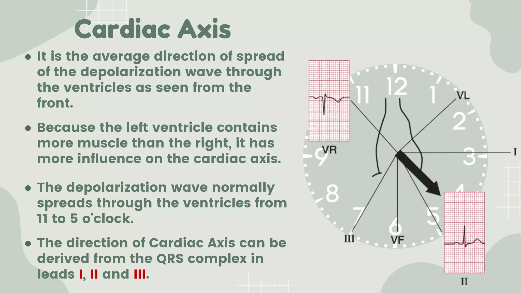
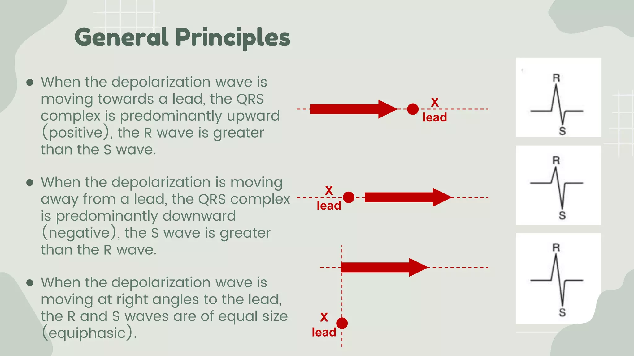
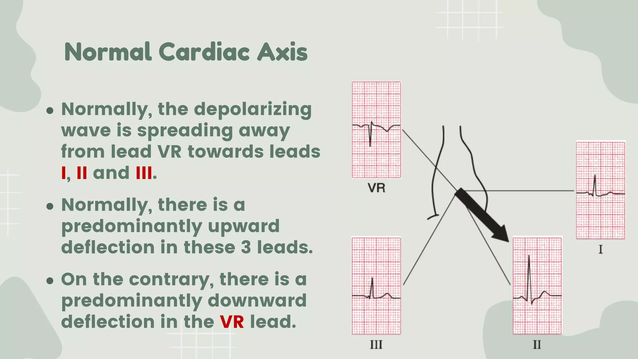
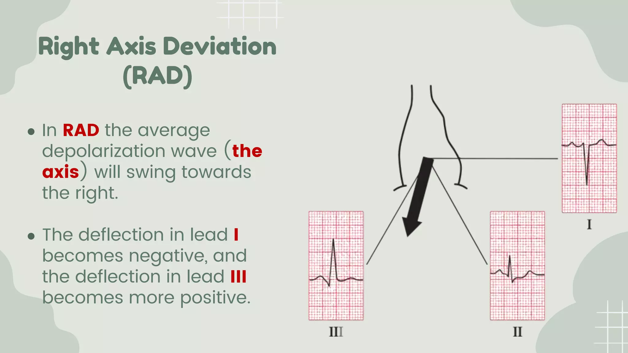
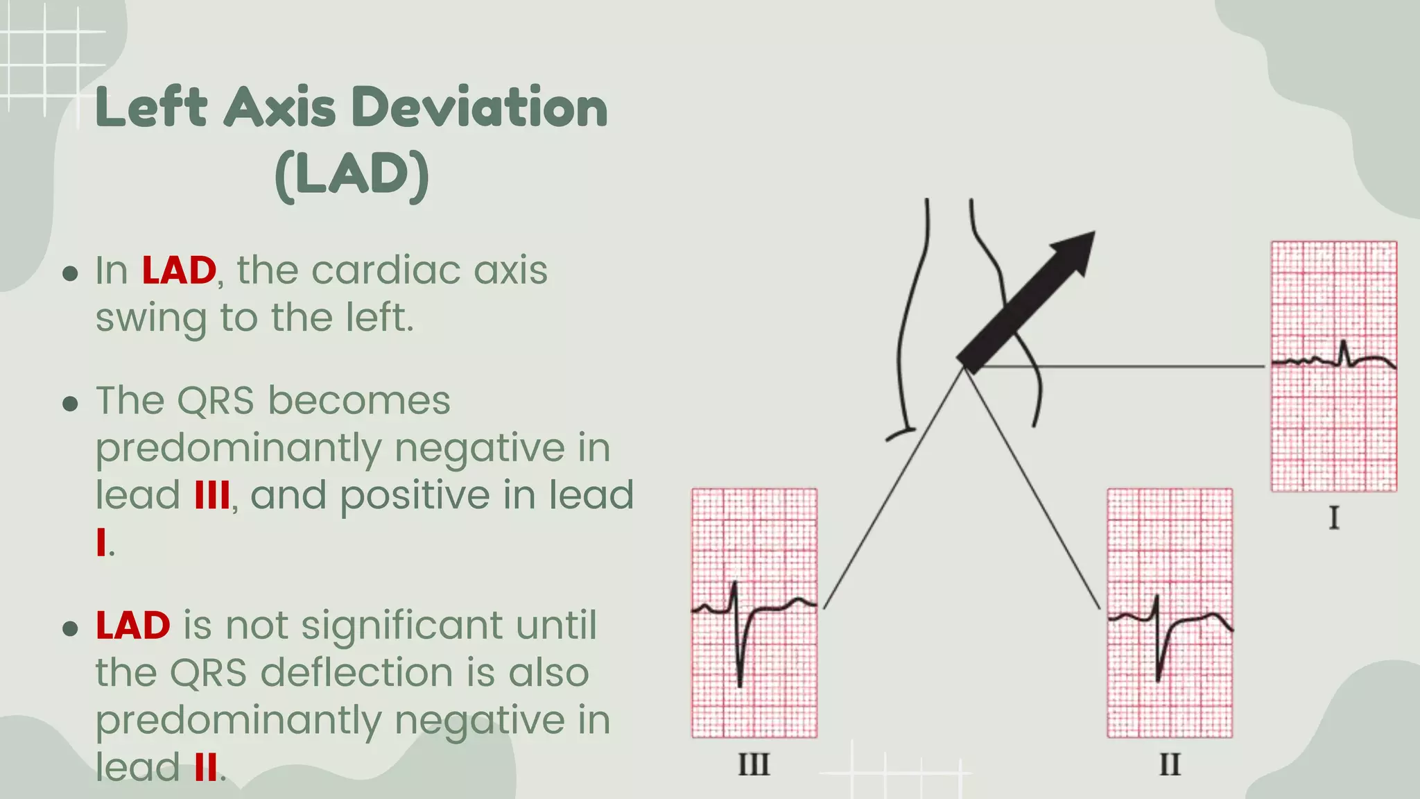
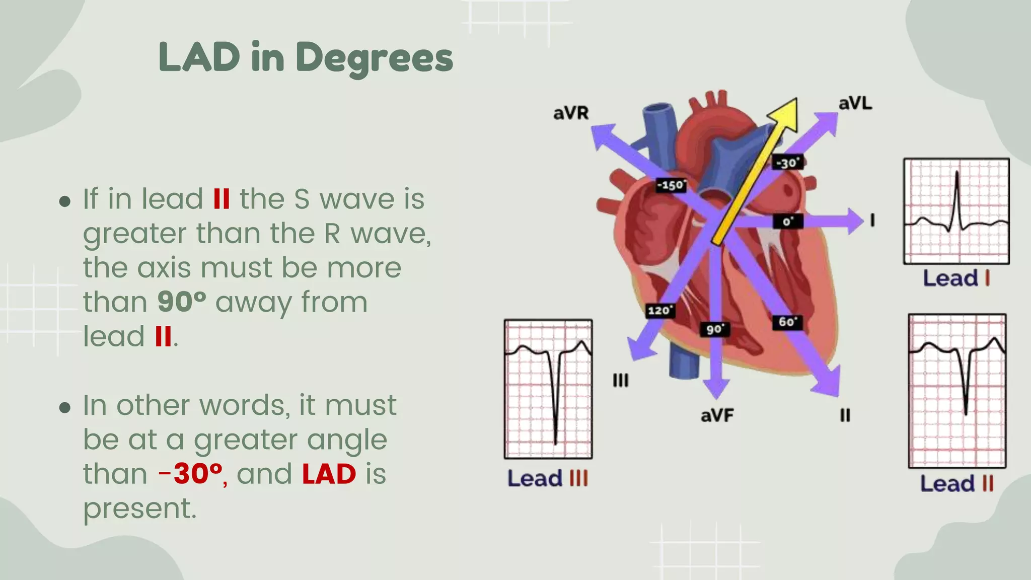
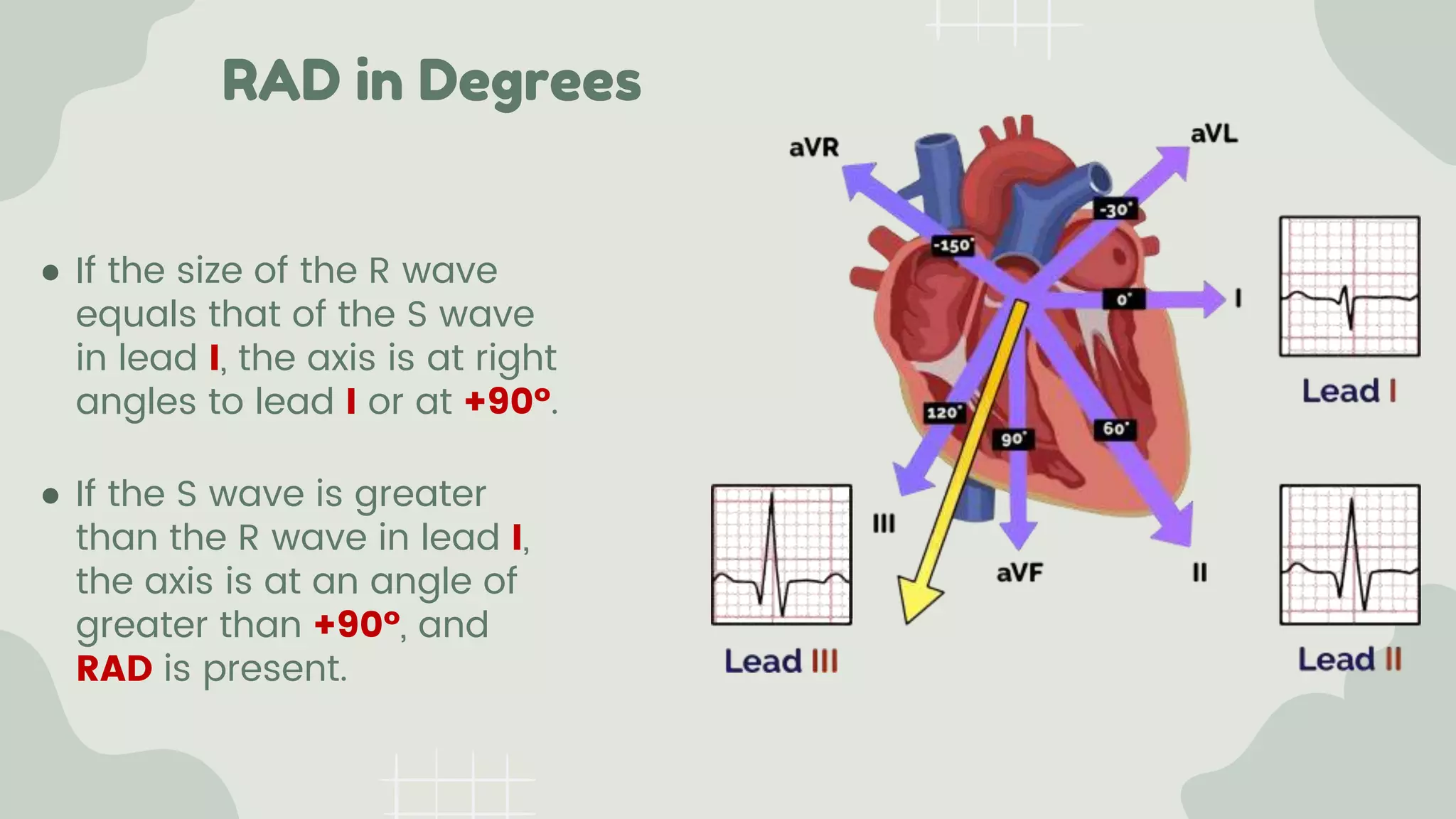
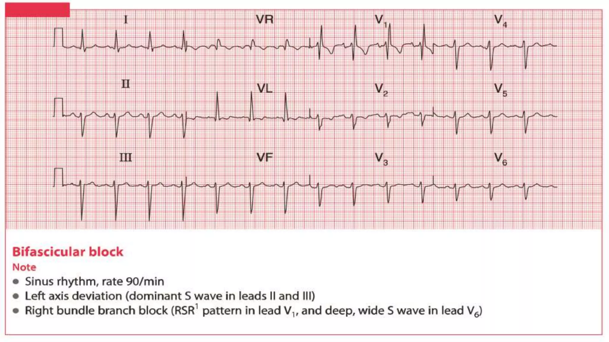
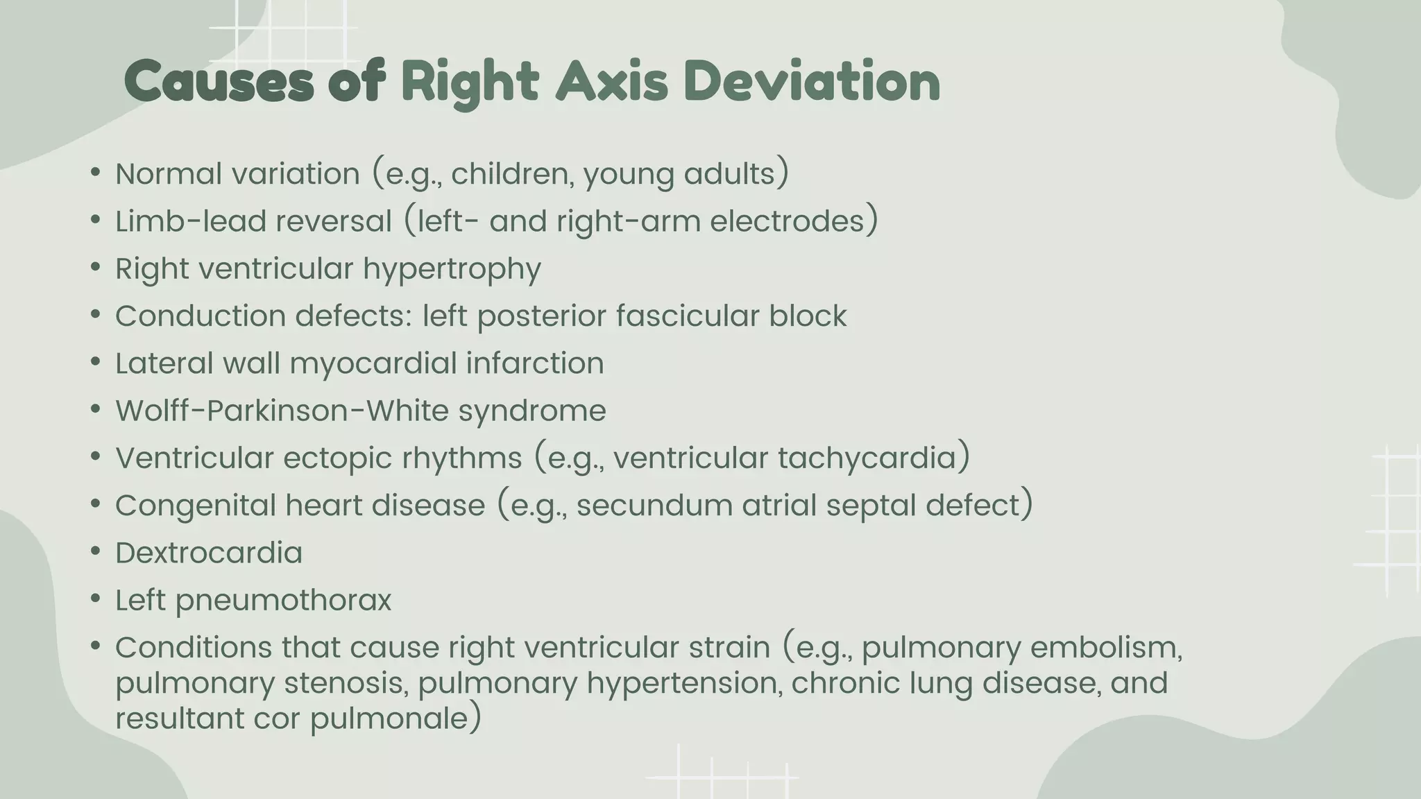
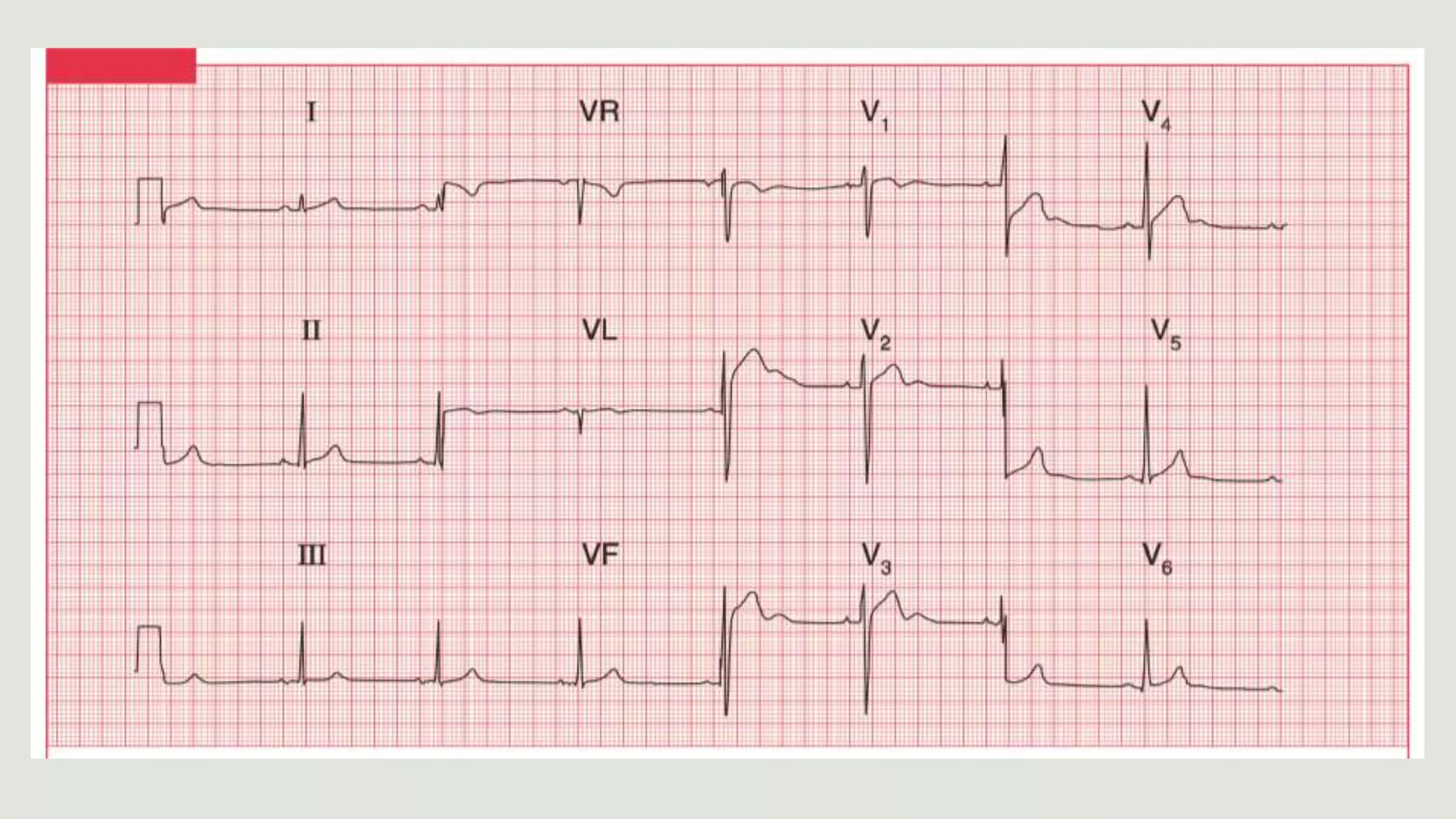
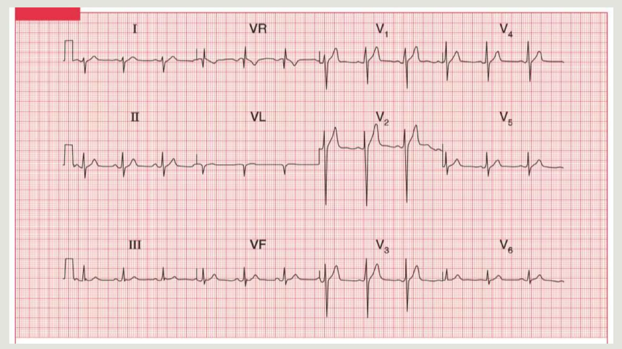
drahmadtemimi@gmail.com
Simplified
Cardiac Axis
● It is the average direction of spread
of the depolarization wave through
the ventricles as seen from the
front.
● Because the left ventricle contains
more muscle than the right, it has
more influence on the cardiac axis.
● The depolarization wave normally
spreads through the ventricles from
11 to 5 o'clock.
● The direction of Cardiac Axis can be
derived from the QRS complex in
leads I, II and III.
General Principles
● When the depolarization wave is
moving towards a lead, the QRS
complex is predominantly upward
(positive), the R wave is greater
than the S wave.
● When the depolarization is moving
away from a lead, the QRS complex
is predominantly downward
(negative), the S wave is greater
than the R wave.
● When the depolarization wave is
moving at right angles to the lead,
the R and S waves are of equal size
(equiphasic).
X
lead
X
lead
X
lead
Normal Cardiac Axis
● Normally, the depolarizing
wave is spreading away
from lead VR towards leads
I, II and III.
● Normally, there is a
predominantly upward
deflection in these 3 leads.
● On the contrary, there is a
predominantly downward
deflection in the VR lead.
Right Axis Deviation
(RAD)
● In RAD the average
depolarization wave (the
axis) will swing towards
the right.
● The deflection in lead I
becomes negative, and
the deflection in lead III
becomes more positive.
Left Axis Deviation
(LAD)
● In LAD, the cardiac axis
swing to the left.
● The QRS becomes
predominantly negative in
lead III, and positive in lead
I.
● LAD is not significant until
the QRS deflection is also
predominantly negative in
lead II.
The Cardiac Axis
in Degrees
● The cardiac axis is sometimes
measured in degrees, though
this is not clinically useful.
● Lead I is taken as looking at
the heart from 0°; lead II from
+60°; lead VF from +90°; and
lead III from +120°. Leads VL
and VR look from-30° and-
150°, respectively.
● The normal cardiac axis is in
the range -30° to +90°.
LAD in Degrees
● If in lead II the S wave is
greater than the R wave,
the axis must be more
than 90° away from
lead II.
● In other words, it must
be at a greater angle
than -30°, and LAD is
present.
RAD in Degrees
● If the size of the R wave
equals that of the S wave
in lead I, the axis is at right
angles to lead I or at +90°.
● If the S wave is greater
than the R wave in lead I,
the axis is at an angle of
greater than +90°, and
RAD is present.
Anatomy
of Conduction System
● The left bundle branch
has two divisions: the
anterior and posterior
'fascicles’.
● The right bundle
branch has no
divisions.
● The depolarization
wave therefore spreads
into the ventricles by
three pathways.
left Anterior Fascicular Block
● If the anterior fascicle of the LBB
fails to conduct, the left ventricle
depolarized through the posterior
fascicle, so the cardiac axis
rotates upwards.
● LAD is therefore due to left
anterior fascicular block, or 'left
anterior hemiblock’.
● If the posterior fascicle blocked
(rare), the ECG shows RAD.
Right Bundle Branch Block
● In RBBB the cardiac axis
usually remains normal,
because there is normal
depolarization of the left
ventricle with its large muscle
mass.
● If both the RBB and the left
anterior fascicle are blocked
(bifascicular block), the ECG
shows RBBB and LAD.
Causes of Right Axis Deviation
• Normal variation (e.g., children, young adults)
• Limb-lead reversal (left- and right-arm electrodes)
• Right ventricular hypertrophy
• Conduction defects: left posterior fascicular block
• Lateral wall myocardial infarction
• Wolff-Parkinson-White syndrome
• Ventricular ectopic rhythms (e.g., ventricular tachycardia)
• Congenital heart disease (e.g., secundum atrial septal defect)
• Dextrocardia
• Left pneumothorax
• Conditions that cause right ventricular strain (e.g., pulmonary embolism,
pulmonary stenosis, pulmonary hypertension, chronic lung disease, and
resultant cor pulmonale)
Causes of Left Axis Deviation
● Normal variation
● Left ventricular hypertrophy
● Conduction defects: left anterior fascicular block, bifascicular block
● Inferior wall myocardial infarction
● Wolff-Parkinson-White syndrome
● Ventricular ectopic rhythms (e.g., ventricular tachycardia)
● Congenital heart disease (e.g., primum atrial septal defect)
● Hyperkalemia
● Mechanical shift, such as with raised diaphragm (e.g., pregnancy,
ascites, abdominal tumor, organomegaly)
● Pacemaker-generated rhythm or paced rhythm
Cardiac Axis Simplified.pptx

Cardiac Axis Simplified.pptx

  • 1.
  • 2.
    Cardiac Axis ● Itis the average direction of spread of the depolarization wave through the ventricles as seen from the front. ● Because the left ventricle contains more muscle than the right, it has more influence on the cardiac axis. ● The depolarization wave normally spreads through the ventricles from 11 to 5 o'clock. ● The direction of Cardiac Axis can be derived from the QRS complex in leads I, II and III.
  • 3.
    General Principles ● Whenthe depolarization wave is moving towards a lead, the QRS complex is predominantly upward (positive), the R wave is greater than the S wave. ● When the depolarization is moving away from a lead, the QRS complex is predominantly downward (negative), the S wave is greater than the R wave. ● When the depolarization wave is moving at right angles to the lead, the R and S waves are of equal size (equiphasic). X lead X lead X lead
  • 4.
    Normal Cardiac Axis ●Normally, the depolarizing wave is spreading away from lead VR towards leads I, II and III. ● Normally, there is a predominantly upward deflection in these 3 leads. ● On the contrary, there is a predominantly downward deflection in the VR lead.
  • 5.
    Right Axis Deviation (RAD) ●In RAD the average depolarization wave (the axis) will swing towards the right. ● The deflection in lead I becomes negative, and the deflection in lead III becomes more positive.
  • 6.
    Left Axis Deviation (LAD) ●In LAD, the cardiac axis swing to the left. ● The QRS becomes predominantly negative in lead III, and positive in lead I. ● LAD is not significant until the QRS deflection is also predominantly negative in lead II.
  • 7.
    The Cardiac Axis inDegrees ● The cardiac axis is sometimes measured in degrees, though this is not clinically useful. ● Lead I is taken as looking at the heart from 0°; lead II from +60°; lead VF from +90°; and lead III from +120°. Leads VL and VR look from-30° and- 150°, respectively. ● The normal cardiac axis is in the range -30° to +90°.
  • 8.
    LAD in Degrees ●If in lead II the S wave is greater than the R wave, the axis must be more than 90° away from lead II. ● In other words, it must be at a greater angle than -30°, and LAD is present.
  • 9.
    RAD in Degrees ●If the size of the R wave equals that of the S wave in lead I, the axis is at right angles to lead I or at +90°. ● If the S wave is greater than the R wave in lead I, the axis is at an angle of greater than +90°, and RAD is present.
  • 11.
    Anatomy of Conduction System ●The left bundle branch has two divisions: the anterior and posterior 'fascicles’. ● The right bundle branch has no divisions. ● The depolarization wave therefore spreads into the ventricles by three pathways.
  • 12.
    left Anterior FascicularBlock ● If the anterior fascicle of the LBB fails to conduct, the left ventricle depolarized through the posterior fascicle, so the cardiac axis rotates upwards. ● LAD is therefore due to left anterior fascicular block, or 'left anterior hemiblock’. ● If the posterior fascicle blocked (rare), the ECG shows RAD.
  • 13.
    Right Bundle BranchBlock ● In RBBB the cardiac axis usually remains normal, because there is normal depolarization of the left ventricle with its large muscle mass. ● If both the RBB and the left anterior fascicle are blocked (bifascicular block), the ECG shows RBBB and LAD.
  • 15.
    Causes of RightAxis Deviation • Normal variation (e.g., children, young adults) • Limb-lead reversal (left- and right-arm electrodes) • Right ventricular hypertrophy • Conduction defects: left posterior fascicular block • Lateral wall myocardial infarction • Wolff-Parkinson-White syndrome • Ventricular ectopic rhythms (e.g., ventricular tachycardia) • Congenital heart disease (e.g., secundum atrial septal defect) • Dextrocardia • Left pneumothorax • Conditions that cause right ventricular strain (e.g., pulmonary embolism, pulmonary stenosis, pulmonary hypertension, chronic lung disease, and resultant cor pulmonale)
  • 16.
    Causes of LeftAxis Deviation ● Normal variation ● Left ventricular hypertrophy ● Conduction defects: left anterior fascicular block, bifascicular block ● Inferior wall myocardial infarction ● Wolff-Parkinson-White syndrome ● Ventricular ectopic rhythms (e.g., ventricular tachycardia) ● Congenital heart disease (e.g., primum atrial septal defect) ● Hyperkalemia ● Mechanical shift, such as with raised diaphragm (e.g., pregnancy, ascites, abdominal tumor, organomegaly) ● Pacemaker-generated rhythm or paced rhythm