Campylobacter & Helicobacter. Medical Importance, Pathogenesis, clinical signs
•Download as DOC, PDF•
3 likes•533 views

This document discusses Campylobacter and Helicobacter bacteria, which are important causes of gastrointestinal disease. Campylobacter jejuni commonly causes diarrhea, while Helicobacter pylori causes chronic gastritis and peptic ulcers. The document covers the morphology, virulence factors, transmission, clinical presentation, and laboratory diagnosis of these bacteria. It also discusses their pathogenesis, epidemiology, and methods for controlling infection.

Recommended
Recommended
More Related Content
What's hot
What's hot (18)
Similar to Campylobacter & Helicobacter. Medical Importance, Pathogenesis, clinical signs
Similar to Campylobacter & Helicobacter. Medical Importance, Pathogenesis, clinical signs (20)
More from Eneutron
More from Eneutron (20)
Recently uploaded
Recently uploaded (20)
Campylobacter & Helicobacter. Medical Importance, Pathogenesis, clinical signs
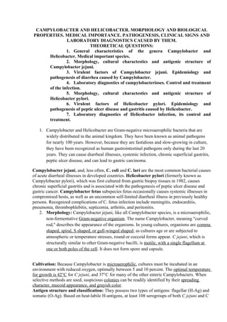
- 1. CAMPYLOBACTER AND HELICOBACTER. MORPHOLOGY AND BIOLOGICAL PROPERTIES. MEDICAL IMPORTANCE. PATHOGENESIS, CLINICAL SIGNS AND LABORATORY DIAGNOSTICS CAUSED BY THEM. THEORETICAL QUESTIONS: 1. General characteristics of the genera Campylobacter and Helicobacter. Medical important species. 2. Morphology, cultural charactestics and antigenic structure of Campylobacter jejuni. 3. Virulent factors of Campylobacter jejuni. Epidemiology and pathogenesis of diarrhea caused by Campylobacter. 4. Laboratory diagnostics of campylobacterioses. Control and treatment of the infection. 5. Morphology, cultural charactestics and antigenic structure of Helicobacter pylori. 6. Virulent factors of Helicobacter pylori. Epidemiology and pathogenesis of peptic ulcer disease and gastritis caused by Helicobacter. 7. Laboratory diagnostics of Helicobacter infection, its control and treatment. 1. Campylobacter and Helicobacter are Gram-negative microaerophilic bacteria that are widely distributed in the animal kingdom. They have been known as animal pathogens for nearly 100 years. However, because they are fastidious and slow-growing in culture, they have been recognized as human gastrointestinal pathogens only during the last 20 years. They can cause diarrheal illnesses, systemic infection, chronic superficial gastritis, peptic ulcer disease, and can lead to gastric carcinoma. Campylobacter jejuni, and, less often, C. coli and C. lari are the most common bacterial causes of acute diarrheal illnesses in developed countries. Helicobacter pylori (formerly known as Campylobacter pylori), which was first cultured from gastric biopsy tissues in 1982, causes chronic superficial gastritis and is associated with the pathogenesis of peptic ulcer disease and gastric cancer. Campylobacter fetus subspecies fetus occasionally causes systemic illnesses in compromised hosts, as well as an uncommon self-limited diarrheal illness in previously healthy persons. Recognized complications of C. fetus infection include meningitis, endocarditis, pneumonia, thrombophlebitis, septicemia, arthritis, and peritonitis. 2. Morphology: Campylobacter jejuni, like all Campylobacter species, is a microaerophilic, non-fermentative Gram-negative organism. The name Campylobacter, meaning "curved rod," describes the appearance of the organisms. In young cultures, organisms are comma shaped, spiral, S shaped, or gull-winged shaped; as cultures age or are subjected to atmospheric or temperature stresses, round or coccoid forms appear. C jejuni, which is structurally similar to other Gram-negative bacilli, is motile, with a single flagellum at one or both poles of the cell. It does not form spore and capsule. Cultivation: Because Campylobacter is microaerophilic, cultures must be incubated in an environment with reduced oxygen, optimally between 5 and 10 percent. The optimal temperature for growth is 42°C for C jejuni, and 37°C for many of the other enteric Campylobacters. When selective methods are used, suspicious colonies can be readily identified by their spreading character, mucoid appearance, and grayish color. Antigen structure and classification: They possess two types of antigens: flagellar (H-Ag) and somatic (O-Ag). Based on heat-labile H-antigens, at least 108 serogroups of both C jejuni and C
- 2. coli have been described. In addition, 47 and 18 different heat-stable somatic (O) antigens have been described among isolates of C jejuni and C coli organisms, respectively. 3. Virulent factors: a. Campylobacter lipopolysaccharide has endotoxin activity similar to that of other Gram-negative bacteria. b. Some C jejuni isolates elaborate very low levels of cytotoxins similar to Shiga toxin. c. Some isolates have been reported to elaborate an enterotoxin similar to cholera toxin. Enterotoxin production has been more frequently observed in isolates from developing countries, where infection by C jejuni has been associated with watery diarrhea. d. A superficial antigen (PEB1) that appears to be the major adhesin is conserved among C jejuni strains. However, the actual in vivo significance of adherence remains undefined. Epidemiology: In developed countries, C jejuni is an important cause of diarrhea, particularly in children and young adults. Between 3 and 14 percent of patients with diarrhea who seek medical attention are infected with C jejuni. Prolonged asymptomatic carriage is rare. The attack rate is highest in children less than 1 year old, and gradually decreases throughout childhood. A second peak occurs in young adults (18 to 29 years old). Although C jejuni enteritis occurs throughout the year, the highest isolation rates occur in summer, as with other enteric pathogens. In contrast, up to 40 percent of healthy children in developing countries may carry the organism at any time. This is an age-related phenomenon, with the highest excretion rates in very young children. The ultimate reservoir for C jejuni is gastrointestinal tract of many wild animals, and a variety of domestic animals, including food animals (cattle, sheep, poultry, swine, and goats). More than 50 percent of poultry sold is contaminated with C jejuni. Transmission from food sources accounts for most human infections. Rodents and pets including dogs, cats, and birds also may transmit infection to humans, and excreta from wild animals may contaminate water supplies. Therefore, C jejuni infection may be transmitted via food, water, or direct contact with infected animals; in rare cases it may be transmitted from person-to-person. Pathogenesis and clinical manifestation: As with other enteric pathogens, the attack rate of C jejuni varies with the ingested dose. In outbreaks of Campylobacter enteritis, the incubation period has ranged from 1-7 days, with most illness developing 2-4 days after infection. Infection leads to multiplication of organisms in the intestines. Patients shed 106 to 109 Campylobacter per gram of feces, concentrations similar to those shed in Salmonella and Shigella enteric infections. The sites of tissue injury include the small and large intestines, and the lesions show an acute exudative and hemorrhagic inflammation. The symptoms and signs of Campylobacter enteritis are not distinctive enough to differentiate it from illness caused by many other enteric pathogens. Symptoms range from mild gastrointestinal distress lasting 24 hours to a fulminating or relapsing colitis that mimics ulcerative colitis or Crohn's disease. The predominant symptoms experienced by individuals in developed countries are diarrhea, abdominal pain, fever, nausea, and vomiting. A cholera-like illness with massive watery diarrhea may also occur. Campylobacter enteritis usually is self-limiting with gradual improvement in symptoms over several days. Most patients recover within a week. Toxic megacolon, pseudomembranous colitis, and massive lower gastrointestinal hemorrhage also have been described. Mesenteric adenitis and appendicitis have been reported in children and young adults. Among populations in developing countries, infection by C jejuni and closely related organisms is associated with much milder illness, without bloody diarrhea, fever or fecal
- 3. leukocytes. Asymptomatic infection is much more common than in the developed countries, especially in older children and adults. Host Defenses Nonspecific defenses such as gastric acidity and intestinal transit time are important. Specific immunity, involving intestinal immunoglobulin (IgA) and systemic antibodies, develops. Persons deficient in humoral immunity develop severe and prolonged illnesses. 4. Laboratory diagnostics: Campylobacter enteritis is hard to distinguish from enteritis caused by other pathogens. The presence of neutrophils or blood in the feces of patients with acute diarrheal illnesses is an important clue to Campylobacter infection. a. Microscopy of the stained and native smears from fresh feces samples: Darting motility in a fresh fecal specimen observed by dark-field or phase- contrast microscopy or characteristic vibrio forms visible after Gram staining permit a presumptive diagnosis. b. Pure culture isolation. The diagnosis is confirmed by isolating the organism from a fecal culture or, rarely, from a blood culture. Because of its growth requirement for microaerobic atmosphere, special laboratory methods are needed to isolate C jejuni. Plating methods must be selective to inhibit the growth of competing microorganisms in the fecal flora. The traditional approach to isolating C jejuni has been to use media that contain antibiotics to which C jejuni is resistant but most members of the usual flora are susceptible. However, owing to their motility and small diameter, Campylobacter organisms have been isolated by filtration methods that do not use antibiotic-containing media. Use of filters (pore size 0.6µm) in conjunction with non-selective media improves stool culture yields of both C jejuni and the atypical enteric Campylobacters. c. Polymerase chain reaction (PCR)-based techniques have been developed for rapid detection, culture confirmation and for typing of C jejuni strains. Prophylaxis: Control of Campylobacter enteritis depends largely on interrupting the transmission of the organism to humans from farm and domestic animals, food of animal origin, or contaminated water. Individuals can reduce the risk of Campylobacter infection by properly cooking and storing meat and dairy products, avoiding contaminated drinking water and unpasteurized milk, and washing their hands after contact with animals or animal products. Helicobacter Pylori and other Gastric Helicobacter-Like Organisms H. pylori differs genetically from members of the genus Campylobacter, and has been reclassified from Campylobacter (where it was initially placed) to the separate genus Helicobacter. Morphology: H. pylori organisms are microaerophilic, nonsporulating, Gram-negative curved rods, 3.5 µm long and 0.5 to 1 µm wide, with a spiral periodicity in fresh cultures and spherical (coccoid) forms present in older cultures. H. pylori further differs from Campylobacter species in having multiple polar sheathed flagellae, a unique composition of cell wall fatty acids, and a smooth surface.
- 4. Cultivation: Unlike most campylobacters, H pylori produces urease and does not grow when incubated below 30°C. Growth is best on chocolate or blood agar plates after incubation for 2 to 5 days; for liquid media, either a blood or a hemin source appears essential. Classification and Antigenic Types. The antigenic nature of H. pylori has not been completely defined. The whole-cell and outer-membrane profiles of all H. pylori isolates have major similarities and are substantially different from those of C jejuni and C fetus. Virulent factors: 1) Urease activity. H. pylori is among the most efficient producers of urease. An important effect of this metabolic activity is the release of ammonia, which buffers acidity. Ammonia, produced by urease, is known to be toxic to eukaryotic cells and may potentiate mucosal injury. 2) High motility. H. pylori is highly motile even in very viscous mucus. This motility may allow organisms to migrate to the most favorable pH gradient. 3) Cytotoxin production. An 87kDa cytotoxin that induces vacuolation of eukaryotic cells is expressed in vitro by about 50% of strains. However, vacA, the gene encoding this toxin, is present in all strains but has substantial variability. Strains from patients with ulcers are more often toxin-producing than are strains from patients with gastritis only. 4) Protease. Isolates cultured in vitro produce an extracellular protease. This proteolytic activity affects the ability of mucus to retard diffusion of hydrogen ions. Epidemiology: H. pylori infection has a worldwide distribution; about 1/3 of the world's population is infected. The prevalence of infection increases with age. The major, if not exclusive, reservoir is humans but the exact modes of transmission are not known. The prevalence of these infections, as documented by both histologic and serologic studies, rises with age, as gastritis. Person-to-person transmission is the major, if not exclusive, source of infection. H. pylori has been isolated from dental plaque, and DNA products may be detected in saliva by PCR. H. pylori has been isolated from feces. These data indicate potential routes of transmission of H pylori. H. pylori is frequently isolated from asymptomatic persons who have no dyspeptic or ulcer-related symptoms. On occasion, transmission occurs from person to person via contaminated endoscopes. Host Defenses: Local and systemic humoral immune responses are essentially universal, but are not able to clear infection. Laboratory diagnostics: Some methods are used to diagnose 1) Microscopy: H. pylori can be presumptively identified in freshly prepared gastric biopsy smears by phase-contrast microscopy, based on the characteristic motility of the microorganisms, and by staining histologic sections from gastric biopsies with Gram (carbol fuchsin counterstain), Warthin-Starry silver, Giemsa, or acridine orange stains. Organisms also can be seen directly in fixed tissue stained with hematoxylin and eosin. 2) Pure culture isolation: H. pylori may be isolated from gastric tissue or from biopsies of esophageal or duodenal tissue containing gastric metaplasia using nonselective media, such as chocolate agar, or antibiotic-containing selective media, such as those of Skirrow or Goodwin. Spiral organisms that are oxidase-, catalase-, and urease-positive can be identified as H. pylori. Culture allows determination of antimicrobial susceptibilities. 3) Chemical method: In gastric biopsies, H.pylori also can be diagnosed presumptively, on the basis of the presence of preformed urease.
- 5. 4) DNA probe and PCR methodologies have been developed as well. 5) All of the above tests require endoscopy and biopsy. A non-invasive technique known as the urea breath test has been developed to diagnose H. pylori infection. 6) Serology: Infection can also be diagnosed accurately by detecting serum antibodies to H. pylori antigens. These methods may be more sensitive than diagnostic methods involving biopsies. Examination of gastric biopsy or stained smears allows presumptive diagnosis; definitive diagnosis is made by culture. Recently, non-invasive techniques such as the urea breath test and serologic tests have been developed to diagnose H pylori infection, with accuracy exceeding 95 percent. Control : Antimicrphobial therapy for treatment of this infection has emerged as the most important means to resolve H pylori infection. Antimicrobial therapy is now one of the primary therapies for duodenal ulceration. Studies to identify the best combinations of antibiotics are being done. However, for most cases of H pylori-associated non-ulcer dyspepsia, data related to efficacy of antimicrobial therapy are not clear.
- 6. 4) DNA probe and PCR methodologies have been developed as well. 5) All of the above tests require endoscopy and biopsy. A non-invasive technique known as the urea breath test has been developed to diagnose H. pylori infection. 6) Serology: Infection can also be diagnosed accurately by detecting serum antibodies to H. pylori antigens. These methods may be more sensitive than diagnostic methods involving biopsies. Examination of gastric biopsy or stained smears allows presumptive diagnosis; definitive diagnosis is made by culture. Recently, non-invasive techniques such as the urea breath test and serologic tests have been developed to diagnose H pylori infection, with accuracy exceeding 95 percent. Control : Antimicrphobial therapy for treatment of this infection has emerged as the most important means to resolve H pylori infection. Antimicrobial therapy is now one of the primary therapies for duodenal ulceration. Studies to identify the best combinations of antibiotics are being done. However, for most cases of H pylori-associated non-ulcer dyspepsia, data related to efficacy of antimicrobial therapy are not clear.