Lecture Notes
on
Campylobacter
Dr VidyaSingh
Senior Scientist
Division of Pathology, ICAR-Indian Veterinary Research
Institute
Izatnagar, Bareilly, UP, India- 243122
2.
Campylobacter
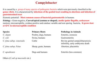
It is causedby a group of many species of pathogenic bacteria which were previously classified in the
genus vibrio. It is characterized by infections of the genital tract resulting in abortions and infection of
gastrointestinal tract
Zoonotic potential: Most common causes of bacterial gastroenteritis in humans
Etiology - Gram-negative, Curved/spiral (comma or shaped), motile (polar flagellla, corkscreew
motion), microaerophilic, oxidase positive and catalase variable and non sporing bacteria, It grows best
at 42o
C in microaerophilic condition
Species Primary Hosts Pathology in Animals
C. Jejuni Poultry, dogs, humans Enteritis, zoonosis
C. coli Swine, humans Gastroenteritis
C. fetus subsp. venerealis Cattle Bovine genital campylobacteriosis
infertility, early embryonic death
C. fetus subsp. Fetus Sheep, goats, humans Abortion, placentitis
C. upsaliensis Dogs and humans Enteritis (less common)
Others (C.sub sp mucosalis etc.)
3.
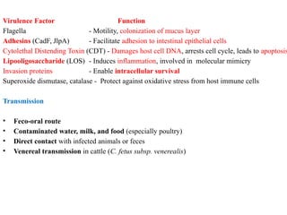
Virulence Factor Function
Flagella- Motility, colonization of mucus layer
Adhesins (CadF, JlpA) - Facilitate adhesion to intestinal epithelial cells
Cytolethal Distending Toxin (CDT) - Damages host cell DNA, arrests cell cycle, leads to apoptosis
Lipooligosaccharide (LOS) - Induces inflammation, involved in molecular mimicry
Invasion proteins - Enable intracellular survival
Superoxide dismutase, catalase - Protect against oxidative stress from host immune cells
Transmission
• Feco-oral route
• Contaminated water, milk, and food (especially poultry)
• Direct contact with infected animals or feces
• Venereal transmission in cattle (C. fetus subsp. venerealis)
4.
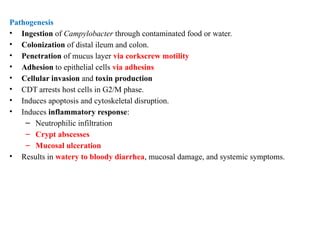
Pathogenesis
• Ingestion ofCampylobacter through contaminated food or water.
• Colonization of distal ileum and colon.
• Penetration of mucus layer via corkscrew motility
• Adhesion to epithelial cells via adhesins
• Cellular invasion and toxin production
• CDT arrests host cells in G2/M phase.
• Induces apoptosis and cytoskeletal disruption.
• Induces inflammatory response:
– Neutrophilic infiltration
– Crypt abscesses
– Mucosal ulceration
• Results in watery to bloody diarrhea, mucosal damage, and systemic symptoms.
5.
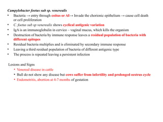
Campylobacter foetus subsp. venerealis
• Bacteria → entry through coitus or AI→ Invade the chorionic epithelium → cause cell death
or cell proliferation
• C. foetus sub sp venerealis shows cyclical antigenic variation
• IgA is an immunoglobulin in cervico – vaginal mucus, which kills the organism
• Destruction of bacteria by immune response leaves a residual population of bacteria with
different epitopes
• Residual bacteria multiplies and is eliminated by secondary immune response
• Leaving a third residual population of bacteria of different antigenic type
• The process is repeated leaving a persistent infection
Lesions and Signs
• Venereal disease in cattle
• Bull do not show any disease but cows suffer from infertility and prolonged oestrus cycle
• Endometritis, abortion at 4-7 months of gestation
6.
Campylobacter foetus subsp. - Foetus
• Infects sheep through oral route
• Birds spread the infection
Pathogenesis
Ingestion of contaminated feed/water → multiply in local lymph nodes →
bacteraemia → localises in the pregnant uterus → haematoma in placenta → separation
of foetal membrane → edema of cotyledons → late abortion, still birth, birth of weak
lambs with necrotic foci in liver
Camylobacter jejuni
• · Normal inhabitant of intestinal tract
• · Causes diarrhoea in animals; abortions in sheep and goats &Mastitis in cattle
Campylobacter sputorum sub sp. mucosalis
• Causes swine proliferative ileitis syndrome or porcine intestinal adenomatosis
(PIA)
7.
Clinical Signs
Clinical Pathologyin Animals
In Poultry (Primary Reservoir)
• Mostly asymptomatic carriers
• Rarely cause diarrhea
• Major public health concern due to contamination of meat
In Dogs and Cats
• Puppies and kittens more susceptible
• Signs: Mucoid or watery diarrhea, vomiting, fever
In Cattle
C. fetus subsp. venerealis:
• Causes bovine genital campylobacteriosis
• Transmitted via natural service or contaminated AI equipment
• Results in early embryonic death, repeat breeding, irregular estrus cycles
C. fetus subsp. fetus:
• Causes abortion in late pregnancy, especially in sheep
• Placentitis, fetal liver necrosis, and peritonitis may be seen
Diagnosis
A. Clinical Diagnosis
B.Laboratory Diagnosis:
• Campylobacter detected in tissue sections of placenta
• Identified by electron microscopy or immunostaining
Stool/Fecal culture : Selective media (Skirrow's, Campy-BAP), grows at 42°C, microaerophilic conditions
Microscopy : Dark field or phase contrast shows darting/corkscrew motility
PCR : Species identification, rapid
Serology : Mainly for herd screening in bovine genital campylobacteriosis
Histopathology : Demonstrates characteristic lesions in tissue
FAT : Used for detection in semen or fetal tissues
10.
Differential Diagnosis
• Salmonellaspp.
• Yersinia enterocolitis
• Escherichia coli (EPEC, EHEC)
• Cryptosporidium spp.
• Rotavirus, coronavirus (in young animals)
Treatment :
• Mild cases: Supportive therapy only (fluids, electrolytes)
• Severe cases or systemic involvement:
– Macrolides (e.g., Azithromycin, Erythromycin) – drug of choice
– Fluoroquinolone – increasing resistance observed
– Tyrosine or gentamicin in veterinary practice (based on susceptibility)
• Note: Treatment in animals should follow antibiotic stewardship principles.
11.
Control & Prevention
•Good hygiene and sanitation
• Proper cooking of meat (especially poultry)
• Pasteurization of milk
• Chlorinated water supply
In cattle:
• Use of artificial insemination
• Testing and culling of infected bulls
• Vaccination in endemic herds (for C. fetus infections)
ZOONOTIC SIGNIFICANCE
• Campylobacter jejuni is a major zoonotic pathogen associated with raw poultry and milk
• Common cause of foodborne illness in humans.
• Guillain-Barré Syndrome (GBS):
• Autoimmune sequela due to molecular mimicry (LOS mimics GM1 gangliosides)
• Leads to ascending paralysis
• Also linked to:
• Reactive arthritis
• Irritable bowel syndrome (IBS) post-infection