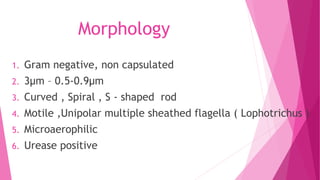
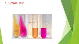
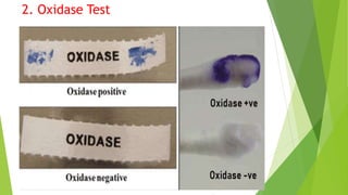
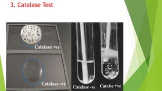
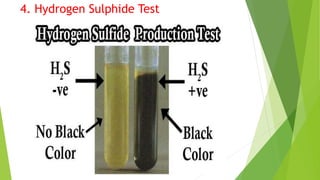
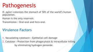
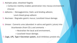
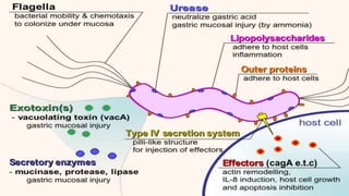
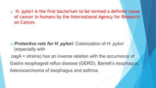
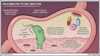
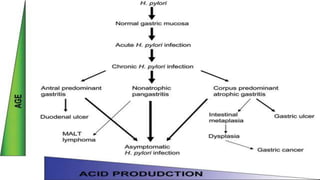
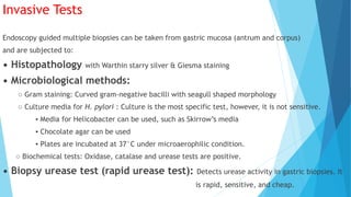
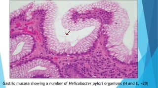
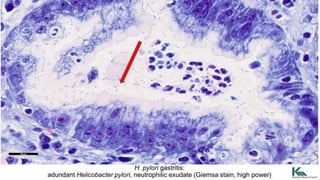
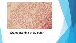
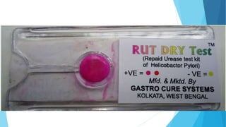
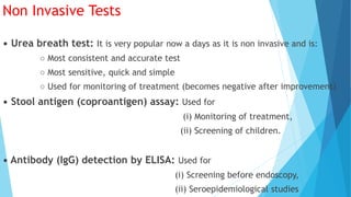
Helicobacter pylori
Made By : Niteesh Kumar
Introduction
H. pylori is a spiral-shaped, gram-negative rod. H pylori is associated
with antral gastritis, duodenal (peptic) ulcer disease, gastric ulcers,
gastric adenocarcinoma, and gastric mucosa-associated lymphoid
tissue (MALT) lymphomas. Previously known as Campylobacter pylori.
Morphology
1. Gram negative, non capsulated
2. 3μm – 0.5-0.9μm
3. Curved , Spiral , S - shaped rod
4. Motile ,Unipolar multiple sheathed flagella ( Lophotrichus )
5. Microaerophilic
6. Urease positive
Cultural Properties
 Microaerophilic.
(5-10% CO2, 2-5% O2 and high humidity).
 34 to 40°C, optimum 37°C.
 Cultures should be inspected from day 3
to day 14 (average 3-7 days).
 H. pylori forms small (0.5-2 mm), round,
convex, translucent smooth colonies.
 Fastidious organism requires enriched &
selective growth media as:
- Blood agar.
- Chocolate agar.
- Modified Skirrow’s blood agar (selective
medium, blood agar with antibiotics) containing
vancomycin, polymyxin & trimethoprim.
Skirrow’s Media containing vancomycin, polymyxin &
trimethoprim
Biochemical Characters
1. Highly urease positive (unlike Campylobacter)
Convert urea to ammonia which make the environment alkaline.
2. Oxidase positive Bacterial cytochrome C oxidase in
respiratory chain is present
3. Catalase positive Convert Hydrogen peroxide into water and
oxygen
4. Hydrogen sulphide production positive.
1. Urease Test
2. Oxidase Test
3. Catalase Test
4. Hydrogen Sulphide Test
Pathogenesis
H. pylori colonizes the stomach of 50% of the world’s human
population.
Human is the only reservoir.
Transmission : Oral-oral and feco-oral.
Virulence Factors
1. Vacuolating cytotoxin : Epithelial cell damage
2. Catalase : Protection from phagocytosis & intracellular killing
by eliminating hydrogen peroxide.
3. Multiple polar, sheathed flagella
• Corkscrew motility enables penetration into viscous environment
(mucus)
4. Adhesins : Hemagglutinins; Sialic acid binding adhesin;
Lewis blood group adhesin
5. Mucinase : Degrades gastric mucus; Localized tissue damage.
6. Urease : Converts urea (abundant in saliva and gastric juices) into
bicarbonate (from CO₂) and ammonia -
• Neutralize the local acid environment,
• Localized tissue damage.
7. CagA, LPS, Type 4 Secretion system, Acid-inhibitory protein etc.
Clinical Manifestations
• Acute gastritis (Antrum is the most common site involved, cardiac
end is not involved).
• Antral gastritis: Predisposes to duodenal ulcers.
• Pan gastritis: Predisposes to adenocarcinoma of stomach.
• Peptic ulcer disease: 80% of duodenal ulcers and 60% of
gastric ulcers are due to H.pylori.
• Chronic atrophic gastritis
• Autoimmune gastritis
• Promotes pernicious anemia
• Adenocarcinoma of stomach Non-Hodgkin’s gastric lymphoma.
 H. pylori is the first bacterium to be termed a definite cause
of cancer in humans by the International Agency for Research
on Cancer.
 Protective role for H. pylori: Colonization of H. pylori
(especially with
cagA + strains) has an inverse relation with the occurrence of
Gastro esophageal reflux disease (GERD), Barrett’s esophagus,
Adenocarcinoma of esophagus and asthma.
The Nobel Prize in Physiology or Medicine 2005 was awarded
jointly to Barry J. Marshall and J. Robin Warren "for their
discovery of the bacterium Helicobacter pylori and its role in
gastritis and peptic ulcer disease."
Risk factors for PUD
H. Pylori infection
• NSAIDs
• Cigarette use
• COPD
• Illicit drugs like cocaine
• Alcoholic cirrhosis
• Psychological stress
• Endocrine cell hyperplasia
• ZE syndrome
• Viral infection (CMV, HSV)
Sign & Symptoms :
1. Epigastric pain
2. Dyspepsia, including belching, bloating, distention,
and fatty food intolerance
3. Heartburn
4. Chest discomfort
5. Hematemesis or melena resulting from
gastrointestinal bleeding
Complications
• Haemorrhage
Blood vessels damaged as ulcer erodes into the muscles GIT leading to Coffee
ground vomitus or occult blood in tarry stools
• Perforation
An ulcer can erode through the entire wall Bacteria and partially digested
food spill into peritoneum : Peritonitis and referred pain in shoulder.
• Narrowing and obstruction (pyloric)
Swelling and scarring can cause obstruction of food leaving stomach leading
to repeated vomiting
Myself JAMES JOYCE ,
famous Irish novelist and
poet died because of
perforated duodenal ulcer
caused by Helicobacter
pylori.
Laboratory Diagnosis
1. Invasive Test
A : Histopathology
B : Microbiological Tests
C : Biopsy Urease Test
2 .Non Invasive Test
A : Urea Breath Test
B : Stool Antigen Testing
C : Antibody Detection
Invasive Tests
Endoscopy guided multiple biopsies can be taken from gastric mucosa (antrum and corpus)
and are subjected to:
• Histopathology with Warthin starry silver & Giesma staining
• Microbiological methods:
○ Gram staining: Curved gram-negative bacilli with seagull shaped morphology
○ Culture media for H. pylori : Culture is the most specific test, however, it is not sensitive.
▪ Media for Helicobacter can be used, such as Skirrow’s media
▪ Chocolate agar can be used
▪ Plates are incubated at 37°C under microaerophilic condition.
○ Biochemical tests: Oxidase, catalase and urease tests are positive.
• Biopsy urease test (rapid urease test): Detects urease activity in gastric biopsies. It
is rapid, sensitive, and cheap.
Gastric mucosa showing a number of Helicobacter pylori organisms (H and E, ×20)
Grams staining of H. pylori
Non Invasive Tests
• Urea breath test: It is very popular now a days as it is non invasive and is:
○ Most consistent and accurate test
○ Most sensitive, quick and simple
○ Used for monitoring of treatment (becomes negative after improvement)
• Stool antigen (coproantigen) assay: Used for
(i) Monitoring of treatment,
(ii) Screening of children.
• Antibody (IgG) detection by ELISA: Used for
(i) Screening before endoscopy,
(ii) Seroepidemiological studies
H. pylori
H. pylori
H. pylori

H. pylori

  • 1.
  • 2.
    Introduction H. pylori isa spiral-shaped, gram-negative rod. H pylori is associated with antral gastritis, duodenal (peptic) ulcer disease, gastric ulcers, gastric adenocarcinoma, and gastric mucosa-associated lymphoid tissue (MALT) lymphomas. Previously known as Campylobacter pylori.
  • 3.
    Morphology 1. Gram negative,non capsulated 2. 3μm – 0.5-0.9μm 3. Curved , Spiral , S - shaped rod 4. Motile ,Unipolar multiple sheathed flagella ( Lophotrichus ) 5. Microaerophilic 6. Urease positive
  • 5.
    Cultural Properties  Microaerophilic. (5-10%CO2, 2-5% O2 and high humidity).  34 to 40°C, optimum 37°C.  Cultures should be inspected from day 3 to day 14 (average 3-7 days).  H. pylori forms small (0.5-2 mm), round, convex, translucent smooth colonies.
  • 6.
     Fastidious organismrequires enriched & selective growth media as: - Blood agar. - Chocolate agar. - Modified Skirrow’s blood agar (selective medium, blood agar with antibiotics) containing vancomycin, polymyxin & trimethoprim.
  • 8.
    Skirrow’s Media containingvancomycin, polymyxin & trimethoprim
  • 9.
    Biochemical Characters 1. Highlyurease positive (unlike Campylobacter) Convert urea to ammonia which make the environment alkaline. 2. Oxidase positive Bacterial cytochrome C oxidase in respiratory chain is present 3. Catalase positive Convert Hydrogen peroxide into water and oxygen 4. Hydrogen sulphide production positive.
  • 10.
  • 11.
  • 12.
  • 13.
  • 14.
    Pathogenesis H. pylori colonizesthe stomach of 50% of the world’s human population. Human is the only reservoir. Transmission : Oral-oral and feco-oral. Virulence Factors 1. Vacuolating cytotoxin : Epithelial cell damage 2. Catalase : Protection from phagocytosis & intracellular killing by eliminating hydrogen peroxide.
  • 15.
    3. Multiple polar,sheathed flagella • Corkscrew motility enables penetration into viscous environment (mucus) 4. Adhesins : Hemagglutinins; Sialic acid binding adhesin; Lewis blood group adhesin 5. Mucinase : Degrades gastric mucus; Localized tissue damage. 6. Urease : Converts urea (abundant in saliva and gastric juices) into bicarbonate (from CO₂) and ammonia - • Neutralize the local acid environment, • Localized tissue damage. 7. CagA, LPS, Type 4 Secretion system, Acid-inhibitory protein etc.
  • 17.
    Clinical Manifestations • Acutegastritis (Antrum is the most common site involved, cardiac end is not involved). • Antral gastritis: Predisposes to duodenal ulcers. • Pan gastritis: Predisposes to adenocarcinoma of stomach. • Peptic ulcer disease: 80% of duodenal ulcers and 60% of gastric ulcers are due to H.pylori. • Chronic atrophic gastritis • Autoimmune gastritis • Promotes pernicious anemia • Adenocarcinoma of stomach Non-Hodgkin’s gastric lymphoma.
  • 18.
     H. pyloriis the first bacterium to be termed a definite cause of cancer in humans by the International Agency for Research on Cancer.  Protective role for H. pylori: Colonization of H. pylori (especially with cagA + strains) has an inverse relation with the occurrence of Gastro esophageal reflux disease (GERD), Barrett’s esophagus, Adenocarcinoma of esophagus and asthma.
  • 19.
    The Nobel Prizein Physiology or Medicine 2005 was awarded jointly to Barry J. Marshall and J. Robin Warren "for their discovery of the bacterium Helicobacter pylori and its role in gastritis and peptic ulcer disease."
  • 22.
    Risk factors forPUD H. Pylori infection • NSAIDs • Cigarette use • COPD • Illicit drugs like cocaine • Alcoholic cirrhosis • Psychological stress • Endocrine cell hyperplasia • ZE syndrome • Viral infection (CMV, HSV)
  • 23.
    Sign & Symptoms: 1. Epigastric pain 2. Dyspepsia, including belching, bloating, distention, and fatty food intolerance 3. Heartburn 4. Chest discomfort 5. Hematemesis or melena resulting from gastrointestinal bleeding
  • 25.
    Complications • Haemorrhage Blood vesselsdamaged as ulcer erodes into the muscles GIT leading to Coffee ground vomitus or occult blood in tarry stools • Perforation An ulcer can erode through the entire wall Bacteria and partially digested food spill into peritoneum : Peritonitis and referred pain in shoulder. • Narrowing and obstruction (pyloric) Swelling and scarring can cause obstruction of food leaving stomach leading to repeated vomiting
  • 26.
    Myself JAMES JOYCE, famous Irish novelist and poet died because of perforated duodenal ulcer caused by Helicobacter pylori.
  • 29.
    Laboratory Diagnosis 1. InvasiveTest A : Histopathology B : Microbiological Tests C : Biopsy Urease Test 2 .Non Invasive Test A : Urea Breath Test B : Stool Antigen Testing C : Antibody Detection
  • 30.
    Invasive Tests Endoscopy guidedmultiple biopsies can be taken from gastric mucosa (antrum and corpus) and are subjected to: • Histopathology with Warthin starry silver & Giesma staining • Microbiological methods: ○ Gram staining: Curved gram-negative bacilli with seagull shaped morphology ○ Culture media for H. pylori : Culture is the most specific test, however, it is not sensitive. ▪ Media for Helicobacter can be used, such as Skirrow’s media ▪ Chocolate agar can be used ▪ Plates are incubated at 37°C under microaerophilic condition. ○ Biochemical tests: Oxidase, catalase and urease tests are positive. • Biopsy urease test (rapid urease test): Detects urease activity in gastric biopsies. It is rapid, sensitive, and cheap.
  • 31.
    Gastric mucosa showinga number of Helicobacter pylori organisms (H and E, ×20)
  • 33.
  • 35.
    Non Invasive Tests •Urea breath test: It is very popular now a days as it is non invasive and is: ○ Most consistent and accurate test ○ Most sensitive, quick and simple ○ Used for monitoring of treatment (becomes negative after improvement) • Stool antigen (coproantigen) assay: Used for (i) Monitoring of treatment, (ii) Screening of children. • Antibody (IgG) detection by ELISA: Used for (i) Screening before endoscopy, (ii) Seroepidemiological studies