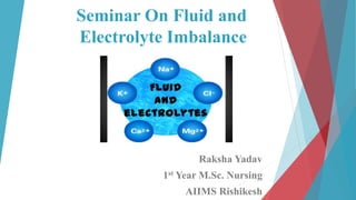
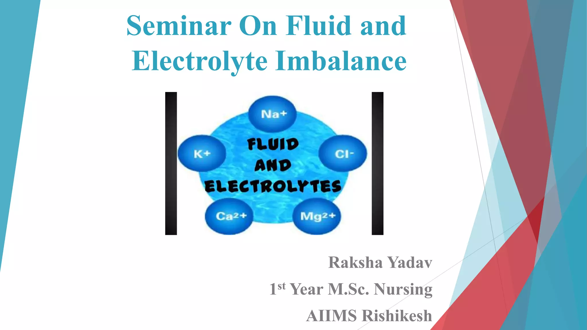
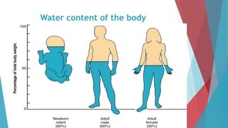
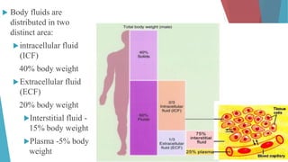
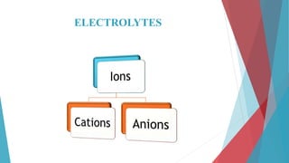
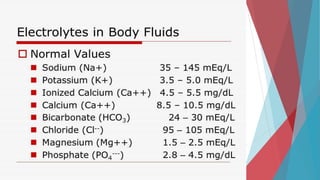
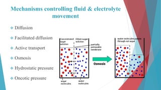
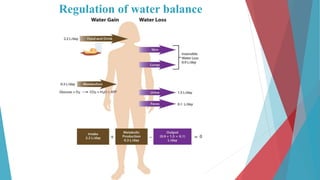
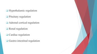
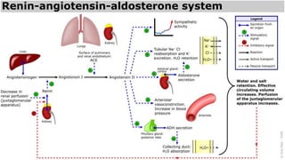
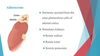
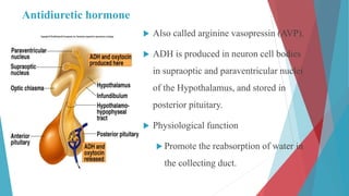
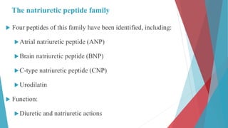
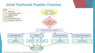
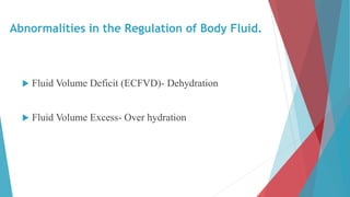
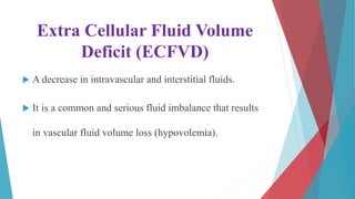
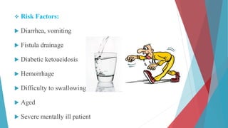
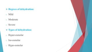
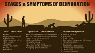
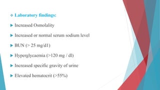
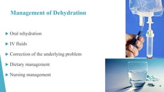
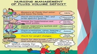
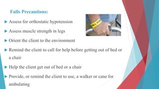
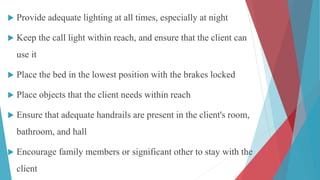
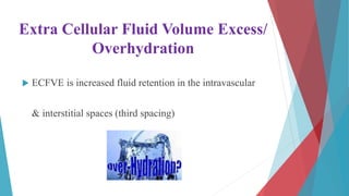
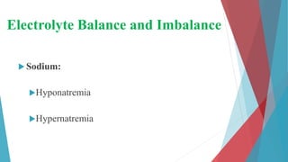
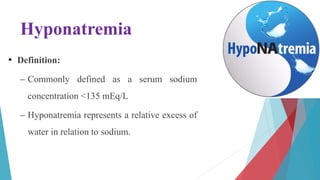
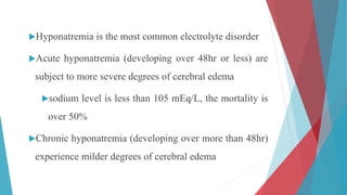
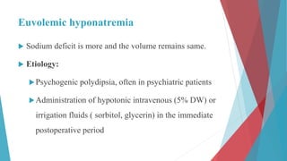
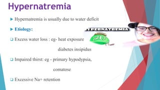
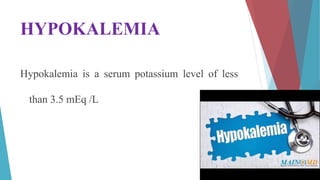
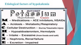
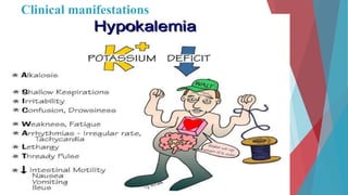
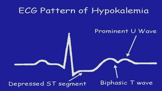
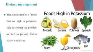
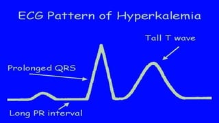
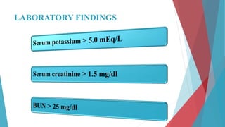
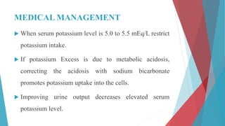
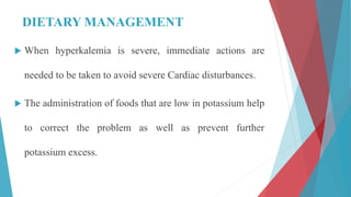
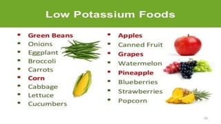
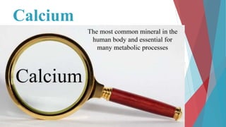
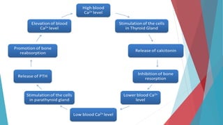
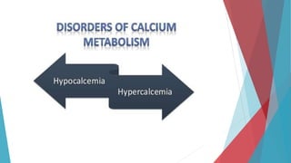
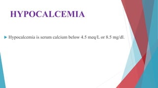
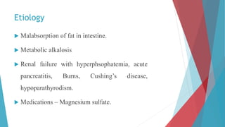
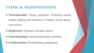
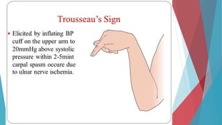
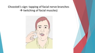
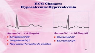
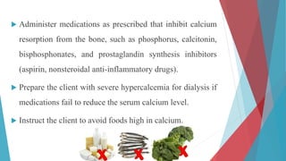
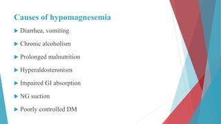
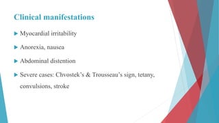
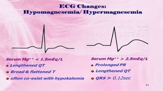
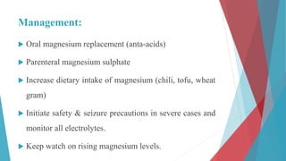
Seminar On Fluid and
Electrolyte Imbalance
Raksha Yadav
1st Year M.Sc. Nursing
AIIMS Rishikesh
INTRODUCTION
HOMEOSTASIS
Water content of the body
 Body fluids are
distributed in two
distinct area:
intracellular fluid
(ICF)
40% body weight
Extracellular fluid
(ECF)
20% body weight
Interstitial fluid -
15% body weight
Plasma -5% body
weight
ELECTROLYTES
Mechanisms controlling fluid & electrolyte
movement
 Diffusion
 Facilitated diffusion
 Active transport
 Osmosis
 Hydrostatic pressure
 Oncotic pressure
Regulation of water balance
 Hypothalamic regulation
 Pituitary regulation
 Adrenal cortical regulation
 Renal regulation
 Cardiac regulation
 Gastro-intestinal regulation
Aldosterone
 Hormone secreted from the
zona glomerulosa cells of
adrenal cortex
 Stimulates kidneys
Retain sodium
Retain water
Secrete potassium
Antidiuretic hormone
 Also called arginine vasopressin (AVP).
 ADH is produced in neuron cell bodies
in supraoptic and paraventricular nuclei
of the Hypothalamus, and stored in
posterior pituitary.
 Physiological function
 Promote the reabsorption of water in
the collecting duct.
The natriuretic peptide family
 Four peptides of this family have been identified, including:
Atrial natriuretic peptide (ANP)
Brain natriuretic peptide (BNP)
C-type natriuretic peptide (CNP)
Urodilatin
 Function:
Diuretic and natriuretic actions
Atrial Natriuretic Peptide: Function
The sensation of thirst
 Conscious desire for water
 Major factor that determines fluid intake
 Initiated by the osmoreceptors in
hypothalamus that are stimulated by
increase in osmotic pressure of body fluids
 Also stimulated by a decrease in the blood
pressure through the baroreceptors.
The regulation of thirsty reaction
 The stimulus sensed by osmoreceptor:
• Not a change in the extracellular fluid osmolality
• But a change in osmoreceptor neuron size or in the some intracellular substance.
Abnormalities in the Regulation of Body Fluid.
 Fluid Volume Deficit (ECFVD)- Dehydration
 Fluid Volume Excess- Over hydration
Extra Cellular Fluid Volume
Deficit (ECFVD)
 A decrease in intravascular and interstitial fluids.
 It is a common and serious fluid imbalance that results
in vascular fluid volume loss (hypovolemia).
 Risk Factors:
 Diarrhea, vomiting
 Fistula drainage
 Diabetic ketoacidosis
 Hemorrhage
 Difficulty to swallowing
 Aged
 Severe mentally ill patient
 Degrees of dehydration:
o Mild
o Moderate
o Severe
 Types of dehydration:
o Hyper-osmolar
o Iso-osmolar
o Hypo-osmolar
 Laboratory findings:
 Increased Osmolality
 Increased or normal serum sodium level
 BUN (> 25 mg/d1)
 Hyperglycaemia (>120 mg / dl)
 Increased specific gravity of urine
 Elevated hematocrit (>55%)
Management of Dehydration
 Oral rehydration
 IV fluids
 Correction of the underlying problem
 Dietary management
 Nursing management
Falls Precautions:
 Assess for orthostatic hypotension
 Assess muscle strength in legs
 Orient the client to the environment
 Remind the client to call for help before getting out of bed or
a chair
 Help the client get out of bed or a chair
 Provide, or remind the client to use, a walker or cane for
ambulating
 Provide adequate lighting at all times, especially at night
 Keep the call light within reach, and ensure that the client can
use it
 Place the bed in the lowest position with the brakes locked
 Place objects that the client needs within reach
 Ensure that adequate handrails are present in the client's room,
bathroom, and hall
 Encourage family members or significant other to stay with the
client
Extra Cellular Fluid Volume Excess/
Overhydration
 ECFVE is increased fluid retention in the intravascular
& interstitial spaces (third spacing)
Etiology:
 Administering fluids rapidly or in a large amount
 Failure to excrete fluids:
o Heart failure
o Renal disorders
o Venous obstructions
o Decreased plasma proteins
o Excessive fluid ingestion
o Increased ADH & Aldosterone
Decreased Excretion
Increased absorption
 Laboratory findings:
 Decreased Osmolality
 Decreased or normal serum sodium level
 BUN (<8 mg/dl)
 Decreased specific gravity of urine
 Decreased hematocrit (<45%)
Management of over hydration
 ICFVE is treated by the addition of solutes to IV fluids.
 Use of D5%, 0.45% Nacl will help to correct ICFVE
when the cause is water excess.
 Oral fluids such as water and soft drinks should be
given in addition to water and ice chips.
 IV therapy should be monitored every hour.
 Monitor vital signs and intake- output
 Weight should be checked daily to measure fluid gain or
loss.
 Administer prescribed antiemetic as needed to allow
food and fluids to be ingested.
 Safety measures are necessary when the client displays
behavioral changes.
NURSING INTERVENTIONS
 Monitor cardiovascular, respiratory, neuromuscular,
renal, integumentary, and gastrointestinal status.
 Prevent further fluid overload and restore normal fluid
balance.
 Administer diuretics; osmotic diuretics typically are
prescribed first to prevent severe electrolyte imbalances.
 Restrict fluid and sodium intake as prescribed.
 Monitor intake and output; monitor weight.
 Monitor electrolyte values, and prepare to administer
medication to treat an imbalance if present.
Electrolyte Balance and Imbalance
 Sodium:
Hyponatremia
Hypernatremia
Hyponatremia
• Definition:
– Commonly defined as a serum sodium
concentration <135 mEq/L
– Hyponatremia represents a relative excess of
water in relation to sodium.
Hyponatremia is the most common electrolyte disorder
Acute hyponatremia (developing over 48hr or less) are
subject to more severe degrees of cerebral edema
sodium level is less than 105 mEq/L, the mortality is
over 50%
Chronic hyponatremia (developing over more than 48hr)
experience milder degrees of cerebral edema
Types of hyponatremia
Hypovolemic hyponatremia
Euvolemic hyponatremia
Hypervolemic hyponatremia
Redistributive hyponatremia
Hypovolemic hyponatremia
 Develops as sodium and free water are lost and/or
replaced by inappropriately hypotonic fluids
 Sodium can be lost through renal or non-renal routes
 Nonrenal loss:
 GI losses
Vomiting, Diarrhea, fistulas, pancreatitis
 Excessive sweating
 Third spacing of fluids
ascites, peritonitis, pancreatitis, and burns
 Cerebral salt-wasting syndrome
traumatic brain injury, aneurysmal subarachnoid
hemorrhage, and intracranial surgery
Must distinguish from SIADH
 Renal Loss:
Acute or chronic renal insufficiency
Diuretics
Euvolemic hyponatremia
 Sodium deficit is more and the volume remains same.
 Etiology:
Psychogenic polydipsia, often in psychiatric patients
Administration of hypotonic intravenous (5% DW) or
irrigation fluids ( sorbitol, glycerin) in the immediate
postoperative period
administration of hypotonic maintenance intravenous
fluids
Infants who may have been given inappropriate
amounts of free water
bowel preparation before colonoscopy or colorectal
surgery
Hypervolemic hyponatremia
 Total body sodium increases, and TBW increases to a greater
extent.
 Can be renal or non-renal
acute or chronic renal failure
dysfunctional kidneys are unable to excrete the ingested
sodium load
cirrhosis, congestive heart failure, or nephrotic syndrome
Redistributive hyponatremia
Water shifts from the intracellular to the extracellular
compartment, with a resultant dilution of sodium. The TBW
and total body sodium are unchanged.
This condition occurs with hyperglycemia
Administration of mannitol
MEDICAL MANAGEMENT
 Determine cause of hyponatremia and to correct it.
 If client has hyponatremia due to fluid volume excess,
intake of fluids will be restricted to allow the sodium to
regain balance.
 If the serum sodium level falls below 125 mEq/L,
sodium replacement is needed.
PHARMACOLOGIC MANAGEMENT
 For client with moderate hyponatremia 125 meq/ L I/V
saline solution (0.9% Nacl) or lactated Ringer solution
may be ordered.
 When the serum sodium level is 115 meq / L or less, a
concentrated saline solution such as 3 % Nacl is
indicated.
NURSING INTERVENTIONS
 Monitor cardiovascular, respiratory, neuromuscular,
cerebral, renal, and gastrointestinal status of the client.
 If hyponatremia is accompanied by a fluid volume
deficit (hypovolemia), IV sodium chloride infusions are
administered to restore sodium content and fluid
volume.
 If hyponatremia is accompanied by fluid volume
excess (hypervolemia), osmotic diuretics are
administered to promote the excretion of water rather
than sodium.
 Instruct the client to increase oral sodium intake and
inform the client about the foods to include in the diet.
 If the client is taking lithium (Lithobid), monitor the
lithium level, because hyponatremia can cause
diminished lithium excretion, resulting in toxicity.
Hypernatremia
 Hypernatremia is usually due to water deficit
 Etiology:
 Excess water loss : eg- heat exposure
diabetes insipidus
 Impaired thirst: eg - primary hypodypsia,
comatose
 Excessive Na+ retention
Clinical features of hypernatremia
 Excessive thirst, polyuria, nausea
 Muscular weakness, neuromuscular
irritability
 Altered mental status,focal
neurological deficit occasionally coma
or seizures
Treatment
 correct water deficit
 Rate of correction :
-Acute hypernatremia- 1mEq/L/hr
-Chronic hypernatremia-1mEq/L/hr or 10mEq/L over 24hr
-rapid correction may lead to cerebral edema
Dietary management
HYPOKALEMIA
Hypokalemia is a serum potassium level of less
than 3.5 mEq /L
Etiological factors of hypokalemia
Clinical manifestations
Medical management
 Determining & correcting the cause of the imbalance.
 Extreme hypokalemia requires cardiac monitoring.
Pharmacological Management
 Oral potassium replacement therapy is usually prescribed for
mild hypokalemia.
 Potassium is extremely irritating to gastric mucosa; therefore
the drug must be taken with Glass of water or juice or during
meals.
 Potassium chloride can be administered intravenously for
moderate or severe hypokalemia & must be diluted in IV
fluids.
 Administration of potassium by IV push may result in
cardiac arrests. Potassium can be given in doses of 10 to
20 mEq/ hour diluted in IV fluid if the client is on heart
monitor.
 High concentration of potassium is irritating to heart
muscle. Thus correcting a potassium deficit may take
several days.
Dietary management
 The administration of foods
that are high in potassium
help to correct the problem
as well as prevent further
potassium losses.
HYPERKALEMIA
Hyperkalemia is an Elevated potassium level over 5.0 mEq/L.
ETIOLOGY
 Retention of Potassium- Renal insufficiency, renal failure,
decreased urine output, potassium sparing diuretics.
 Excessive release of Cellular Potassium - severe traumatic
injuries. Severe burns, severe infection, metabolic
acidosis.
 Excessive IV infusions or Oral administration of
potassium.
CLINICAL MANIFESTATIONS
LABORATORY FINDINGS
MEDICAL MANAGEMENT
 When serum potassium level is 5.0 to 5.5 mEq/L restrict
potassium intake.
 If potassium Excess is due to metabolic acidosis,
correcting the acidosis with sodium bicarbonate
promotes potassium uptake into the cells.
 Improving urine output decreases elevated serum
potassium level.
DIETARY MANAGEMENT
 When hyperkalemia is severe, immediate actions are
needed to be taken to avoid severe Cardiac disturbances.
 The administration of foods that are low in potassium help
to correct the problem as well as prevent further
potassium excess.
NURSING INTERVENTIONS
 Monitor cardiovascular, respiratory, neuromuscular,
renal, and gastrointestinal status; place the client on a
cardiac monitor.
 Discontinue IV potassium and hold oral potassium
supplements.
 Prepare to administer potassium-excreting diuretics if
renal function is not impaired.
 Initiate a potassium-restricted diet.
 If renal function is impaired, prepare to administer
sodium polystyrene sulfonate (Kayexalate).
 Prepare the client for dialysis if potassium levels are
critically high.
 Prepare for the IV administration of hypertonic glucose
with regular insulin to move excess potassium into the
cells.
 Monitor renal function.
 Teach the client to avoid foods high in potassium.
 Instruct the client to avoid the use of salt substitutes or
other potassium-containing substances.
Calcium
HYPOCALCEMIA
 Hypocalcemia is serum calcium below 4.5 meq/L or 8.5 mg/dl.
Etiology
 Malabsorption of fat in intestine.
 Metabolic alkalosis
 Renal failure with hyperphsophatemia, acute
pancreatitis, Burns, Cushing’s disease,
hypoparathyrodism.
 Medications – Magnesium sulfate.
CLINICAL MANIFESTATIONS
 Neuromuscular: Tetany symptoms: Twitching around
mouth, tingling and numbness of fingers, facial spasm,
convulsions.
 Respiratory: Dyspnea, laryngeal spasm.
 Gastrointestinal: increased peristalsis, diarrhea.
 Cardiovascular: Dysrhythmias, palpitations
MEDICAL MANAGEMENT
 Determining & correcting the cause of hypocalcemia.
 Asymptomatic hypocalcemia is usually corrected with oral
calcium gluconate, calcium lactate or calcium chloride.
 Administer calcium supplements 30 minutes before meals
for better absorption and with glass of milk because vitamin
D is necessary for absorption of calcium from the intestine.
 Intravenous calcium chloride or calcium gluconate
(10%) is given slowly to avoid hypertension,
bradycardia & other arrhythmias.
 Chronic or mild hypocalcemia can be treated in part by
having the client consume a diet high in calcium.
NURSING INTERVENTIONS
 Monitor cardiovascular, respiratory, neuromuscular, and
gastrointestinal status; place the client on a cardiac monitor.
 Administer calcium supplements orally or calcium
intravenously.
 When administering calcium intravenously, warm the injection
solution to body temperature before administration and
administer slowly, monitor for electrocardiographic changes,
and monitor for hypercalcemia.
 Administer medications that increase calcium
absorption. i.e. Vitamin D aids in the absorption of
calcium from the intestinal tract.
 Initiate seizure precautions.
 Keep 10% calcium gluconate available for treatment of
acute calcium deficit.
 Instruct the client to consume foods high in calcium.
HYPERCALCEMIA
 Hypercalcemia is a serum level over 5.5 meq/L or 11 mg/L
ETIOLOGY
 Metastatic malignancy-lung, breast, Ovarian, Prostatic,
bladder, leukemia, Kidney.
 Hyperparathyroidism.
 Thiazide diuretic therapy.
 Prolong immobilization.
 Excessive intake of calcium supplements and vitamin D.
CLINICAL MANIFESTATIONS
 Gastrointestinal: Anorexia, Vomiting, Constipation
 Neuromuscular:
Mild to moderate hypercalcemia state –weakness
Severe hypercalcemic state-Extreme lethargy
 Cardiovascular: Dysrhythmias
 Electro-cardiographic Changes: Shortened ST Segment and
lengthened QT interval.
 Musculoskeletal: Bone pain, fracture.
LABORATORY FINDINGS
MEDICAL MANAGEMENT
 Treatment consists of correcting the underlying cause.
 Intravenous normal saline (0.9% Nacl) given rapidly
with furosemide to prevent fluid overload, Promote
urinary calcium excretion.
 Corticosteroid drugs decrease calcium levels by
competing with vitamin D thus resulting in decreased
intestinal absorption of calcium.
 If the cause is excessive use of calcium or vitamin D
supplements or calcium containing antacids these agents
should be either avoided or used in reduced dosage.
 A newer form of drug therapy is etidronate di-sodium.
This drug reduces serum calcium by reducing normal
and abnormal bone reabsorption of calcium and
secondarily by reducing bone formation.
NURSING INTERVENTIONS
 Monitor cardiovascular, respiratory, neuromuscular, renal,
and gastrointestinal status; place the client on a cardiac
monitor.
 Discontinue IV infusions of solutions containing calcium and
oral medications containing calcium or vitamin D.
 Discontinue thiazide diuretics and replace with diuretics that
enhance the excretion of calcium.
 Administer medications as prescribed that inhibit calcium
resorption from the bone, such as phosphorus, calcitonin,
bisphosphonates, and prostaglandin synthesis inhibitors
(aspirin, nonsteroidal anti-inflammatory drugs).
 Prepare the client with severe hypercalcemia for dialysis if
medications fail to reduce the serum calcium level.
 Instruct the client to avoid foods high in calcium.
Phosphorus
 Hypophosphatemia
 Hyperphosphatemia
Hypophosphatemia
 Hypophosphatemia is defined as plasma phosphorus
<1.2mEq/L
Causes of hypophosphatemia
 Malabsorption syndrome
 TPN
 Alcohol withdrawal
 Phosphate binding anta-acids
 Respiratory alkalosis
 Recovery from DKA
Clinical manifestations
 Muscle weakness
 Osteomalacia
 Rhabdomyolysis
 Cadiac problems- Dysrhythmias, decreased stroke
volume
 CNS dysfunction
Management:
 Mild cases- treated with dietary restrictions
 Teach client about balanced diet
 Severe cases: phosphate supplementation
Hyperphosphatemia
 Hyperphosphatemia is defined as plasma
phosphorus >3mEq/L
Causes of hyperphosphatemia
 Excess intake of high phosphate foods
 Excess vitamine D supplementation especially in renal
insufficiency
 Impaired colonic motility
 Hypoparathyroidism
 Addison’s disease
Clinical manifestations
 Tachycardia, palpitations
 Restlessness
 Anorexia, nausea, vomiting
 Hyper-reflexia, tetany
 Dysrhythmias
Management:
 Mild cases- limit phosphate rich foods ( milk, ice-cream,
cheese, meat, fish)
 Giving calcium, aluminium products that promotes
binding & excretion of phosphate.
 Dialysis is the TOC in case of hyperphosphatemia with
renal failure.
Magnesium
Hypomagnesemia
Hypermagnesemia
Hypomagnesemia
 Hypomagnesemia is defined as plasma magnesium
<1.8mg/dl
Causes of hypomagnesemia
 Diarrhea, vomiting
 Chronic alcoholism
 Prolonged malnutrition
 Hyperaldosteronism
 Impaired GI absorption
 NG suction
 Poorly controlled DM
Clinical manifestations
 Myocardial irritability
 Anorexia, nausea
 Abdominal distention
 Severe cases: Chvostek’s & Trousseau’s sign, tetany,
convulsions, stroke
Management:
 Oral magnesium replacement (anta-acids)
 Parenteral magnesium sulphate
 Increase dietary intake of magnesium (chili, tofu, wheat
gram)
 Initiate safety & seizure precautions in severe cases and
monitor all electrolytes.
 Keep watch on rising magnesium levels.
Hypermagnesemia
 Hypermagnesemia is defined as plasma magnesium
>3mg/dl
Causes of hypermagnesemia
 Renal insufficiency
 Excessive anta-acid use
 Adrenal insufficiency
 Ketoacidosis
Clinical manifestations
Clinical amnifestations are related to blocked release of
Acetylcholine from myoneronal junction which affects muscle
cell activity.
 Hypotension
 Muscle weakness
 Loss of DTR
 Prolonged QT, PR interval
 Lethargy, drowiness
 Respiratory paralysis, loss of consiosness
Management:
 Low magnesium diet (eat chicken, eggs, green peas, white
bread, hamburger)
 Decrease magnesium sulphate use
 In severe cases saline infusion with diuretics is give to
promote magnesium excretion
 IV calcium (antagonistic action)
 Drugs: Albuterol
 If renal failure is also present than hemodialysis is done in
severe cases
Bibliography
 Priscilla Lemone, Karen Burke. “Medical surgical nursing, critical thinking in client care”.
4th edition (2008). Page no. 185-198
 Ignatavicius Workman. “Medical surgical nursing, Patient centred collaborative care”. 6th
edition (2010). Elsevier publication. Page no. 1022-1024
 Brunner and Suddharth. “Text book of medical surgical nursing”. Page no. 249-255
 Guyyton and Hall. “Text book of medical physiology”. 11th edition (2006). Elsevier
publications. Page no. 642-650
 Gerard.J.Tortora. “Principles of anatomy and physiology”. 11th edition (2006). Wiley
publication. Page no. 1122-1130
 K. Sembulingam, Prema Sembulingam. “Essentials of medical
physiology”. 5thedition (2010). Jaypee publications. Page no. 302-
309
1. The type of fluid used to manipulate fluid shifts
among compartments states is:
A. Whole blood
B. TPN
C. Albumin
D. Normal saline
2. The balance of anions and cations as it occurs
across cell membranes is known as:
A. osmotic activity
B. Electrical neutrality
C. Electrical stability
D. Sodium potassium pump
3. A diet containing the minimum
daily sodium requirement for an adult would
be:
A. no-salt diet
B. diet including 2 gm sodium
C. diet including 4 gm sodium
D. 1500 calorie weight-loss diet
THANK YOU…………

Fluid and electrolyte imbalance and management

  • 1.
    Seminar On Fluidand Electrolyte Imbalance Raksha Yadav 1st Year M.Sc. Nursing AIIMS Rishikesh
  • 2.
  • 3.
  • 4.
  • 5.
     Body fluidsare distributed in two distinct area: intracellular fluid (ICF) 40% body weight Extracellular fluid (ECF) 20% body weight Interstitial fluid - 15% body weight Plasma -5% body weight
  • 6.
  • 8.
    Mechanisms controlling fluid& electrolyte movement  Diffusion  Facilitated diffusion  Active transport  Osmosis  Hydrostatic pressure  Oncotic pressure
  • 9.
  • 10.
     Hypothalamic regulation Pituitary regulation  Adrenal cortical regulation  Renal regulation  Cardiac regulation  Gastro-intestinal regulation
  • 12.
    Aldosterone  Hormone secretedfrom the zona glomerulosa cells of adrenal cortex  Stimulates kidneys Retain sodium Retain water Secrete potassium
  • 13.
    Antidiuretic hormone  Alsocalled arginine vasopressin (AVP).  ADH is produced in neuron cell bodies in supraoptic and paraventricular nuclei of the Hypothalamus, and stored in posterior pituitary.  Physiological function  Promote the reabsorption of water in the collecting duct.
  • 14.
    The natriuretic peptidefamily  Four peptides of this family have been identified, including: Atrial natriuretic peptide (ANP) Brain natriuretic peptide (BNP) C-type natriuretic peptide (CNP) Urodilatin  Function: Diuretic and natriuretic actions
  • 15.
  • 16.
    The sensation ofthirst  Conscious desire for water  Major factor that determines fluid intake  Initiated by the osmoreceptors in hypothalamus that are stimulated by increase in osmotic pressure of body fluids  Also stimulated by a decrease in the blood pressure through the baroreceptors.
  • 17.
    The regulation ofthirsty reaction  The stimulus sensed by osmoreceptor: • Not a change in the extracellular fluid osmolality • But a change in osmoreceptor neuron size or in the some intracellular substance.
  • 21.
    Abnormalities in theRegulation of Body Fluid.  Fluid Volume Deficit (ECFVD)- Dehydration  Fluid Volume Excess- Over hydration
  • 22.
    Extra Cellular FluidVolume Deficit (ECFVD)  A decrease in intravascular and interstitial fluids.  It is a common and serious fluid imbalance that results in vascular fluid volume loss (hypovolemia).
  • 23.
     Risk Factors: Diarrhea, vomiting  Fistula drainage  Diabetic ketoacidosis  Hemorrhage  Difficulty to swallowing  Aged  Severe mentally ill patient
  • 24.
     Degrees ofdehydration: o Mild o Moderate o Severe  Types of dehydration: o Hyper-osmolar o Iso-osmolar o Hypo-osmolar
  • 26.
     Laboratory findings: Increased Osmolality  Increased or normal serum sodium level  BUN (> 25 mg/d1)  Hyperglycaemia (>120 mg / dl)  Increased specific gravity of urine  Elevated hematocrit (>55%)
  • 27.
    Management of Dehydration Oral rehydration  IV fluids  Correction of the underlying problem  Dietary management  Nursing management
  • 29.
    Falls Precautions:  Assessfor orthostatic hypotension  Assess muscle strength in legs  Orient the client to the environment  Remind the client to call for help before getting out of bed or a chair  Help the client get out of bed or a chair  Provide, or remind the client to use, a walker or cane for ambulating
  • 30.
     Provide adequatelighting at all times, especially at night  Keep the call light within reach, and ensure that the client can use it  Place the bed in the lowest position with the brakes locked  Place objects that the client needs within reach  Ensure that adequate handrails are present in the client's room, bathroom, and hall  Encourage family members or significant other to stay with the client
  • 31.
    Extra Cellular FluidVolume Excess/ Overhydration  ECFVE is increased fluid retention in the intravascular & interstitial spaces (third spacing)
  • 32.
    Etiology:  Administering fluidsrapidly or in a large amount  Failure to excrete fluids: o Heart failure o Renal disorders o Venous obstructions o Decreased plasma proteins o Excessive fluid ingestion o Increased ADH & Aldosterone Decreased Excretion Increased absorption
  • 35.
     Laboratory findings: Decreased Osmolality  Decreased or normal serum sodium level  BUN (<8 mg/dl)  Decreased specific gravity of urine  Decreased hematocrit (<45%)
  • 36.
    Management of overhydration  ICFVE is treated by the addition of solutes to IV fluids.  Use of D5%, 0.45% Nacl will help to correct ICFVE when the cause is water excess.  Oral fluids such as water and soft drinks should be given in addition to water and ice chips.  IV therapy should be monitored every hour.
  • 37.
     Monitor vitalsigns and intake- output  Weight should be checked daily to measure fluid gain or loss.  Administer prescribed antiemetic as needed to allow food and fluids to be ingested.  Safety measures are necessary when the client displays behavioral changes.
  • 38.
    NURSING INTERVENTIONS  Monitorcardiovascular, respiratory, neuromuscular, renal, integumentary, and gastrointestinal status.  Prevent further fluid overload and restore normal fluid balance.  Administer diuretics; osmotic diuretics typically are prescribed first to prevent severe electrolyte imbalances.
  • 39.
     Restrict fluidand sodium intake as prescribed.  Monitor intake and output; monitor weight.  Monitor electrolyte values, and prepare to administer medication to treat an imbalance if present.
  • 41.
    Electrolyte Balance andImbalance  Sodium: Hyponatremia Hypernatremia
  • 42.
    Hyponatremia • Definition: – Commonlydefined as a serum sodium concentration <135 mEq/L – Hyponatremia represents a relative excess of water in relation to sodium.
  • 43.
    Hyponatremia is themost common electrolyte disorder Acute hyponatremia (developing over 48hr or less) are subject to more severe degrees of cerebral edema sodium level is less than 105 mEq/L, the mortality is over 50% Chronic hyponatremia (developing over more than 48hr) experience milder degrees of cerebral edema
  • 44.
    Types of hyponatremia Hypovolemichyponatremia Euvolemic hyponatremia Hypervolemic hyponatremia Redistributive hyponatremia
  • 45.
    Hypovolemic hyponatremia  Developsas sodium and free water are lost and/or replaced by inappropriately hypotonic fluids  Sodium can be lost through renal or non-renal routes
  • 46.
     Nonrenal loss: GI losses Vomiting, Diarrhea, fistulas, pancreatitis  Excessive sweating  Third spacing of fluids ascites, peritonitis, pancreatitis, and burns  Cerebral salt-wasting syndrome traumatic brain injury, aneurysmal subarachnoid hemorrhage, and intracranial surgery Must distinguish from SIADH
  • 47.
     Renal Loss: Acuteor chronic renal insufficiency Diuretics
  • 48.
    Euvolemic hyponatremia  Sodiumdeficit is more and the volume remains same.  Etiology: Psychogenic polydipsia, often in psychiatric patients Administration of hypotonic intravenous (5% DW) or irrigation fluids ( sorbitol, glycerin) in the immediate postoperative period
  • 49.
    administration of hypotonicmaintenance intravenous fluids Infants who may have been given inappropriate amounts of free water bowel preparation before colonoscopy or colorectal surgery
  • 50.
    Hypervolemic hyponatremia  Totalbody sodium increases, and TBW increases to a greater extent.  Can be renal or non-renal acute or chronic renal failure dysfunctional kidneys are unable to excrete the ingested sodium load cirrhosis, congestive heart failure, or nephrotic syndrome
  • 51.
    Redistributive hyponatremia Water shiftsfrom the intracellular to the extracellular compartment, with a resultant dilution of sodium. The TBW and total body sodium are unchanged. This condition occurs with hyperglycemia Administration of mannitol
  • 52.
    MEDICAL MANAGEMENT  Determinecause of hyponatremia and to correct it.  If client has hyponatremia due to fluid volume excess, intake of fluids will be restricted to allow the sodium to regain balance.  If the serum sodium level falls below 125 mEq/L, sodium replacement is needed.
  • 53.
    PHARMACOLOGIC MANAGEMENT  Forclient with moderate hyponatremia 125 meq/ L I/V saline solution (0.9% Nacl) or lactated Ringer solution may be ordered.  When the serum sodium level is 115 meq / L or less, a concentrated saline solution such as 3 % Nacl is indicated.
  • 54.
    NURSING INTERVENTIONS  Monitorcardiovascular, respiratory, neuromuscular, cerebral, renal, and gastrointestinal status of the client.  If hyponatremia is accompanied by a fluid volume deficit (hypovolemia), IV sodium chloride infusions are administered to restore sodium content and fluid volume.
  • 55.
     If hyponatremiais accompanied by fluid volume excess (hypervolemia), osmotic diuretics are administered to promote the excretion of water rather than sodium.  Instruct the client to increase oral sodium intake and inform the client about the foods to include in the diet.  If the client is taking lithium (Lithobid), monitor the lithium level, because hyponatremia can cause diminished lithium excretion, resulting in toxicity.
  • 56.
    Hypernatremia  Hypernatremia isusually due to water deficit  Etiology:  Excess water loss : eg- heat exposure diabetes insipidus  Impaired thirst: eg - primary hypodypsia, comatose  Excessive Na+ retention
  • 57.
    Clinical features ofhypernatremia  Excessive thirst, polyuria, nausea  Muscular weakness, neuromuscular irritability  Altered mental status,focal neurological deficit occasionally coma or seizures
  • 58.
    Treatment  correct waterdeficit  Rate of correction : -Acute hypernatremia- 1mEq/L/hr -Chronic hypernatremia-1mEq/L/hr or 10mEq/L over 24hr -rapid correction may lead to cerebral edema
  • 59.
  • 61.
    HYPOKALEMIA Hypokalemia is aserum potassium level of less than 3.5 mEq /L
  • 62.
  • 63.
  • 65.
    Medical management  Determining& correcting the cause of the imbalance.  Extreme hypokalemia requires cardiac monitoring.
  • 66.
    Pharmacological Management  Oralpotassium replacement therapy is usually prescribed for mild hypokalemia.  Potassium is extremely irritating to gastric mucosa; therefore the drug must be taken with Glass of water or juice or during meals.  Potassium chloride can be administered intravenously for moderate or severe hypokalemia & must be diluted in IV fluids.
  • 67.
     Administration ofpotassium by IV push may result in cardiac arrests. Potassium can be given in doses of 10 to 20 mEq/ hour diluted in IV fluid if the client is on heart monitor.  High concentration of potassium is irritating to heart muscle. Thus correcting a potassium deficit may take several days.
  • 68.
    Dietary management  Theadministration of foods that are high in potassium help to correct the problem as well as prevent further potassium losses.
  • 69.
    HYPERKALEMIA Hyperkalemia is anElevated potassium level over 5.0 mEq/L.
  • 70.
    ETIOLOGY  Retention ofPotassium- Renal insufficiency, renal failure, decreased urine output, potassium sparing diuretics.  Excessive release of Cellular Potassium - severe traumatic injuries. Severe burns, severe infection, metabolic acidosis.  Excessive IV infusions or Oral administration of potassium.
  • 71.
  • 73.
  • 74.
    MEDICAL MANAGEMENT  Whenserum potassium level is 5.0 to 5.5 mEq/L restrict potassium intake.  If potassium Excess is due to metabolic acidosis, correcting the acidosis with sodium bicarbonate promotes potassium uptake into the cells.  Improving urine output decreases elevated serum potassium level.
  • 75.
    DIETARY MANAGEMENT  Whenhyperkalemia is severe, immediate actions are needed to be taken to avoid severe Cardiac disturbances.  The administration of foods that are low in potassium help to correct the problem as well as prevent further potassium excess.
  • 77.
    NURSING INTERVENTIONS  Monitorcardiovascular, respiratory, neuromuscular, renal, and gastrointestinal status; place the client on a cardiac monitor.  Discontinue IV potassium and hold oral potassium supplements.  Prepare to administer potassium-excreting diuretics if renal function is not impaired.
  • 78.
     Initiate apotassium-restricted diet.  If renal function is impaired, prepare to administer sodium polystyrene sulfonate (Kayexalate).  Prepare the client for dialysis if potassium levels are critically high.  Prepare for the IV administration of hypertonic glucose with regular insulin to move excess potassium into the cells.
  • 79.
     Monitor renalfunction.  Teach the client to avoid foods high in potassium.  Instruct the client to avoid the use of salt substitutes or other potassium-containing substances.
  • 80.
  • 83.
    HYPOCALCEMIA  Hypocalcemia isserum calcium below 4.5 meq/L or 8.5 mg/dl.
  • 84.
    Etiology  Malabsorption offat in intestine.  Metabolic alkalosis  Renal failure with hyperphsophatemia, acute pancreatitis, Burns, Cushing’s disease, hypoparathyrodism.  Medications – Magnesium sulfate.
  • 85.
    CLINICAL MANIFESTATIONS  Neuromuscular:Tetany symptoms: Twitching around mouth, tingling and numbness of fingers, facial spasm, convulsions.  Respiratory: Dyspnea, laryngeal spasm.  Gastrointestinal: increased peristalsis, diarrhea.  Cardiovascular: Dysrhythmias, palpitations
  • 89.
    MEDICAL MANAGEMENT  Determining& correcting the cause of hypocalcemia.  Asymptomatic hypocalcemia is usually corrected with oral calcium gluconate, calcium lactate or calcium chloride.  Administer calcium supplements 30 minutes before meals for better absorption and with glass of milk because vitamin D is necessary for absorption of calcium from the intestine.
  • 90.
     Intravenous calciumchloride or calcium gluconate (10%) is given slowly to avoid hypertension, bradycardia & other arrhythmias.  Chronic or mild hypocalcemia can be treated in part by having the client consume a diet high in calcium.
  • 92.
    NURSING INTERVENTIONS  Monitorcardiovascular, respiratory, neuromuscular, and gastrointestinal status; place the client on a cardiac monitor.  Administer calcium supplements orally or calcium intravenously.  When administering calcium intravenously, warm the injection solution to body temperature before administration and administer slowly, monitor for electrocardiographic changes, and monitor for hypercalcemia.
  • 93.
     Administer medicationsthat increase calcium absorption. i.e. Vitamin D aids in the absorption of calcium from the intestinal tract.  Initiate seizure precautions.  Keep 10% calcium gluconate available for treatment of acute calcium deficit.  Instruct the client to consume foods high in calcium.
  • 94.
    HYPERCALCEMIA  Hypercalcemia isa serum level over 5.5 meq/L or 11 mg/L
  • 95.
    ETIOLOGY  Metastatic malignancy-lung,breast, Ovarian, Prostatic, bladder, leukemia, Kidney.  Hyperparathyroidism.  Thiazide diuretic therapy.  Prolong immobilization.  Excessive intake of calcium supplements and vitamin D.
  • 96.
    CLINICAL MANIFESTATIONS  Gastrointestinal:Anorexia, Vomiting, Constipation  Neuromuscular: Mild to moderate hypercalcemia state –weakness Severe hypercalcemic state-Extreme lethargy  Cardiovascular: Dysrhythmias  Electro-cardiographic Changes: Shortened ST Segment and lengthened QT interval.  Musculoskeletal: Bone pain, fracture.
  • 97.
  • 98.
    MEDICAL MANAGEMENT  Treatmentconsists of correcting the underlying cause.  Intravenous normal saline (0.9% Nacl) given rapidly with furosemide to prevent fluid overload, Promote urinary calcium excretion.  Corticosteroid drugs decrease calcium levels by competing with vitamin D thus resulting in decreased intestinal absorption of calcium.
  • 99.
     If thecause is excessive use of calcium or vitamin D supplements or calcium containing antacids these agents should be either avoided or used in reduced dosage.  A newer form of drug therapy is etidronate di-sodium. This drug reduces serum calcium by reducing normal and abnormal bone reabsorption of calcium and secondarily by reducing bone formation.
  • 100.
    NURSING INTERVENTIONS  Monitorcardiovascular, respiratory, neuromuscular, renal, and gastrointestinal status; place the client on a cardiac monitor.  Discontinue IV infusions of solutions containing calcium and oral medications containing calcium or vitamin D.  Discontinue thiazide diuretics and replace with diuretics that enhance the excretion of calcium.
  • 101.
     Administer medicationsas prescribed that inhibit calcium resorption from the bone, such as phosphorus, calcitonin, bisphosphonates, and prostaglandin synthesis inhibitors (aspirin, nonsteroidal anti-inflammatory drugs).  Prepare the client with severe hypercalcemia for dialysis if medications fail to reduce the serum calcium level.  Instruct the client to avoid foods high in calcium.
  • 102.
  • 103.
    Hypophosphatemia  Hypophosphatemia isdefined as plasma phosphorus <1.2mEq/L
  • 104.
    Causes of hypophosphatemia Malabsorption syndrome  TPN  Alcohol withdrawal  Phosphate binding anta-acids  Respiratory alkalosis  Recovery from DKA
  • 105.
    Clinical manifestations  Muscleweakness  Osteomalacia  Rhabdomyolysis  Cadiac problems- Dysrhythmias, decreased stroke volume  CNS dysfunction
  • 106.
    Management:  Mild cases-treated with dietary restrictions  Teach client about balanced diet  Severe cases: phosphate supplementation
  • 107.
    Hyperphosphatemia  Hyperphosphatemia isdefined as plasma phosphorus >3mEq/L
  • 108.
    Causes of hyperphosphatemia Excess intake of high phosphate foods  Excess vitamine D supplementation especially in renal insufficiency  Impaired colonic motility  Hypoparathyroidism  Addison’s disease
  • 109.
    Clinical manifestations  Tachycardia,palpitations  Restlessness  Anorexia, nausea, vomiting  Hyper-reflexia, tetany  Dysrhythmias
  • 110.
    Management:  Mild cases-limit phosphate rich foods ( milk, ice-cream, cheese, meat, fish)  Giving calcium, aluminium products that promotes binding & excretion of phosphate.  Dialysis is the TOC in case of hyperphosphatemia with renal failure.
  • 111.
  • 112.
    Hypomagnesemia  Hypomagnesemia isdefined as plasma magnesium <1.8mg/dl
  • 113.
    Causes of hypomagnesemia Diarrhea, vomiting  Chronic alcoholism  Prolonged malnutrition  Hyperaldosteronism  Impaired GI absorption  NG suction  Poorly controlled DM
  • 114.
    Clinical manifestations  Myocardialirritability  Anorexia, nausea  Abdominal distention  Severe cases: Chvostek’s & Trousseau’s sign, tetany, convulsions, stroke
  • 116.
    Management:  Oral magnesiumreplacement (anta-acids)  Parenteral magnesium sulphate  Increase dietary intake of magnesium (chili, tofu, wheat gram)  Initiate safety & seizure precautions in severe cases and monitor all electrolytes.  Keep watch on rising magnesium levels.
  • 117.
    Hypermagnesemia  Hypermagnesemia isdefined as plasma magnesium >3mg/dl
  • 118.
    Causes of hypermagnesemia Renal insufficiency  Excessive anta-acid use  Adrenal insufficiency  Ketoacidosis
  • 119.
    Clinical manifestations Clinical amnifestationsare related to blocked release of Acetylcholine from myoneronal junction which affects muscle cell activity.  Hypotension  Muscle weakness  Loss of DTR  Prolonged QT, PR interval  Lethargy, drowiness  Respiratory paralysis, loss of consiosness
  • 120.
    Management:  Low magnesiumdiet (eat chicken, eggs, green peas, white bread, hamburger)  Decrease magnesium sulphate use  In severe cases saline infusion with diuretics is give to promote magnesium excretion  IV calcium (antagonistic action)  Drugs: Albuterol  If renal failure is also present than hemodialysis is done in severe cases
  • 124.
    Bibliography  Priscilla Lemone,Karen Burke. “Medical surgical nursing, critical thinking in client care”. 4th edition (2008). Page no. 185-198  Ignatavicius Workman. “Medical surgical nursing, Patient centred collaborative care”. 6th edition (2010). Elsevier publication. Page no. 1022-1024  Brunner and Suddharth. “Text book of medical surgical nursing”. Page no. 249-255  Guyyton and Hall. “Text book of medical physiology”. 11th edition (2006). Elsevier publications. Page no. 642-650  Gerard.J.Tortora. “Principles of anatomy and physiology”. 11th edition (2006). Wiley publication. Page no. 1122-1130
  • 125.
     K. Sembulingam,Prema Sembulingam. “Essentials of medical physiology”. 5thedition (2010). Jaypee publications. Page no. 302- 309
  • 132.
    1. The typeof fluid used to manipulate fluid shifts among compartments states is: A. Whole blood B. TPN C. Albumin D. Normal saline
  • 133.
    2. The balanceof anions and cations as it occurs across cell membranes is known as: A. osmotic activity B. Electrical neutrality C. Electrical stability D. Sodium potassium pump
  • 134.
    3. A dietcontaining the minimum daily sodium requirement for an adult would be: A. no-salt diet B. diet including 2 gm sodium C. diet including 4 gm sodium D. 1500 calorie weight-loss diet
  • 135.