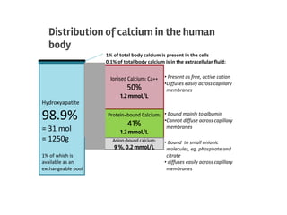
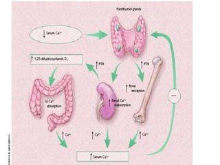
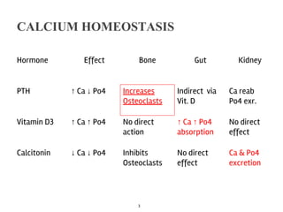
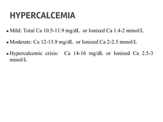
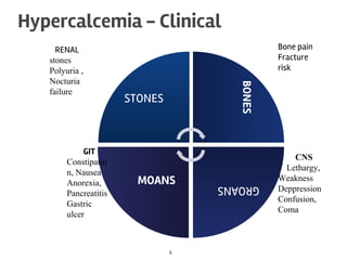
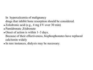
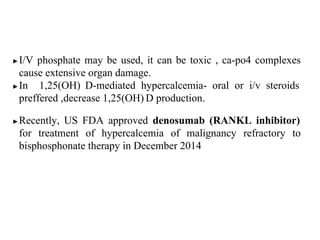
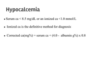
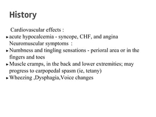
The document discusses calcium homeostasis in the human body. It notes that 98.9% of calcium in the body is found in bones and teeth in the form of hydroxyapatite. The remaining 1.1% is divided between the extracellular fluid and cells. Extracellular calcium is present as ionized calcium and protein-bound or anion-bound forms, while intracellular calcium is primarily protein-bound. The document also summarizes calcium regulation by parathyroid hormone, vitamin D, and calcitonin, as well as causes, symptoms, diagnosis and treatment of hypercalcemia and hypocalcemia.
1. Distribution of calcium in the human
body
Hydroxyapatite
98.9%
= 31 mol
= 1250g
1% of which is
available as an
exchangeable pool
0.1% of total body calcium is in the extracellular fluid:
Ionised Calcium: Ca++
50%
1.2 mmol/L
1% of total body calcium is present in the cells
Protein-bound Calcium:
41%
1.2 mmol/L
Anion-bound calcium:
9 %, 0.2 mmol/L
• Present as free, active cation
•Diffuses easily across capillary
membranes
• Bound mainly to albumin
•Cannot diffuse across capillary
membranes
• Bound to small anionic
molecules, eg. phosphate and
citrate
• diffuses easily across capillary
membranes
2.
3. CALCIUM HOMEOSTASIS
Hormone Effect Bone Gut Kidney
PTH ↑ Ca ↓ Po4 Increases
Osteoclasts
Indirect via
Vit. D
Ca reab
Po4 exr.
Vitamin D3 ↑ Ca ↑ Po4 No direct
action
↑ Ca ↑ Po4
absorption
No direct
effect
Calcitonin ↓ Ca ↓ Po4 Inhibits
Osteoclasts
No direct
effect
Ca & Po4
excretion
3
4. ▶Mild: Total Ca 10.5-11.9 mg/dL or Ionized Ca 1.4-2 mmol/L
▶Moderate: Ca 12-13.9 mg/dL or Ionized Ca 2-2.5 mmol/L
▶Hypercalcemic crisis: Ca 14-16 mg/dL or Ionized Ca 2.5-3
mmol/L
HYPERCALCEMIA
8. ▶Mild, asymptomatic hypercalcemia do not require immediate
therapy, and manage underlying cause.
▶ Symptomatic hypercalcemia requires therapeutic intervention
▶ Initial therapy - volume expansion because hypercalcemia
invariably leads to dehydration
▶ 4–6 L of i/v saline may be required over the first 24 h
▶ Loop diuretics - enhance sodium and calcium excretion.
.
Treatment
9. In hypercalcemia of malignancy
drugs that inhibit bone resorption should be considered.
▶Zoledronic acid (e.g., 4 mg I/V over 30 min)
▶Pamidronate ,Etidronate
▶Onset of action is within 1–3 days.
Because of their effectiveness, bisphosphonates have replaced
calcitonin widely
▶In rare instances, dialysis may be necessary.
10. ▶I/V phosphate may be used, it can be toxic , ca-po4 complexes
cause extensive organ damage.
▶In 1,25(OH) D-mediated hypercalcemia- oral or i/v steroids
preffered ,decrease 1,25(OH) D production.
▶Recently, US FDA approved denosumab (RANKL inhibitor)
for treatment of hypercalcemia of malignancy refractory to
bisphosphonate therapy in December 2014
11. ▶Serum ca < 8.5 mg/dL or an ionized ca <1.0 mmol/L
▶ Ionized ca is the definitive method for diagnosis
▶ Corrected ca(mg%) = serum ca + (4.0 - albumin g%) x 0.8
Hypocalcemia
12. Cardiovascular effects :
▶acute hypocalcemia - syncope, CHF, and angina
Neuromuscular symptoms :
▶Numbness and tingling sensations - perioral area or in the
fingers and toes
▶Muscle cramps, in the back and lower extremities; may
progress to carpopedal spasm (ie, tetany)
▶Wheezing ,Dysphagia,Voice changes
History
14. ▶ Hair may appear coarse, and alopecia may be present.
▶ In chronic hypocalcemia - dental caries,enamel hypoplasia.
▶ Eye examination-subcapsular cataract or papilledema
▶ Patient may appear confused, disoriented, Irritabile
▶ Hallucinations, dementia, and seizures may occur.
▶On R/S-wheezes , laryngeal stridor
▶ On CVS- bradycardia, tachycardia, S3
, signs of HF.
PHYSICAL EXAMINATION
15. ▶Chvostek sign - tapping the skin over the facial nerve about 2 cm
anterior to the external auditory meatus.
▶Ipsilateral contraction of the facial muscles is a positive sign.
▶Depending on the ca level, a graded response will occur:
twitching first at the angle of the mouth, then by nose, eye and
the facial muscles.
▶ 10% of the population will have a positive Chvostek sign in the
absence of hypocalcemia so test is not diagnostic
16. ▶ Trousseau sign- placing a blood pressure cuff on the patient’s
arm and inflating to 20 mm Hg above SBP for 3-5 minutes.
▶flexion of wrist and MCP joints ,extension of IP joints and
adduction of the thumb (carpal spasm).
▶The Trousseau sign is more specific than the Chvostek sign
Extra pyramidal symptom :
▶Choreoathetosis ,Parkinsonism,Hemiballism
18. CAUSES and Diagnostic Approach
LOW PARATHYROID
HORMONE LEVEL
HIGH PARATHYROID HORMONE
LEVEL
PARATHYROID
• AGENESIS-digeorge
•DESTRUCTION
surgical
radiation
infiltration
autoimmune
•HYPOFUNCTION-mg,CaSR
•VIT D related
Deficiency
Renal insufficiency
Resistance
•PTH horm resistance syndromes
•DRUGS
Chelators,biphosphanates
Miscellaneous
Acute pancreatitis
Acute rhabdomyolisis
Hungry bone
Osteoblastic metastasis
19. ▶On X-RAY, rickets or osteomalacia may present with the
pathognomonic Looser zones, better observed in the pubic
ramus, upper femoral bone, and ribs.
▶CT scans of head may show basal ganglia calcification
(extrapyramidal neurologic symptoms )
20. ▶Mild to Moderate Hypocalcemia,asymptomatic needs only ca
supplement
▶IV replacement is recommended in symptomatic or severe
hypocalcemia.
▶Doses of 100-300 mg of elemental ca in 50-100 mL of 5% D
should be given over 5-10 minutes.
▶10 mL of calcium gluconate contains 90 mg elemental ca
▶10 mL of calcium chloride contains 272 mg elemental ca
Treatment
21. ▶Measure serum calcium every 4-6 hours to maintain at levels of
8-9 mg/dL.
▶Start oral calcium and vitamin D treatment early
▶oral calcium supplements must be given between meals
otherwise, they will act as phosphate binders
▶ Vit D deficiency - vitamin D supplementation (50,000 U, 2–3
times per week for several months)
▶ Vit D deficiency due to malabsorption- higher doses (100,000
U/d or more).