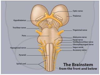
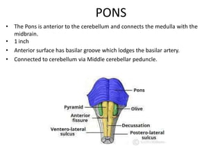
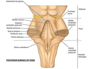
THE BRAINSTEM
Mr.SAJID HUSSAIN
(Asst. Professor)
S.R. Institute of Pharmacy
Bhuta, Bareilly (UP)
PONS
MIDBRAIN
MEDULLA
PARTS OF THE BRAIN STEM
DORSAL VIEW
VENTRAL VIEW
LATERAL VIEW
3 PARTS
• MID BRAIN
• PONS
• MEDULLA
• Present in the Posterior Cranial fossa.
• Connects narrow spinal cord with the
expanded Forebrain.
Broad functions
1. It serves as a conduit for the ascending tracts
and descending tracts connecting the spinal
cord to the different parts of the higher
centers in the forebrain
2. Inportant reflex centers present - respiration,
cvs and control of consiousness
3. Nuclei for CN 3 to 12
MEDULLA OBLONGATA
• Connects Pons with Spinal cord
• Conical in shape
• The central canal in lower half continues as that of
spinal cord while the upper half expands as the
cavity of fourth ventricle
• The region between the anterior median sulcus
and the antero lateral sulcus is occupied (on
either side of the midline) by an elevation called
the pyramid.
• The elevation is caused by a large bundle of fibres
that descend from the cerebral cortex to the spinal
Cord (corticospinal fibres)
• Some of these fibres cross from one side to the
other in the lower part of the medulla,
obliterating the anterior median fissure. These
crossing fibres constitute the decussation of the
pyramids.
• The posterior surface of the superior half of
medulla forms the lower part of the floor of the
fourth ventricle
• The posterior surface of the inferior half of the
medulla is continuous with posterior aspect of the
spinal cord and has POSTERIOR MEDIAN SULCUS
Hypoglossal nerve
pyramid
olive Vagus
Glossopharyngeal nerve
Inferior cerebellar peduncle
Spinal accessory nerve
PONS
• The Pons is anterior to the cerebellum and connects the medulla with the
midbrain.
• 1 inch
• Anterior surface has basilar groove which lodges the basilar artery.
• Connected to cerebellum via Middle cerebellar peduncle.
• On either side of the lower part of the Pons there is a region called the
cerebello-pontine angle. This region lies near the lateral aperture of the
fourth ventricle. The facial, vestibulocochlear and Glossopharyngeal
nerves, the nervus intermedius, and sometimes the labyrinthine arteries
lie in this region.
• The pons is divisible into a ventral part and a dorsal part
• The ventral (or basilar) part contains numerous transverse and vertical
fibres. Amongst the fibres are groups of cells that constitute the pontine
nuclei.
• When traced laterally the transverse fibres form the middle cerebellar
peduncle.
• The vertical fibres are of two types. Some of them descend from the
cerebral cortex to end in the pontine nuclei. Others are corticospinal fibres
that descend through the pons into the medulla where they form the
pyramids.
POSTERIOR SURFACE OF PONS
MIDBRAIN
• 0.8 inch in length
• Connects Pons and cerebellum with the forebrain
• The midbrain is traversed by NARROW CHANNEL, THE CEREBRAL
AQUEDUCT, filled with CSF
• Posterior surface- 4 colliculi- Corpora Quadrigemina
• Superior colliculi- Visual
• Inferior colliculi- auditory
• In the midline below the Inferior colliculus- trochlear nerves emerge
• When the midbrain is viewed from the anterior aspect, we see two large
bundles of fibres, one on each side of the middle line. These are the crura
of the midbrain
• The crura are separated by a deep fissure. Near the pons the fissure is narrow,
but broadens as the crura diverge to enter the corresponding cerebral
hemispheres. The parts of the crura just below the cerebrum form the
posterior boundary of a space called the interpeduncular fossa. The
oculomotor nerve emerges from the medial aspect of the crus of the same
side.
• Each colliculus is related laterally to a ridge called the brachium.
• The superior brachium (also called the superior quadrigeminal brachium, or
brachium of superior colliculus) connects the superior colliculus to the lateral
geniculate body.
• Similarly, the inferior brachium (also called the inferior quadrigeminal
brachium or brachium of inferior colliculus) connects the inferior colliculus to
the medial geniculate body.
• Just below the colliculi, there is the uppermost part of a membrane, the
superior medullary velum, which stretches between the two superior
cerebellar peduncles, and helps to form the roof of the fourth ventricle.
• For convenience of description, the midbrain may be divided as follows
A. The part lying behind a transverse line drawn through the cerebral
aqueduct is called the tectum. It consists of the superior and inferior
colliculi of the two sides.
B. The part lying in front of the transverse line is made up of right and left
halves called the cerebral peduncles.
• Each peduncle consists of three parts. From anterior to posterior side
these are the
1. crus cerebri (or basis pedunculi),
2. the substantia nigra and
3. the tegmentum.
Brain stem.pdf
Brain stem.pdf
Brain stem.pdf

Brain stem.pdf

  • 1.
    THE BRAINSTEM Mr.SAJID HUSSAIN (Asst.Professor) S.R. Institute of Pharmacy Bhuta, Bareilly (UP)
  • 2.
  • 3.
  • 6.
    3 PARTS • MIDBRAIN • PONS • MEDULLA • Present in the Posterior Cranial fossa. • Connects narrow spinal cord with the expanded Forebrain.
  • 7.
    Broad functions 1. Itserves as a conduit for the ascending tracts and descending tracts connecting the spinal cord to the different parts of the higher centers in the forebrain 2. Inportant reflex centers present - respiration, cvs and control of consiousness 3. Nuclei for CN 3 to 12
  • 8.
    MEDULLA OBLONGATA • ConnectsPons with Spinal cord • Conical in shape • The central canal in lower half continues as that of spinal cord while the upper half expands as the cavity of fourth ventricle • The region between the anterior median sulcus and the antero lateral sulcus is occupied (on either side of the midline) by an elevation called the pyramid. • The elevation is caused by a large bundle of fibres that descend from the cerebral cortex to the spinal Cord (corticospinal fibres)
  • 9.
    • Some ofthese fibres cross from one side to the other in the lower part of the medulla, obliterating the anterior median fissure. These crossing fibres constitute the decussation of the pyramids. • The posterior surface of the superior half of medulla forms the lower part of the floor of the fourth ventricle • The posterior surface of the inferior half of the medulla is continuous with posterior aspect of the spinal cord and has POSTERIOR MEDIAN SULCUS
  • 10.
    Hypoglossal nerve pyramid olive Vagus Glossopharyngealnerve Inferior cerebellar peduncle Spinal accessory nerve
  • 11.
    PONS • The Ponsis anterior to the cerebellum and connects the medulla with the midbrain. • 1 inch • Anterior surface has basilar groove which lodges the basilar artery. • Connected to cerebellum via Middle cerebellar peduncle.
  • 12.
    • On eitherside of the lower part of the Pons there is a region called the cerebello-pontine angle. This region lies near the lateral aperture of the fourth ventricle. The facial, vestibulocochlear and Glossopharyngeal nerves, the nervus intermedius, and sometimes the labyrinthine arteries lie in this region. • The pons is divisible into a ventral part and a dorsal part • The ventral (or basilar) part contains numerous transverse and vertical fibres. Amongst the fibres are groups of cells that constitute the pontine nuclei. • When traced laterally the transverse fibres form the middle cerebellar peduncle. • The vertical fibres are of two types. Some of them descend from the cerebral cortex to end in the pontine nuclei. Others are corticospinal fibres that descend through the pons into the medulla where they form the pyramids.
  • 13.
  • 14.
    MIDBRAIN • 0.8 inchin length • Connects Pons and cerebellum with the forebrain • The midbrain is traversed by NARROW CHANNEL, THE CEREBRAL AQUEDUCT, filled with CSF • Posterior surface- 4 colliculi- Corpora Quadrigemina • Superior colliculi- Visual • Inferior colliculi- auditory • In the midline below the Inferior colliculus- trochlear nerves emerge • When the midbrain is viewed from the anterior aspect, we see two large bundles of fibres, one on each side of the middle line. These are the crura of the midbrain
  • 15.
    • The cruraare separated by a deep fissure. Near the pons the fissure is narrow, but broadens as the crura diverge to enter the corresponding cerebral hemispheres. The parts of the crura just below the cerebrum form the posterior boundary of a space called the interpeduncular fossa. The oculomotor nerve emerges from the medial aspect of the crus of the same side. • Each colliculus is related laterally to a ridge called the brachium. • The superior brachium (also called the superior quadrigeminal brachium, or brachium of superior colliculus) connects the superior colliculus to the lateral geniculate body. • Similarly, the inferior brachium (also called the inferior quadrigeminal brachium or brachium of inferior colliculus) connects the inferior colliculus to the medial geniculate body. • Just below the colliculi, there is the uppermost part of a membrane, the superior medullary velum, which stretches between the two superior cerebellar peduncles, and helps to form the roof of the fourth ventricle.
  • 16.
    • For convenienceof description, the midbrain may be divided as follows A. The part lying behind a transverse line drawn through the cerebral aqueduct is called the tectum. It consists of the superior and inferior colliculi of the two sides. B. The part lying in front of the transverse line is made up of right and left halves called the cerebral peduncles. • Each peduncle consists of three parts. From anterior to posterior side these are the 1. crus cerebri (or basis pedunculi), 2. the substantia nigra and 3. the tegmentum.