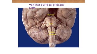
Development of the
Brain
o The brain develops from the cranial part of
neural tube*.
o The cranial part is divided into 3 parts:
*recall from embryology the caudal 2/3 forms the spinal
cord and the cranial or upper 1/3 forms the brain
FOREBRAIN MIDBRAIN HINDBRAIN
Cavity
2 lateral
ventricles
3rd ventricle cerebral
aqueduct
4th ventricle
Subdivided
into
Two cerebral
hemispheres
Diencephalon:
1. thalamus,
2. hypothalamus,
3. epithalamus &
4. subthalamus
The midbrain is
also called
mesencephalon
1 Pons.
2 Cerebellum.
3Medulla
oblongata.
Note: the brain stem develops from 2 different parts. The
pons and medulla oblongata develop from the hindbrain
where as the midbrain develops from the midbrain.
Located between the cerebrum
and the spinal cord
Provides a pathway for tracts running
between higher and lower neural
centers.
Consists of the
midbrain, pons, and medulla
oblongata.
Each region is about an inch in
length.
Microscopically, it consists of deep
gray matter surrounded by white
matter fiber tracts.
Produce automatic behaviors
necessary for survival.
Brainste
m
Midbrain
Pons
Medulla
obongat
a
3 PARTS
• MID BRAIN
• PONS
• MEDULLA
• Present in the Posterior Cranial fossa.
• Connects narrow spinal cord with the
expanded Forebrain.
3 broad functions
1. It serves as a conduit for the ascending tracts
and descending tracts connecting the spinal
cord to the different parts of the higher
centers in the forebrain
2. Inportant reflex centers present - respiration,
cvs and control of consiousness
3. Nuclei for CN 3 to 12
DORSAL VIEW
VENTRAL VIEW
LATERAL VIEW
Midbrain
Pons
Medulla
oblongata
Ve n t r a l surface o f brain
stem
Midbrain
Cerebral
peduncles
Pons
Basis
pontis
Medull
a
Ventral – Lateral
View
POSTERIOR SURFACE OF PONS
PONS
MIDBRAIN
MEDULLA
PARTS OF THE BRAIN STEM
OLIVES
DECUSSATION OF
PYRAMIDS
PYRAMIDS
MEDULLA
THE FOURTH
VENTRICLE
FOURTH VENTRICLE
VENTRAL ASPECT
DORSAL ASPECT
Vertical Columns of Cranial Nerves
Internal Columns of Nuclei
Brain
Stem
Clivus
o The brainstem is the region of the brain that
connects the cerebrum with the spinal cord
o SITE:
It lies on the basilar part of occipital bone
(clivus).
o PARTS: From above downwards:
Mid brain, pons & medulla oblongata
o CONNECTIONS WITH CEREBELLUM:
Each part of brain stem is connected to
cerebellum by cerebellar peduncles
(superior, middle & inferior).
Superior peduncle connects midbrain with cerebellum
Middle peduncle connects pons with cerebellum
Inferior peduncle connects medulla oblongata with
cerebellum
Sagittal Section Of
Brain
Cerebellum
Mid brain
Pons
Medulla
Superior
cerebellar
peduncle
Thalamus
Functions of the Brain
Stem
1. Pathway of tracts between cerebral
cortex & spinal cord (ascending and descending
tracts).
2. Site of origin of nuclei of cranial nerves
(from 3rd to 12th).
3. Site of emergence of cranial nerves
(from 3rd to 12th).
4. Contains groups of nuclei & related
fibers known as reticular formation*
responsible for: control of level of
consciousness, perception of pain,
regulation of cardiovascular &
respiratory systems.
*Complex matrix of
nuclei
and related
fibers/axons
1 2 & 3
4
Brain Stem – Ventral
Surface
Note: the numbers refer to the cranial nerves
Outline of the lecture
We will discuss the ventral surface of each part (medulla, pons, midbrain) then
we will discuss the dorsal.
In each side (ventral/dorsal) we will see the general feature and the nerves
coming out.
Medulla – Ventral
Surface
o Ventral median fissure:
• Continuation of ventral median fissure
of spinal cord.
• Divides the medulla into 2 halves
• Its lower part is marked by decussation
of most of pyramidal (corticospinal)
fibers (75%-90%).
o Pyramid:
• An elevation, lies on either (lateral) side
of ventral median fissure
• Produced by corticospinal tract.
Medulla – Ventral
Surface
o Olive:
• An elevation, lies lateral to the
pyramid.
• Produced by inferior olivary nucleus*
(important in control of movement).
o Nerves emerging from Medulla (4
nerves):
• Hypoglossal (12th): from sulcus
between pyramid & olive
• Glossopharyngeal (9th), vagus
(10th) &
Extra
Extra
cranial part of accessory (11th): from
sulcus dorsolateral to olive (from above
downwards)
*Convoluted mass of grey matter
Pons – Ventral
Surface
o Basilar sulcus:
• Divides the pons into 2 halves,
occupied by basilar artery.
o Transverse pontine
(pontocerebellar) fibers:
• Originate from pontine nuclei,
• They cross the midline & pass
through the contralateral middle
cerebellar peduncle to enter the
opposite cerebellar
hemisphere.
*
*
Extra
Extra
Pons – Ventral
Surface
o Nerves emerging from Pons (4 nerves):
• Trigeminal (5th): from the middle of
ventrolateral aspect of pons, as 2 roots:
a small medial motor root & a large lateral
sensory root*.
• Abducent (6th): from sulcus/junction
between pons & pyramid.
• Facial (7th) & Vestibulocochlear (8th): at
cerebellopontine angle (junction between
medulla, pons & cerebellum). Both nerves
emerge as 2 roots: from medial to lateral:
motor root of 7th, sensory root of 7th
vestibular part of 8th & cochlear part of
8th.
Vestibulo 1st since its first
in the name then cochlear
*to remember
small medial motor root & a
large lateral sensory root
Extra
Midbrain – Ventral
Surface
o It is formed of a large column
of descending fibers (crus
cerebri or basis pedunculi), on
either side.
o The 2 crura cerebri are separated
by a depression called the
interpeduncular fossa.
o Nerve emerging from Midbrain
(one):
• Occulomotor (3rd): from medial
aspect of crus cerebri.
*
Crus
Cerebri
Medulla – Dorsal
Surface
open medulla closed medulla
o The features differ in the
caudal part (closed medulla) and
the cranial part (open medulla).
Extra
The caudal part closes around the fourth ventricle
forming the central canal so it is called closed medulla.
• Dorsal median sulcus: divides the closed
medulla into 2 halves.
• Fasciculus gracilis: on either side of dorsal
median sulcus.
• Gracile tubercle: an elevation produced at the upper
part of fasciculus gracilis, marks the site of gracile
nucleus.
• Fasciculus cuneatus: on either side of
fasciculus gracilis.
• Cuneate tubercle: an elevation produced at the
upper part of fasciculus cuneatus, marks the site of
cuneate nucleus.
Medulla – Dorsal
Surface
Closed Medulla
o Cavity: central canal.
o Composed of:
Recall: Fasciculus gracilis and fasciculus cuneatus are
ascending tracts in the dorsal white column which
terminate on their respective nuclei: gracile nucleus
and cuneate nucleus.
o Cavity: 4th ventricle
o On either side, an inverted V-shaped
sulcus divides the area into 3 parts
(from medial to lateral):
1. Hypoglossal triangle: overlies
hypoglossal nucleus.
2. Vagal triangle: overlies dorsal vagal
nucleus.
3. Vestibular area: overlies vestibular
nuclei.
Medulla – Dorsal
Surface
Open
Medulla
Extra
Pons – Dorsal
Surface
o Separated from open medulla by an
imaginary line passing between the
caudal margins of middle cerebellar
peduncle.
o On either side of median sulcus, it
divides
into 2 parts (from medial to lateral):
• Medial eminence & facial colliculus:
overlies abducent nucleus
The abducent nucleus lies medially, and below it is the fiber of the
facial nerve which goes above and around it and forms the facial
colliculus.
• Vestibular area: overlies vestibular
nuclei.
**
Extra
o The dorsal surfaces of open medulla and pons lie in the caudal 1/3rd
and the rostral 2/3rd of the floor of the 4th ventricle respectively.
Dorsal surface of pons  rostral or cranial 2/3rd of 4th ventricle Dorsal
surface of open medulla  caudal 1/3rd of 4th ventricle
P
MO
pons
Midbrain – Dorsal
Surface
o Marked by 4 elevations/colliculi:
• Two superior colliculi: concerned
with visual reflexes*.
• Two inferior colliculi: forms part of
auditory pathway.
o Nerve emerging from Midbrain (one):
• Trochlear (4th): just caudal to inferior
colliculus (The only cranial nerve
emerging from dorsal surface of brain
stem, the rest were from the ventral).
To remember:
The trochlear is the only cranial nerve that emerges
from the dorsal / posterior surface.
*To remember: he superior colliculi is concerned with the eyes
(auditory) & the eyes are at the top of the face (superior).
SUMMARY
o The brain stem is composed (from above downwards) of: midbrain,
pons & medulla oblongata which are continuous with each other, and with
diencephalon above & with spinal cord below.
o The brain stem is connected with cerebellum through three pair of cerebellar
peduncles.
o The brain stem is the site of (1) cranial nuclei, (2) the pathway of important
ascending & descending tracts & (3) the site of emergence of cranial
nerves (from 3rd to 12th).
o Cranial nerves (with the exception of 4th) emerge from ventral surface of brain
stem.
MIDBRAIN
• 0.8 inch in length
• Connects Pons and cerebellum with the forebrain
• The midbrain is traversed by NARROW CHANNEL, THE
CEREBRAL AQUEDUCT, filled with CSF
• Posterior surface- 4 colliculi- Corpora Quadrigemina
• Superior colliculi- Visual
• Inferior colliculi- auditory
• In the midline below the Inferior colliculus- trochlear nerves
emerge
• When the midbrain is viewed from the anterior aspect, we see two
large bundles of fibres, one on each side of the middle line. These are
the crura of the midbrain
• For convenience of description, the midbrain may be divided as follows
A. The part lying behind a transverse line drawn through the cerebral
aqueduct is called the tectum. It consists of the superior and
inferior colliculi of the two sides.
B. The part lying in front of the transverse line is made up of right and
left halves called the cerebral peduncles.
• Each peduncle consists of three parts. From anterior to posterior side
these are the
1. crus cerebri (or basis pedunculi),
2. the substantia nigra and
3. the tegmentum.
Connects pons and cerebrum with forebrain
Shortest brain stem,not more than
2cm in length,lies in the posterior
cranial Fossa.
For descriptive purpose,divided into
Dorsal tectum and right and left
cerebral Peduncles.
Each cerebral peduncles divide further
into ventral crus cerebri and a dorsal
Tegmentum by a pigmented lamina
“ Substantia nigra”
Cerebral peduncles contains:
-Descending fibers that go to
the cerebellum via the pons
-Descending pyramidal tracts
Running through the midbrain is the
hollow c e r e b r a l aqueduct which
connects the 3rd and 4th ventricles of the
brain.
Midbrain
Crus cerebri
Basis Tegmentu
m
Tectu
m
Ventral – Dorsal Organization
Patterning of the Midbrain
The roof of the aqueduct
( the tectum) contains the
corpora quadrigemina
2 superior colliculi that control
reflex movements of the eyes, head
and neck in response to visual
stimuli
2 inferior colliculi that control
reflex movements of the head,
neck, and trunk in response to
auditory stimuli
Corpora
quadregemina
Superior and inferior colliculi seperated by
cruciform sulcus
Superior colliculi larger and darker than inferior
colliculi,the difference In colour due to superficial
neurons in Superior colliculi
Superior
colliculi
Inferior
colliculi
Internal Structure of
Midbrain
Cross section at two levels
• Level of inferior colliculus
• Level of superior colliculus
INTERNAL STRUCTURE
Transverse section of midbrain
Common to both at inferior and superior colliculus:
Crus cerebri (or basis pedunculi):
- Consists of fibres descending from cerebral cortex.
- Its medial one-sixth is occupied by coticopontine
fibres from frontal lobe,lateral one-sixth fibres
from temporal,occipital and parietal lobes,the
intermediate two third by corticospinal and
cortico- nuclear fibres.
Substantia nigra :
- Present immediately behind and medial to basis
pedunculi.
- It appears dark as neuron within it contain
pigment.
( neuromelanin )
The main features to be seen through a section of
upper part of the Midbrain
The main features to be seen through a section of
lower part of the Midbrain
Basis
pedunculi
Substantia
nigra
Crus cerebri
(cerebral peduncle)
Cerebral
aqueduct
PONS
• The Pons is anterior to the cerebellum and connects the medulla with the
midbrain.
• 1 inch
• Anterior surface has basilar groove which lodges the basilar artery.
• Connected to cerebellum via Middle cerebellar peduncle.
• On either side of the lower part of the Pons there is a region called
the cerebello-pontine angle. This region lies near the lateral
aperture of the fourth ventricle. The facial, vestibulocochlear and
Glossopharyngeal nerves, the nervus intermedius, and sometimes
the labyrinthine arteries lie in this region.
• The pons is divisible into a ventral part and a dorsal part
• The ventral (or basilar) part contains numerous transverse and
vertical fibres. Amongst the fibres are groups of cells that constitute
the pontine nuclei.
• When traced laterally the transverse fibres form the middle
cerebellar peduncle.
• The vertical fibres are of two types. Some of them descend from the
cerebral cortex to end in the pontine nuclei. Others are corticospinal
fibres that descend through the pons into the medulla where they
form the pyramids.
Pons
The pons shows a convex anterior
surface with prominent transversely
running fibres. These fibres collect to
form bundles,the middle cerebellar
peduncles.
Trigeminal nerve emerges from the
anterior surface,at the junction
between pons and middle cerebellar
peduncle.
The anterior surface of pons is marked
in the midline by a shallow
groove,the sulcus basilaris which
lodges the basilar artery.
Pons
s
Sulcus basilaris
POSTERIOR SURFACE OF PONS
INTERNAL STRUCTURE OF PONS
• Divided into
• Studied at two levels :
1. At level of facial colliculus(caudal part)
2. At level of trigeminal nuclei(cranial part)
Trapezoid body (anterior)
Tegmentum (posterior)
Subdivided into ventral and dorsal part
Ventral part of the pons contains
Pontine nuclei:
•Recieves corticopontine fibres from
frontal, temporal,parietal and occipital
lobes of cerebrum
•The efferent fibres form the transverse
fibres of pons.
•It has been estimated that there are
about twenty million neurons in pontine
nuclei. Most of them are glutaminergic.
Vertically running corticospinal
and corticopontine
fibres.
Transversely running fibres arising in
pontine nuclei
Pontine nuclei
Transverse section through cranial part
Transverse section through caudal part
INTERNAL STRUCTURE OF THE MEDULLA
• Considered at 4 levels
1. Level of decussation of Pyramids
2. Level of decussation of lemnisci
3. Level of the olives
4. Level just inferior to the Pons
Cross section at three levels
Level of pyramidal decussation
Internal Structure of Medulla
Level of decussation of Pyramids
Pyramidal
tract
Lateral corticospinal
tract
75 – 90%
spinal
nucleus of V
From pons to
C4
Gracile
nucleus
Anterior corticospinal
tract innervate muscles of
trunk
-- fibers to
Levelof Pyramidal Decussation
Cross section at level of
lemniscal decussation
Internal Structure of Medulla
Sensory decussation / level of lemnisci
Medial lemniscus
Carries 2nd order sensory
neurons to VPL
thalamus
Internal arcuate
fibers
Cuneate
nucleus
Gracile
nucleus
Medial longitudinal
fasciculus
Level of Lemniscal Decussation
Cross section at level of
Level of inferior olivary nuclei
Level of the olives
Inferior olivary
nuclei Relay between
cortex, vestibular
nuclei, cerebellum, basal
ganglia, and dorsal column
nuclei
Inferior
cerebellar
peduncle =
Restiform body
Hypoglossal nucleus CN
XII
Vestibular
nuclei
Medial
Inferior
Level of Inferior Olives
BRAINSTEM-SUMMARY WITH CLINICAL IMPORTANCE.pptx

BRAINSTEM-SUMMARY WITH CLINICAL IMPORTANCE.pptx

  • 1.
    Development of the Brain oThe brain develops from the cranial part of neural tube*. o The cranial part is divided into 3 parts: *recall from embryology the caudal 2/3 forms the spinal cord and the cranial or upper 1/3 forms the brain FOREBRAIN MIDBRAIN HINDBRAIN Cavity 2 lateral ventricles 3rd ventricle cerebral aqueduct 4th ventricle Subdivided into Two cerebral hemispheres Diencephalon: 1. thalamus, 2. hypothalamus, 3. epithalamus & 4. subthalamus The midbrain is also called mesencephalon 1 Pons. 2 Cerebellum. 3Medulla oblongata. Note: the brain stem develops from 2 different parts. The pons and medulla oblongata develop from the hindbrain where as the midbrain develops from the midbrain.
  • 2.
    Located between thecerebrum and the spinal cord Provides a pathway for tracts running between higher and lower neural centers. Consists of the midbrain, pons, and medulla oblongata. Each region is about an inch in length. Microscopically, it consists of deep gray matter surrounded by white matter fiber tracts. Produce automatic behaviors necessary for survival. Brainste m Midbrain Pons Medulla obongat a
  • 3.
    3 PARTS • MIDBRAIN • PONS • MEDULLA • Present in the Posterior Cranial fossa. • Connects narrow spinal cord with the expanded Forebrain.
  • 4.
    3 broad functions 1.It serves as a conduit for the ascending tracts and descending tracts connecting the spinal cord to the different parts of the higher centers in the forebrain 2. Inportant reflex centers present - respiration, cvs and control of consiousness 3. Nuclei for CN 3 to 12
  • 5.
  • 6.
    Midbrain Pons Medulla oblongata Ve n tr a l surface o f brain stem
  • 7.
  • 10.
  • 12.
  • 13.
  • 14.
  • 15.
  • 16.
  • 17.
    Vertical Columns ofCranial Nerves
  • 18.
  • 19.
    Brain Stem Clivus o The brainstemis the region of the brain that connects the cerebrum with the spinal cord o SITE: It lies on the basilar part of occipital bone (clivus). o PARTS: From above downwards: Mid brain, pons & medulla oblongata o CONNECTIONS WITH CEREBELLUM: Each part of brain stem is connected to cerebellum by cerebellar peduncles (superior, middle & inferior). Superior peduncle connects midbrain with cerebellum Middle peduncle connects pons with cerebellum Inferior peduncle connects medulla oblongata with cerebellum
  • 20.
    Sagittal Section Of Brain Cerebellum Midbrain Pons Medulla Superior cerebellar peduncle Thalamus
  • 21.
    Functions of theBrain Stem 1. Pathway of tracts between cerebral cortex & spinal cord (ascending and descending tracts). 2. Site of origin of nuclei of cranial nerves (from 3rd to 12th). 3. Site of emergence of cranial nerves (from 3rd to 12th). 4. Contains groups of nuclei & related fibers known as reticular formation* responsible for: control of level of consciousness, perception of pain, regulation of cardiovascular & respiratory systems. *Complex matrix of nuclei and related fibers/axons 1 2 & 3 4
  • 22.
    Brain Stem –Ventral Surface Note: the numbers refer to the cranial nerves Outline of the lecture We will discuss the ventral surface of each part (medulla, pons, midbrain) then we will discuss the dorsal. In each side (ventral/dorsal) we will see the general feature and the nerves coming out.
  • 23.
    Medulla – Ventral Surface oVentral median fissure: • Continuation of ventral median fissure of spinal cord. • Divides the medulla into 2 halves • Its lower part is marked by decussation of most of pyramidal (corticospinal) fibers (75%-90%). o Pyramid: • An elevation, lies on either (lateral) side of ventral median fissure • Produced by corticospinal tract.
  • 24.
    Medulla – Ventral Surface oOlive: • An elevation, lies lateral to the pyramid. • Produced by inferior olivary nucleus* (important in control of movement). o Nerves emerging from Medulla (4 nerves): • Hypoglossal (12th): from sulcus between pyramid & olive • Glossopharyngeal (9th), vagus (10th) & Extra Extra cranial part of accessory (11th): from sulcus dorsolateral to olive (from above downwards) *Convoluted mass of grey matter
  • 25.
    Pons – Ventral Surface oBasilar sulcus: • Divides the pons into 2 halves, occupied by basilar artery. o Transverse pontine (pontocerebellar) fibers: • Originate from pontine nuclei, • They cross the midline & pass through the contralateral middle cerebellar peduncle to enter the opposite cerebellar hemisphere. * * Extra Extra
  • 26.
    Pons – Ventral Surface oNerves emerging from Pons (4 nerves): • Trigeminal (5th): from the middle of ventrolateral aspect of pons, as 2 roots: a small medial motor root & a large lateral sensory root*. • Abducent (6th): from sulcus/junction between pons & pyramid. • Facial (7th) & Vestibulocochlear (8th): at cerebellopontine angle (junction between medulla, pons & cerebellum). Both nerves emerge as 2 roots: from medial to lateral: motor root of 7th, sensory root of 7th vestibular part of 8th & cochlear part of 8th. Vestibulo 1st since its first in the name then cochlear *to remember small medial motor root & a large lateral sensory root
  • 27.
    Extra Midbrain – Ventral Surface oIt is formed of a large column of descending fibers (crus cerebri or basis pedunculi), on either side. o The 2 crura cerebri are separated by a depression called the interpeduncular fossa. o Nerve emerging from Midbrain (one): • Occulomotor (3rd): from medial aspect of crus cerebri. * Crus Cerebri
  • 28.
    Medulla – Dorsal Surface openmedulla closed medulla o The features differ in the caudal part (closed medulla) and the cranial part (open medulla). Extra The caudal part closes around the fourth ventricle forming the central canal so it is called closed medulla.
  • 29.
    • Dorsal mediansulcus: divides the closed medulla into 2 halves. • Fasciculus gracilis: on either side of dorsal median sulcus. • Gracile tubercle: an elevation produced at the upper part of fasciculus gracilis, marks the site of gracile nucleus. • Fasciculus cuneatus: on either side of fasciculus gracilis. • Cuneate tubercle: an elevation produced at the upper part of fasciculus cuneatus, marks the site of cuneate nucleus. Medulla – Dorsal Surface Closed Medulla o Cavity: central canal. o Composed of: Recall: Fasciculus gracilis and fasciculus cuneatus are ascending tracts in the dorsal white column which terminate on their respective nuclei: gracile nucleus and cuneate nucleus.
  • 30.
    o Cavity: 4thventricle o On either side, an inverted V-shaped sulcus divides the area into 3 parts (from medial to lateral): 1. Hypoglossal triangle: overlies hypoglossal nucleus. 2. Vagal triangle: overlies dorsal vagal nucleus. 3. Vestibular area: overlies vestibular nuclei. Medulla – Dorsal Surface Open Medulla Extra
  • 31.
    Pons – Dorsal Surface oSeparated from open medulla by an imaginary line passing between the caudal margins of middle cerebellar peduncle. o On either side of median sulcus, it divides into 2 parts (from medial to lateral): • Medial eminence & facial colliculus: overlies abducent nucleus The abducent nucleus lies medially, and below it is the fiber of the facial nerve which goes above and around it and forms the facial colliculus. • Vestibular area: overlies vestibular nuclei. ** Extra
  • 32.
    o The dorsalsurfaces of open medulla and pons lie in the caudal 1/3rd and the rostral 2/3rd of the floor of the 4th ventricle respectively. Dorsal surface of pons  rostral or cranial 2/3rd of 4th ventricle Dorsal surface of open medulla  caudal 1/3rd of 4th ventricle P MO pons
  • 33.
    Midbrain – Dorsal Surface oMarked by 4 elevations/colliculi: • Two superior colliculi: concerned with visual reflexes*. • Two inferior colliculi: forms part of auditory pathway. o Nerve emerging from Midbrain (one): • Trochlear (4th): just caudal to inferior colliculus (The only cranial nerve emerging from dorsal surface of brain stem, the rest were from the ventral). To remember: The trochlear is the only cranial nerve that emerges from the dorsal / posterior surface. *To remember: he superior colliculi is concerned with the eyes (auditory) & the eyes are at the top of the face (superior).
  • 34.
    SUMMARY o The brainstem is composed (from above downwards) of: midbrain, pons & medulla oblongata which are continuous with each other, and with diencephalon above & with spinal cord below. o The brain stem is connected with cerebellum through three pair of cerebellar peduncles. o The brain stem is the site of (1) cranial nuclei, (2) the pathway of important ascending & descending tracts & (3) the site of emergence of cranial nerves (from 3rd to 12th). o Cranial nerves (with the exception of 4th) emerge from ventral surface of brain stem.
  • 35.
    MIDBRAIN • 0.8 inchin length • Connects Pons and cerebellum with the forebrain • The midbrain is traversed by NARROW CHANNEL, THE CEREBRAL AQUEDUCT, filled with CSF • Posterior surface- 4 colliculi- Corpora Quadrigemina • Superior colliculi- Visual • Inferior colliculi- auditory • In the midline below the Inferior colliculus- trochlear nerves emerge • When the midbrain is viewed from the anterior aspect, we see two large bundles of fibres, one on each side of the middle line. These are the crura of the midbrain
  • 38.
    • For convenienceof description, the midbrain may be divided as follows A. The part lying behind a transverse line drawn through the cerebral aqueduct is called the tectum. It consists of the superior and inferior colliculi of the two sides. B. The part lying in front of the transverse line is made up of right and left halves called the cerebral peduncles. • Each peduncle consists of three parts. From anterior to posterior side these are the 1. crus cerebri (or basis pedunculi), 2. the substantia nigra and 3. the tegmentum.
  • 41.
    Connects pons andcerebrum with forebrain Shortest brain stem,not more than 2cm in length,lies in the posterior cranial Fossa. For descriptive purpose,divided into Dorsal tectum and right and left cerebral Peduncles. Each cerebral peduncles divide further into ventral crus cerebri and a dorsal Tegmentum by a pigmented lamina “ Substantia nigra” Cerebral peduncles contains: -Descending fibers that go to the cerebellum via the pons -Descending pyramidal tracts Running through the midbrain is the hollow c e r e b r a l aqueduct which connects the 3rd and 4th ventricles of the brain. Midbrain Crus cerebri
  • 42.
  • 43.
  • 44.
    The roof ofthe aqueduct ( the tectum) contains the corpora quadrigemina 2 superior colliculi that control reflex movements of the eyes, head and neck in response to visual stimuli 2 inferior colliculi that control reflex movements of the head, neck, and trunk in response to auditory stimuli Corpora quadregemina Superior and inferior colliculi seperated by cruciform sulcus Superior colliculi larger and darker than inferior colliculi,the difference In colour due to superficial neurons in Superior colliculi Superior colliculi Inferior colliculi
  • 45.
    Internal Structure of Midbrain Crosssection at two levels • Level of inferior colliculus • Level of superior colliculus
  • 46.
    INTERNAL STRUCTURE Transverse sectionof midbrain Common to both at inferior and superior colliculus: Crus cerebri (or basis pedunculi): - Consists of fibres descending from cerebral cortex. - Its medial one-sixth is occupied by coticopontine fibres from frontal lobe,lateral one-sixth fibres from temporal,occipital and parietal lobes,the intermediate two third by corticospinal and cortico- nuclear fibres. Substantia nigra : - Present immediately behind and medial to basis pedunculi. - It appears dark as neuron within it contain pigment. ( neuromelanin )
  • 48.
    The main featuresto be seen through a section of upper part of the Midbrain
  • 51.
    The main featuresto be seen through a section of lower part of the Midbrain
  • 53.
  • 55.
    PONS • The Ponsis anterior to the cerebellum and connects the medulla with the midbrain. • 1 inch • Anterior surface has basilar groove which lodges the basilar artery. • Connected to cerebellum via Middle cerebellar peduncle.
  • 56.
    • On eitherside of the lower part of the Pons there is a region called the cerebello-pontine angle. This region lies near the lateral aperture of the fourth ventricle. The facial, vestibulocochlear and Glossopharyngeal nerves, the nervus intermedius, and sometimes the labyrinthine arteries lie in this region. • The pons is divisible into a ventral part and a dorsal part • The ventral (or basilar) part contains numerous transverse and vertical fibres. Amongst the fibres are groups of cells that constitute the pontine nuclei. • When traced laterally the transverse fibres form the middle cerebellar peduncle. • The vertical fibres are of two types. Some of them descend from the cerebral cortex to end in the pontine nuclei. Others are corticospinal fibres that descend through the pons into the medulla where they form the pyramids.
  • 57.
    Pons The pons showsa convex anterior surface with prominent transversely running fibres. These fibres collect to form bundles,the middle cerebellar peduncles. Trigeminal nerve emerges from the anterior surface,at the junction between pons and middle cerebellar peduncle. The anterior surface of pons is marked in the midline by a shallow groove,the sulcus basilaris which lodges the basilar artery. Pons s Sulcus basilaris
  • 60.
  • 61.
    INTERNAL STRUCTURE OFPONS • Divided into • Studied at two levels : 1. At level of facial colliculus(caudal part) 2. At level of trigeminal nuclei(cranial part) Trapezoid body (anterior) Tegmentum (posterior)
  • 62.
    Subdivided into ventraland dorsal part Ventral part of the pons contains Pontine nuclei: •Recieves corticopontine fibres from frontal, temporal,parietal and occipital lobes of cerebrum •The efferent fibres form the transverse fibres of pons. •It has been estimated that there are about twenty million neurons in pontine nuclei. Most of them are glutaminergic. Vertically running corticospinal and corticopontine fibres. Transversely running fibres arising in pontine nuclei Pontine nuclei
  • 66.
  • 70.
  • 76.
    INTERNAL STRUCTURE OFTHE MEDULLA • Considered at 4 levels 1. Level of decussation of Pyramids 2. Level of decussation of lemnisci 3. Level of the olives 4. Level just inferior to the Pons
  • 83.
    Cross section atthree levels Level of pyramidal decussation Internal Structure of Medulla
  • 86.
  • 87.
    Pyramidal tract Lateral corticospinal tract 75 –90% spinal nucleus of V From pons to C4 Gracile nucleus Anterior corticospinal tract innervate muscles of trunk -- fibers to Levelof Pyramidal Decussation
  • 88.
    Cross section atlevel of lemniscal decussation Internal Structure of Medulla
  • 90.
    Sensory decussation /level of lemnisci
  • 91.
    Medial lemniscus Carries 2ndorder sensory neurons to VPL thalamus Internal arcuate fibers Cuneate nucleus Gracile nucleus Medial longitudinal fasciculus Level of Lemniscal Decussation
  • 93.
    Cross section atlevel of Level of inferior olivary nuclei
  • 95.
  • 96.
    Inferior olivary nuclei Relaybetween cortex, vestibular nuclei, cerebellum, basal ganglia, and dorsal column nuclei Inferior cerebellar peduncle = Restiform body Hypoglossal nucleus CN XII Vestibular nuclei Medial Inferior Level of Inferior Olives