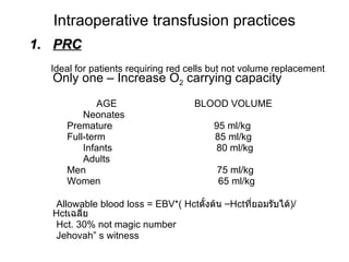
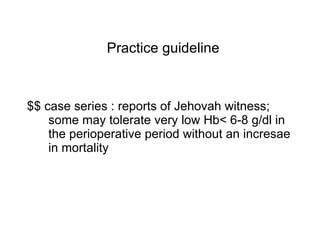
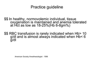
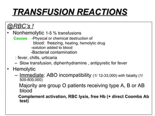
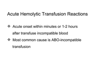
Blood transfusion พญ . เพชรรัตน์ วิสุทธิเมธีกร พ . บ .,  ป .  ชั้นสูงสาขาวิสัญญีวิทยา ,  วว . ( วิสัญญี ) ภาควิชาวิสัญญีวิทยา  วิทยาลัยแพทยศาสตร์กรุงเทพมหานคร และวชิรพยาบาล
Topic modules Blood blank practices Indication to blood transfusion Complication Alternative strategies for management of blood loss during surgery
Blood blank practices Human red cell membrane : least 300 different antigen fortunately, only the ABO and the Rh systems are important in the majority of blood transfusion History Hct. Infection : Hepatitis B,C  syphillis  HIV-1,2  HTLV-I,II
#Crossmatching (50 min) Confirms ABO and Rh typing Detects antibodies to the other blood group systems Detects antibodies in low titers or those that do not agglutinate easily Blood blank practices
Blood blank practices # Antibody screen : Indirect Coombs test  (45 mins) the subject serum  +  red cells  (  antigenic composition)   ----- red cell agglutination # Type&screen # Emergency transfusion
T&S -determines ABO and Rh status and the presence of most commonly encountered antibodies – risk of adverse rxn is 1:1000 -takes about 5 mins T&C -determines ABO and Rh status as well as adverse rxn to even low incidence antigens – risk of rxn is 1:10,000 -takes about 45 mins Type and screen vs Type and crossmatch
Type and screen vs Type and crossmatch T&S: Type O red cells are mixed with pt serum Antibody screen T&C  Type O red cells are mixed with pt serum Antibody screen Donor red cells are then mixed with the pt’s serum to determine possible incompatibility :
Blood blank practices All units – RBC @ PRC 1unit (250 ml Hct.70%) --platelet@ 1 unit (50-70 ml, stored at 20-24c for 5 days) --plasma @ FFP --cryoprecipitate @ high conc. Of factor VII, fibrinogen
Intraoperative transfusion practices PRC Ideal for patients requiring red cells but not volume replacement  Only one – Increase O 2  carrying capacity   AGE  BLOOD VOLUME Neonates Premature  95 ml/kg Full-term  85 ml/kg Infants  80 ml/kg Adults Men  75 ml/kg Women  65 ml/kg Allowable blood loss = EBV*( Hct ตั้งต้น  – Hct ที่ยอมรับได้ )/ Hct เฉลี่ย Hct. 30% not magic number Jehovah” s witness
Practice guideline $$ case series : reports of Jehovah witness; some may tolerate very low Hb< 6-8 g/dl in the perioperative period without an incresae in mortality
Practice guideline $$   In healthy, normovolemic individual, tissue oxygenation is maintained and anemia tolerated at Hct as low as 18-25%(Hb 6-8gm%) $$ RBC transfusion is rarely indicated when Hb> 10 g/dl and is almost always indicated when Hb< 6 g/dl American Society Anesthesiologist : 1996
คุณหมอขาหนูหน้าซีดแล้วต้องให้เลือดหนูหรือเปล่าคะ
Intraoperative transfusion practices 2.  FFP  ( initial therapeutic dose : 10-15 ml/kg ) isolated factor deficiencies reverse warfarin therapy correction of coagulopathy associated with liver disease used in patients who are received massive blood transfusion   with  microvascular  bleeding  Complications (PATCH)  Platelets – dec,Potassium – inc., ARDS, Acidosis,Temp dec., Citrate intoxication, Hepatiti >1 BV/ 24 HR> 50 % BV within 3 hrs > 150 ml/min antithrombin III deficiency TTP ( Thrombotic thrombocytopenic purpura )  Do not use for volume
Intraoperative transfusion practices 3.   PLATELETS **thrombocytopenia or dysfunction platelets in  the presence bleeding * prophylactic : plt.counts below 10,000-20,000 * prophylactic   preoperative : plt.counts below  50,000  *Microvascular bleeding in surgical patient with platelets < 50,000  *Neuro/ ocular surgery > 75,000
Intraoperative transfusion practices 3.   PLATELETS *Massive transfusion  with  microvascular bleeding with platelets < 100,000 2 BVs = 50,000 *Qualitative dysfunction  with  microvascular bleeding (may be > 100,000)
Intraoperative transfusion practices 3.   PLATELETS 50 ml: 0.5- 0.6 x 10  9  platelets (some RBC’s and WBC’s)  Single donor apheresis OR Random donor (x 6)
Intraoperative transfusion practices 4.  CRYOPRECIPITATE  10 ml: fibrinogen (150-250 mg),  VIII (80-145 U),  fibronectin, XIII 1U/ 10kg    fibrinogen 50 mg/dL (usually a 6- pack) Hypofibrinogenemia (congenital or acquired) Microvascular bleeding with massive BT (fibrinogen < 80-100mg/dL) 2 BVs = < 100 mg/dL  Bleeding  patients with vWD (or unresponsive to DDAVP)
Alternative strategies for management of blood loss during surgery Autologous transfusion Blood salvage & refusion Normovolemic hemodilution
 
“ Blood is still the best possible thing to have in our veins” - Woody Allen Blood transfusion is a lot like marriage.  It should not be entered upon lightly, unadvisedly or wantonly, or more often than is absolutely necessary” - Beal
คุณหมอขาตัวหนูแดงทั้งตัวแล้ว แล้วคุณหมอเป็นไงบ้าง หัวบวมหรือยังคะ
TRANSFUSION REACTIONS is any unfavorable transfusion-related event occurring in a patient during or after transfusion of blood components
TRANSFUSION REACTIONS @RBC’s ! Nonhemolytic  1-5 % transfusions  Causes  - Physical or chemical destruction of blood:  freezing,  heating, hemolytic drug  -solution added to blood -Bacterial contamination : fever, chills, urticaria Slow transfusion, diphenhydramine , antipyretic for fever Hemolytic Immediate : ABO incompatibility  (1/ 12-33,000) with fatality (1/ 500-800,000) Majority are group O patients receiving type A, B or AB blood  Complement activation, RBC lysis, free Hb (+ direct Coombs Ab test)
Acute Hemolytic Transfusion Reaction   Ab ( in recipient serum ) + Ag ( on RBC donor ) - Neuroendocrine responses -Complement Activation -Coagulation Activation - Cytokines Effects Acute hemolytic transfusion reaction Pathophysiology
Acute Hemolytic Transfusion Reactions Acute onset within minutes or 1-2 hours after transfuse incompatible blood Most common cause is ABO-incompatible transfusion
Signs and Symptoms of AHTR Chills ,  fever Facial flushing Hypotension Renal failure DIC Chest pain Dyspnea Generalized bleeding Hemoglobinemia Hemoglobinuria Shock Nausea Vomitting Back pain Pain along infusion vein
Anesthesia: hypotension, urticaria, abnormal bleeding Stop infusion, blood and urine to blood bank, coagulation screen (urine/plasma Hb, haptoglobin) Fluid therapy and osmotic diuresis Alkalinization of urine (increase solubility of Hb degradation products) Correct bleeding, Rx. DIC
Laboratory investigation for AHTR sample from blood bag  Repeat ABO, Rh, Ab screening Patient sample  Pre Tx sample  Repeat ABO, Rh, Ab screening Post Tx sample  Repeat ABO, Rh, Ab screening, DAT, CBC, UA, Bilirubin, BUN, Cr, Coagulation screening Repeat compatibility test - Pre Tx sample  &  Donor unit - Post Tx sample &  Donor unit
Delayed : (extravascular immune)1/ 5-10,000 Hemolysis 1-2 weeks after transfusion (reappearance of Ab against donor Ag from previous exposure)  Fever, anemia, jaundice Alloimmunization Recipient produces Ab’s against RBC membrane Ag Related to future delayed hemolytic reactions and difficulty crossmatching
@WBC’s! Europe: All products leukodepleted USA: Initial FDA recommendation now reversed pending objective data (NOT    length of stay for    expense)  Febrile reactions Recipient Ab reacts with donor Ag, stimulates pyrogens  (1-2 % transfusions)  20 - 30% of platelet transfusions Slow transfusion, antipyretic, meperidine for shivering
TRALI ( Transfusion related acute lung injury) Donor Ab reacts with recipient Ag  (1/ 10,000)   noncardiogenic pulmonary edema Supportive therapy
Transfusion-related Acute Lung Injury  (TRALI) Pathophysiology   Leukocyte Ab in donor react with pt. leukocytes Activate complements Adherence of granulocytes to pulmonary endothelium with release of proteolytic enz.& toxic O 2  metabolites Endothelial damage Interstitial edema and fluid in alveoli
Transfusion-related Acute Lung Injury  (TRALI) Acute and severe type of transfusion reaction Symptoms and signs Fever Hypotension Tachypnea Dyspnea Diffuse pulmonary infiltration on X-rays Clinical of noncardiogenic pumonary edema
Transfusion-related Acute Lung Injury   (TRALI) Therapy and Prevention Adequate respiratory and hemodynamic supportive treatment If TRALI is caused by pt. Ab    use LPB If TRALI is caused by donor Ab   no special blood components
Transfusion-associated Graft-versus-Host   Disease ( TA-GVHD) Rare: immunocompromised patients  Suggestion that more common with designated donors BMT, LBW neonates, Hodgkin's disease, exchange Tx in neonates
Transfusion-associated Graft-versus-Host   Disease  ( TA-GVHD)   Pathophysiology Infusion of Immunocompetent Cells (Lymphocyte) Patient at risk proliferation of donor T lymphocytes attack against patient tissue
Graft-versus-Host Reaction Signs & Symptoms Onset ~ 3 to 30 days after transfusion Clinical significant – pancytopenia Other effects include fever,  liver enzyme, copious watery diarrhea,  erythematous skin  erythroderma and desquamation
@Platelets! Alloimmunization 50 % of repeated platelet transfusions Ab-dependent elimination of platelets with lack of  response Use single donor apheresis  Signs & Symptoms mild    slight fever and  Hb severe    platelet refractoriness with bleeding Post-transfusion purpura Recipient Ab leads to sudden destruction of platelets 1-2 weeks after transfusion (sudden onset) Rare complication
Immunomodulatory effects of transfusion Wound infection: circumstantial evidence (? leukocyte filters for immunocompromised) Beneficial effects on renal graft survival (now < NB with CyA) 97: 9% graft survival advantage after 5 years  Nonspecific overload of RES     lymphocytes, APCs Modification T helper/suppressor ratio Allogeneic lymphocytes may circulate for years after transfusion
Cancer recurrence (mostly  retrospective )  Colon: 90 % studies suggest increased recurrence Breast: 70 % studies  Head and neck: 75 % studies “ Allogeneic blood products increase cancer recurrence after potentially curative surgical resection” - Landers  Evidence circumstantial NOT causal
INFECTIOUS COMPLICATIONS I.  Viral  (Hepatitis 88% of per unit viral risk)   Hepatitis B  Risk 1/ 200,000 due to HBsAg, antiHBc screening (7-17 % of PTH)  Per unit risk 1/63-66,000 0.002% residual HBV remains in ‘negative’ donors (window 2-16 weeks) Anti-HBc testing retained as surrogate marker for HIV
NANB and Hepatitis C Risk now 1/ 103,000 (NEJM 96) with 2nd/ 1/ 125,000 with 3rd generation HCV Ab/ HVC RNA tests  Window 4 weeks 70 % patients become chronic carriers, 10-20 % develop cirrhosis
HIV Current risk 1/ 450- 660,000 (95)  With current screening (Abs to HIV I, II and p24 Ag), window 6-8 weeks (third generation ELISA tests in Europe)    sero -ve window to < 16 days
HTLV I, II Only in cellular components (not FFP, cryo) Risk 1/ 641,000 (window period unknown) Screening for antibody I may not pick up II  CJD (and variant CJD)
CMV Cellular components only Problem in immunocompromised, although 80 % adults have serum Ab WBC filtration decreases risk of transmission CMV -ve blood:  CMV -ve pregnant patients, LBW neonates, CMV -ve transplant recipient,  CMV-ve/ HIV +ve
II. Bacterial Contamination unlikely in products stored for > 72 hours at 1-6  0  C  gram –ve, gram +ve bacteria most frequent  –  Yersinia enterocolitica  Produced  endotoxin Platelets stored at room temperature for 5 days, with infection rate of 0.25%  III. Protozoal Trypanosoma cruzi (Chaga’s disease)  Malaria Toxoplasmosis Leishmaniasis
Serological Testing for Infectious markers HIV – Ag Anti – HIV HBsAg Anti – HCV Test for syphilis
METABOLIC COMPLICATIONS Citrate toxicity Citrate (3G/ unit WB) binds Ca 2+  /   Mg + Metabolized liver, mobilization bone stores Hypocalcemia ONLY if > 1 unit/ 5 min or hepatic dysfunction Hypotension more likely due to    cardiac output/ perfusion than    calcium (except neonates)  Worse with hypothermia/ hepatic dysfunction
Hyperkalemia  After 3 weeks, K +  is 25- 30 mmol/l  Only 8- 15 mmol per unit PRBC/ WB Concern with > 1 unit/5 min @ infants
Acidosis Acid load after after 3 weeks 30-40 mmol/l (pH 6.6 - 6.9) Metabolic acidosis more likely due to decreased perfusion, hepatic impairment, hypothermia NaHCO 3  or THAM if base deficit > 7-10 mEq/l
2, 3 DPG Depleted within 96 hours of storage O 2  Hb DC to left Restored within 8- 24 hours of transfusion
E. REFERENCES Practice Guidelines for Blood Component Therapy (ASA Task Force). Anesthesiology 1996; 84: 732-47. Safety of the Blood Supply. JAMA 1995; 274:1368--73. Infectious Disease Testing for Blood Transfusions (NIH Consensus Conference). JAMA 1995; 274: 1374-9.

Blood Transfusion

  • 1.
    Blood transfusion พญ. เพชรรัตน์ วิสุทธิเมธีกร พ . บ ., ป . ชั้นสูงสาขาวิสัญญีวิทยา , วว . ( วิสัญญี ) ภาควิชาวิสัญญีวิทยา วิทยาลัยแพทยศาสตร์กรุงเทพมหานคร และวชิรพยาบาล
  • 2.
    Topic modules Bloodblank practices Indication to blood transfusion Complication Alternative strategies for management of blood loss during surgery
  • 3.
    Blood blank practicesHuman red cell membrane : least 300 different antigen fortunately, only the ABO and the Rh systems are important in the majority of blood transfusion History Hct. Infection : Hepatitis B,C syphillis HIV-1,2 HTLV-I,II
  • 4.
    #Crossmatching (50 min)Confirms ABO and Rh typing Detects antibodies to the other blood group systems Detects antibodies in low titers or those that do not agglutinate easily Blood blank practices
  • 5.
    Blood blank practices# Antibody screen : Indirect Coombs test (45 mins) the subject serum + red cells ( antigenic composition) ----- red cell agglutination # Type&screen # Emergency transfusion
  • 6.
    T&S -determines ABOand Rh status and the presence of most commonly encountered antibodies – risk of adverse rxn is 1:1000 -takes about 5 mins T&C -determines ABO and Rh status as well as adverse rxn to even low incidence antigens – risk of rxn is 1:10,000 -takes about 45 mins Type and screen vs Type and crossmatch
  • 7.
    Type and screenvs Type and crossmatch T&S: Type O red cells are mixed with pt serum Antibody screen T&C Type O red cells are mixed with pt serum Antibody screen Donor red cells are then mixed with the pt’s serum to determine possible incompatibility :
  • 8.
    Blood blank practicesAll units – RBC @ PRC 1unit (250 ml Hct.70%) --platelet@ 1 unit (50-70 ml, stored at 20-24c for 5 days) --plasma @ FFP --cryoprecipitate @ high conc. Of factor VII, fibrinogen
  • 9.
    Intraoperative transfusion practicesPRC Ideal for patients requiring red cells but not volume replacement Only one – Increase O 2 carrying capacity AGE BLOOD VOLUME Neonates Premature 95 ml/kg Full-term 85 ml/kg Infants 80 ml/kg Adults Men 75 ml/kg Women 65 ml/kg Allowable blood loss = EBV*( Hct ตั้งต้น – Hct ที่ยอมรับได้ )/ Hct เฉลี่ย Hct. 30% not magic number Jehovah” s witness
  • 10.
    Practice guideline $$case series : reports of Jehovah witness; some may tolerate very low Hb< 6-8 g/dl in the perioperative period without an incresae in mortality
  • 11.
    Practice guideline $$ In healthy, normovolemic individual, tissue oxygenation is maintained and anemia tolerated at Hct as low as 18-25%(Hb 6-8gm%) $$ RBC transfusion is rarely indicated when Hb> 10 g/dl and is almost always indicated when Hb< 6 g/dl American Society Anesthesiologist : 1996
  • 12.
  • 13.
    Intraoperative transfusion practices2. FFP ( initial therapeutic dose : 10-15 ml/kg ) isolated factor deficiencies reverse warfarin therapy correction of coagulopathy associated with liver disease used in patients who are received massive blood transfusion with microvascular bleeding Complications (PATCH) Platelets – dec,Potassium – inc., ARDS, Acidosis,Temp dec., Citrate intoxication, Hepatiti >1 BV/ 24 HR> 50 % BV within 3 hrs > 150 ml/min antithrombin III deficiency TTP ( Thrombotic thrombocytopenic purpura ) Do not use for volume
  • 14.
    Intraoperative transfusion practices3. PLATELETS **thrombocytopenia or dysfunction platelets in the presence bleeding * prophylactic : plt.counts below 10,000-20,000 * prophylactic preoperative : plt.counts below 50,000 *Microvascular bleeding in surgical patient with platelets < 50,000 *Neuro/ ocular surgery > 75,000
  • 15.
    Intraoperative transfusion practices3. PLATELETS *Massive transfusion with microvascular bleeding with platelets < 100,000 2 BVs = 50,000 *Qualitative dysfunction with microvascular bleeding (may be > 100,000)
  • 16.
    Intraoperative transfusion practices3. PLATELETS 50 ml: 0.5- 0.6 x 10 9 platelets (some RBC’s and WBC’s) Single donor apheresis OR Random donor (x 6)
  • 17.
    Intraoperative transfusion practices4. CRYOPRECIPITATE 10 ml: fibrinogen (150-250 mg), VIII (80-145 U), fibronectin, XIII 1U/ 10kg  fibrinogen 50 mg/dL (usually a 6- pack) Hypofibrinogenemia (congenital or acquired) Microvascular bleeding with massive BT (fibrinogen < 80-100mg/dL) 2 BVs = < 100 mg/dL Bleeding patients with vWD (or unresponsive to DDAVP)
  • 18.
    Alternative strategies formanagement of blood loss during surgery Autologous transfusion Blood salvage & refusion Normovolemic hemodilution
  • 19.
  • 20.
    “ Blood isstill the best possible thing to have in our veins” - Woody Allen Blood transfusion is a lot like marriage. It should not be entered upon lightly, unadvisedly or wantonly, or more often than is absolutely necessary” - Beal
  • 21.
  • 22.
    TRANSFUSION REACTIONS isany unfavorable transfusion-related event occurring in a patient during or after transfusion of blood components
  • 23.
    TRANSFUSION REACTIONS @RBC’s! Nonhemolytic 1-5 % transfusions Causes - Physical or chemical destruction of blood: freezing, heating, hemolytic drug -solution added to blood -Bacterial contamination : fever, chills, urticaria Slow transfusion, diphenhydramine , antipyretic for fever Hemolytic Immediate : ABO incompatibility (1/ 12-33,000) with fatality (1/ 500-800,000) Majority are group O patients receiving type A, B or AB blood Complement activation, RBC lysis, free Hb (+ direct Coombs Ab test)
  • 24.
    Acute Hemolytic TransfusionReaction Ab ( in recipient serum ) + Ag ( on RBC donor ) - Neuroendocrine responses -Complement Activation -Coagulation Activation - Cytokines Effects Acute hemolytic transfusion reaction Pathophysiology
  • 25.
    Acute Hemolytic TransfusionReactions Acute onset within minutes or 1-2 hours after transfuse incompatible blood Most common cause is ABO-incompatible transfusion
  • 26.
    Signs and Symptomsof AHTR Chills , fever Facial flushing Hypotension Renal failure DIC Chest pain Dyspnea Generalized bleeding Hemoglobinemia Hemoglobinuria Shock Nausea Vomitting Back pain Pain along infusion vein
  • 27.
    Anesthesia: hypotension, urticaria,abnormal bleeding Stop infusion, blood and urine to blood bank, coagulation screen (urine/plasma Hb, haptoglobin) Fluid therapy and osmotic diuresis Alkalinization of urine (increase solubility of Hb degradation products) Correct bleeding, Rx. DIC
  • 28.
    Laboratory investigation forAHTR sample from blood bag Repeat ABO, Rh, Ab screening Patient sample Pre Tx sample Repeat ABO, Rh, Ab screening Post Tx sample Repeat ABO, Rh, Ab screening, DAT, CBC, UA, Bilirubin, BUN, Cr, Coagulation screening Repeat compatibility test - Pre Tx sample & Donor unit - Post Tx sample & Donor unit
  • 29.
    Delayed : (extravascularimmune)1/ 5-10,000 Hemolysis 1-2 weeks after transfusion (reappearance of Ab against donor Ag from previous exposure) Fever, anemia, jaundice Alloimmunization Recipient produces Ab’s against RBC membrane Ag Related to future delayed hemolytic reactions and difficulty crossmatching
  • 30.
    @WBC’s! Europe: Allproducts leukodepleted USA: Initial FDA recommendation now reversed pending objective data (NOT  length of stay for  expense) Febrile reactions Recipient Ab reacts with donor Ag, stimulates pyrogens (1-2 % transfusions) 20 - 30% of platelet transfusions Slow transfusion, antipyretic, meperidine for shivering
  • 31.
    TRALI ( Transfusionrelated acute lung injury) Donor Ab reacts with recipient Ag (1/ 10,000) noncardiogenic pulmonary edema Supportive therapy
  • 32.
    Transfusion-related Acute LungInjury (TRALI) Pathophysiology Leukocyte Ab in donor react with pt. leukocytes Activate complements Adherence of granulocytes to pulmonary endothelium with release of proteolytic enz.& toxic O 2 metabolites Endothelial damage Interstitial edema and fluid in alveoli
  • 33.
    Transfusion-related Acute LungInjury (TRALI) Acute and severe type of transfusion reaction Symptoms and signs Fever Hypotension Tachypnea Dyspnea Diffuse pulmonary infiltration on X-rays Clinical of noncardiogenic pumonary edema
  • 34.
    Transfusion-related Acute LungInjury (TRALI) Therapy and Prevention Adequate respiratory and hemodynamic supportive treatment If TRALI is caused by pt. Ab  use LPB If TRALI is caused by donor Ab  no special blood components
  • 35.
    Transfusion-associated Graft-versus-Host Disease ( TA-GVHD) Rare: immunocompromised patients Suggestion that more common with designated donors BMT, LBW neonates, Hodgkin's disease, exchange Tx in neonates
  • 36.
    Transfusion-associated Graft-versus-Host Disease ( TA-GVHD) Pathophysiology Infusion of Immunocompetent Cells (Lymphocyte) Patient at risk proliferation of donor T lymphocytes attack against patient tissue
  • 37.
    Graft-versus-Host Reaction Signs& Symptoms Onset ~ 3 to 30 days after transfusion Clinical significant – pancytopenia Other effects include fever, liver enzyme, copious watery diarrhea, erythematous skin erythroderma and desquamation
  • 38.
    @Platelets! Alloimmunization 50% of repeated platelet transfusions Ab-dependent elimination of platelets with lack of response Use single donor apheresis Signs & Symptoms mild  slight fever and Hb severe  platelet refractoriness with bleeding Post-transfusion purpura Recipient Ab leads to sudden destruction of platelets 1-2 weeks after transfusion (sudden onset) Rare complication
  • 39.
    Immunomodulatory effects oftransfusion Wound infection: circumstantial evidence (? leukocyte filters for immunocompromised) Beneficial effects on renal graft survival (now < NB with CyA) 97: 9% graft survival advantage after 5 years Nonspecific overload of RES  lymphocytes, APCs Modification T helper/suppressor ratio Allogeneic lymphocytes may circulate for years after transfusion
  • 40.
    Cancer recurrence (mostly retrospective ) Colon: 90 % studies suggest increased recurrence Breast: 70 % studies Head and neck: 75 % studies “ Allogeneic blood products increase cancer recurrence after potentially curative surgical resection” - Landers Evidence circumstantial NOT causal
  • 41.
    INFECTIOUS COMPLICATIONS I. Viral (Hepatitis 88% of per unit viral risk) Hepatitis B Risk 1/ 200,000 due to HBsAg, antiHBc screening (7-17 % of PTH) Per unit risk 1/63-66,000 0.002% residual HBV remains in ‘negative’ donors (window 2-16 weeks) Anti-HBc testing retained as surrogate marker for HIV
  • 42.
    NANB and HepatitisC Risk now 1/ 103,000 (NEJM 96) with 2nd/ 1/ 125,000 with 3rd generation HCV Ab/ HVC RNA tests Window 4 weeks 70 % patients become chronic carriers, 10-20 % develop cirrhosis
  • 43.
    HIV Current risk1/ 450- 660,000 (95) With current screening (Abs to HIV I, II and p24 Ag), window 6-8 weeks (third generation ELISA tests in Europe)  sero -ve window to < 16 days
  • 44.
    HTLV I, IIOnly in cellular components (not FFP, cryo) Risk 1/ 641,000 (window period unknown) Screening for antibody I may not pick up II CJD (and variant CJD)
  • 45.
    CMV Cellular componentsonly Problem in immunocompromised, although 80 % adults have serum Ab WBC filtration decreases risk of transmission CMV -ve blood: CMV -ve pregnant patients, LBW neonates, CMV -ve transplant recipient, CMV-ve/ HIV +ve
  • 46.
    II. Bacterial Contaminationunlikely in products stored for > 72 hours at 1-6 0 C gram –ve, gram +ve bacteria most frequent – Yersinia enterocolitica Produced endotoxin Platelets stored at room temperature for 5 days, with infection rate of 0.25% III. Protozoal Trypanosoma cruzi (Chaga’s disease) Malaria Toxoplasmosis Leishmaniasis
  • 47.
    Serological Testing forInfectious markers HIV – Ag Anti – HIV HBsAg Anti – HCV Test for syphilis
  • 48.
    METABOLIC COMPLICATIONS Citratetoxicity Citrate (3G/ unit WB) binds Ca 2+ / Mg + Metabolized liver, mobilization bone stores Hypocalcemia ONLY if > 1 unit/ 5 min or hepatic dysfunction Hypotension more likely due to  cardiac output/ perfusion than  calcium (except neonates) Worse with hypothermia/ hepatic dysfunction
  • 49.
    Hyperkalemia After3 weeks, K + is 25- 30 mmol/l Only 8- 15 mmol per unit PRBC/ WB Concern with > 1 unit/5 min @ infants
  • 50.
    Acidosis Acid loadafter after 3 weeks 30-40 mmol/l (pH 6.6 - 6.9) Metabolic acidosis more likely due to decreased perfusion, hepatic impairment, hypothermia NaHCO 3 or THAM if base deficit > 7-10 mEq/l
  • 51.
    2, 3 DPGDepleted within 96 hours of storage O 2 Hb DC to left Restored within 8- 24 hours of transfusion
  • 52.
    E. REFERENCES PracticeGuidelines for Blood Component Therapy (ASA Task Force). Anesthesiology 1996; 84: 732-47. Safety of the Blood Supply. JAMA 1995; 274:1368--73. Infectious Disease Testing for Blood Transfusions (NIH Consensus Conference). JAMA 1995; 274: 1374-9.