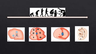
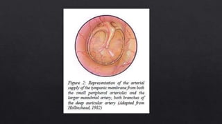
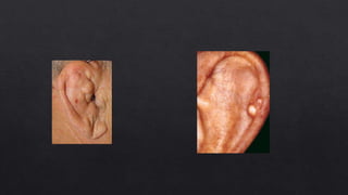
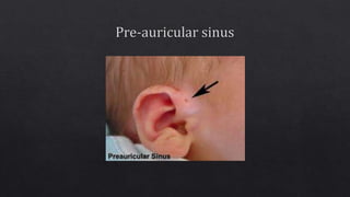
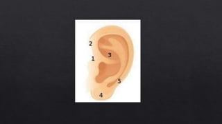
1. The pinna develops from six mesenchymal condensations called the hillocks of His around the first pharyngeal cleft.
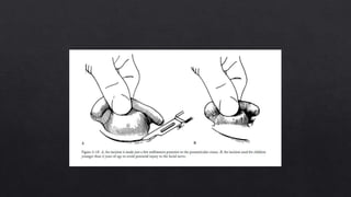
2. The cartilage of the auricle is connected to the temporal bone by two extrinsic ligaments - the anterior and posterior ligaments. Intrinsic ligaments also connect parts of the auricular cartilage.
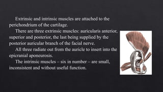
3. Three extrinsic and six intrinsic muscles are attached to the auricular cartilage and ligaments. The extrinsic muscles include the auricularis anterior, superior, and posterior.