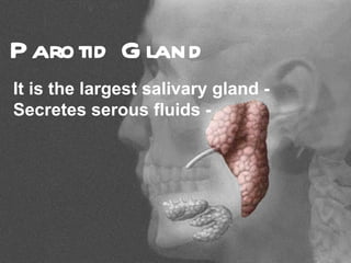
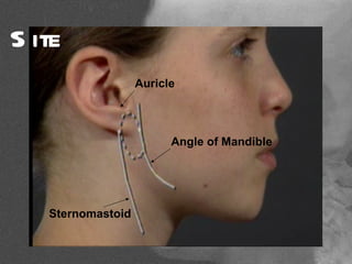
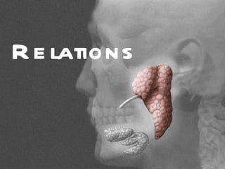
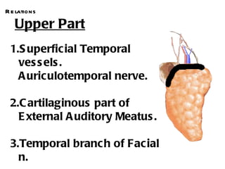
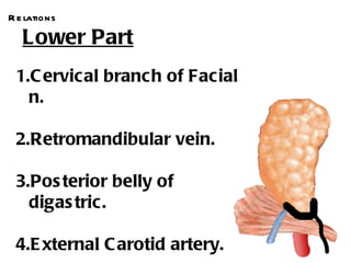
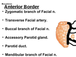
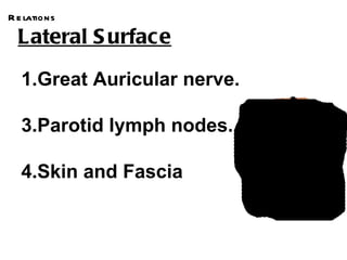
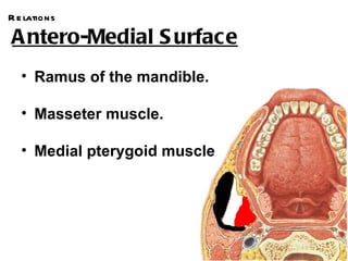
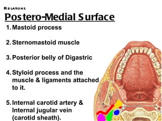
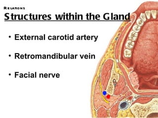
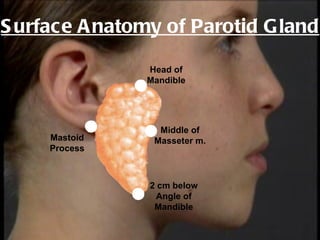
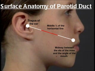
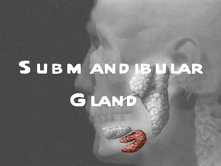
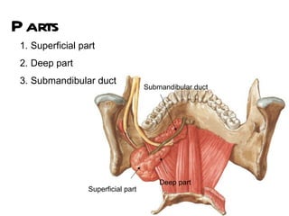
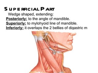
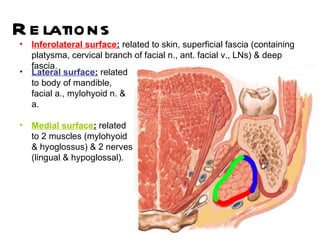
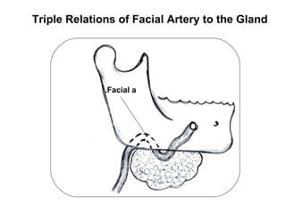
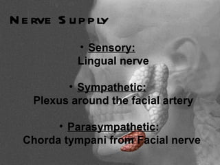
Anatomy of
S alivary
Glands
P arotid G land
It is the largest salivary gland -
Secretes serous fluids -
S ite
                   Auricle


                         Angle of Mandible


   Sternomastoid
R e lations
R e lations

   Upper Part
 1.S uperficial Temporal
   ves s els .
   A uriculotemporal nerve.

 2.C artilaginous part of
   E xternal A uditory Meatus .

 3.Temporal branc h of Facial
   n.
R e lations

   Lower Part
 1.C ervical branch of Facial
   n.

 2.Retromandibular vein.

 3.Pos terior belly of
   digas tric.

 4.E xternal C arotid artery.
R e lations
  A nterior B order
• Zygomatic branch of Facial n.

• Transverse Facial artery.

• Buccal branch of Facial n.

• Accessory Parotid gland.

• Parotid duct.

• Mandibular branch of Facial n.
R e lations

  Lateral S urface
   1.Great Auricular nerve.

   3.Parotid lymph nodes.

   4.Skin and Fascia
R e lations

A ntero-Medial S urface
   • Ramus of the mandible.

   • Masseter muscle.

   • Medial pterygoid muscle
R e lations

Pos tero-Medial S urface
 1. Mastoid process

 2. Sternomastoid muscle

 3. Posterior belly of Digastric

 4. Styloid process and the
    muscle & ligaments attached
    to it.

 5. Internal carotid artery &
    Internal jugular vein
    (carotid sheath).
R e lations

S tructures within the G land

  • External carotid artery

  • Retromandibular vein

  • Facial nerve
C aps ule
1.False Capsule
C aps ule
1.False Capsule

2.True Capsule
A cces ory Part
    Semidetached part of the gland,
    which lies just above the parotid
                  duct
Parotid Duct
 5 cm long &
 emerges from
  the anterior
 border & runs
 superficial to
 masseter m.
Parotid Duct
           Then pierces the buccinator to
               open in the vestibule of
             mouth, opposite the upper
                   2nd molar tooth
A rterial S upply
External Carotid Artery

 Venous Drainage
Retromandibular Vein
Nerve S upply
 1. Sensory:
   a. Gland:

      Auriculotemporal nerve

    a. Capsule:

        Great Auricular nerve
Nerve S upply
 2. Sympathetic:
      From the sympathetic plexus around
             .the external carotid
Nerve S upply
    3. Parasympathetic:
The secretomotor fibers arise from the inferior salivary nucleus in the medulla
oblongata.
Joins the glossopharyngeal nerve (9th cranial)
Leaves the 9th nerve through its tympanic branch which enters the middle ear where
it joins the tympanic plexus.
Lesser Petrosal nerve arises from the tympanic plexus as a continuation of the
tympanic branch
                                                                     Otic                      Medulla
Enters the foramen ovale to relay in                               Ganglion                   Oblangata
the otic ganglion                          .Auriculotemporal n


Joins auriculotemporal nerve
 through which they reach the gland
                                                                                            Inferior
                                                              Lesser                        Salivary
                                                                                            Nucleus
                                                             Petrosal n.
                                                                           Tympanic
                                                                            Plexus    .Glossopharyngeal n
S urface A natomy of Parotid G land

                Head of
                Mandible


                  Middle of
     Mastoid     Masseter m.
     Process


                2 cm below
                 Angle of
                 Mandible
S urface A natomy of Parotid Duct

         Tragus of
          the ear
                     Middle ⅓ of the
                     horizontal line


                            Midway between
                           the ala of the nose
                          and the angle of the
                                 mouth
S u b m and ib u lar
      G land
S ite
  Digastric triangle,
 partly below & partly
deep to the mandible
P arts
 1. Superficial part
 2. Deep part
 3. Submandibular duct
                                     Submandibular duct


                                         Deep part
                  Superficial part
S u p e rficial P art
 Wedge shaped, extending:
Posteriorly: to the angle of mandible.
Superiorly: to mylohyoid line of mandible.
Inferiorly: it overlaps the 2 bellies of digastric m
R e lations
•   Inferolateral surface: related to skin, superficial fascia (containing
    platysma, cervical branch of facial n., ant. facial v., LNs) & deep
    fascia.
•   Lateral surface: related
    to body of mandible,
    facial a., mylohyoid n. &
    a.

•   Medial surface: related
    to 2 muscles (mylohyoid
    & hyoglossus) & 2 nerves
    (lingual & hypoglossal).
Triple Relations of Facial Artery to the Gland


              .Facial a
Triple Relations of Lingual Nerve to the Duct


           .Lingual n


                                   .Hypoglossal n
D e e p P art
Small part lying deep to mylohyoid
Superficial to hyoglossus
Between lingual n. above & hypoglossal n. below

                  .Lingual n


            Mylohyoid
S u b m and ib u lar D u ct
5cm long
Triple relations with the lingual nerve


                   .Lingual n
S u b m and ib u lar D u ct
5cm long
Triple relations with the lingual nerve
Opening into the floor of mouth close to frenulum of
  the tongue


          Frenulum
N e rve S u p p ly
            • Sensory:
            Lingual nerve

          • Sympathetic:
    Plexus around the facial artery

        • Parasympathetic:
  Chorda tympani from Facial nerve
Thank
 You

Salivary glands 0

  • 1.
  • 2.
    P arotid Gland It is the largest salivary gland - Secretes serous fluids -
  • 3.
    S ite Auricle Angle of Mandible Sternomastoid
  • 4.
  • 5.
    R e lations Upper Part 1.S uperficial Temporal ves s els . A uriculotemporal nerve. 2.C artilaginous part of E xternal A uditory Meatus . 3.Temporal branc h of Facial n.
  • 6.
    R e lations Lower Part 1.C ervical branch of Facial n. 2.Retromandibular vein. 3.Pos terior belly of digas tric. 4.E xternal C arotid artery.
  • 7.
    R e lations A nterior B order • Zygomatic branch of Facial n. • Transverse Facial artery. • Buccal branch of Facial n. • Accessory Parotid gland. • Parotid duct. • Mandibular branch of Facial n.
  • 8.
    R e lations Lateral S urface 1.Great Auricular nerve. 3.Parotid lymph nodes. 4.Skin and Fascia
  • 9.
    R e lations Antero-Medial S urface • Ramus of the mandible. • Masseter muscle. • Medial pterygoid muscle
  • 10.
    R e lations Postero-Medial S urface 1. Mastoid process 2. Sternomastoid muscle 3. Posterior belly of Digastric 4. Styloid process and the muscle & ligaments attached to it. 5. Internal carotid artery & Internal jugular vein (carotid sheath).
  • 11.
    R e lations Structures within the G land • External carotid artery • Retromandibular vein • Facial nerve
  • 12.
  • 13.
    C aps ule 1.FalseCapsule 2.True Capsule
  • 14.
    A cces oryPart Semidetached part of the gland, which lies just above the parotid duct
  • 15.
    Parotid Duct 5cm long & emerges from the anterior border & runs superficial to masseter m.
  • 16.
    Parotid Duct Then pierces the buccinator to open in the vestibule of mouth, opposite the upper 2nd molar tooth
  • 17.
    A rterial Supply External Carotid Artery Venous Drainage Retromandibular Vein
  • 18.
    Nerve S upply 1. Sensory: a. Gland: Auriculotemporal nerve a. Capsule: Great Auricular nerve
  • 19.
    Nerve S upply 2. Sympathetic: From the sympathetic plexus around .the external carotid
  • 20.
    Nerve S upply 3. Parasympathetic: The secretomotor fibers arise from the inferior salivary nucleus in the medulla oblongata. Joins the glossopharyngeal nerve (9th cranial) Leaves the 9th nerve through its tympanic branch which enters the middle ear where it joins the tympanic plexus. Lesser Petrosal nerve arises from the tympanic plexus as a continuation of the tympanic branch Otic Medulla Enters the foramen ovale to relay in Ganglion Oblangata the otic ganglion .Auriculotemporal n Joins auriculotemporal nerve through which they reach the gland Inferior Lesser Salivary Nucleus Petrosal n. Tympanic Plexus .Glossopharyngeal n
  • 21.
    S urface Anatomy of Parotid G land Head of Mandible Middle of Mastoid Masseter m. Process 2 cm below Angle of Mandible
  • 22.
    S urface Anatomy of Parotid Duct Tragus of the ear Middle ⅓ of the horizontal line Midway between the ala of the nose and the angle of the mouth
  • 23.
    S u bm and ib u lar G land
  • 24.
    S ite Digastric triangle, partly below & partly deep to the mandible
  • 25.
    P arts 1.Superficial part 2. Deep part 3. Submandibular duct Submandibular duct Deep part Superficial part
  • 26.
    S u pe rficial P art Wedge shaped, extending: Posteriorly: to the angle of mandible. Superiorly: to mylohyoid line of mandible. Inferiorly: it overlaps the 2 bellies of digastric m
  • 27.
    R e lations • Inferolateral surface: related to skin, superficial fascia (containing platysma, cervical branch of facial n., ant. facial v., LNs) & deep fascia. • Lateral surface: related to body of mandible, facial a., mylohyoid n. & a. • Medial surface: related to 2 muscles (mylohyoid & hyoglossus) & 2 nerves (lingual & hypoglossal).
  • 28.
    Triple Relations ofFacial Artery to the Gland .Facial a
  • 29.
    Triple Relations ofLingual Nerve to the Duct .Lingual n .Hypoglossal n
  • 30.
    D e ep P art Small part lying deep to mylohyoid Superficial to hyoglossus Between lingual n. above & hypoglossal n. below .Lingual n Mylohyoid
  • 31.
    S u bm and ib u lar D u ct 5cm long Triple relations with the lingual nerve .Lingual n
  • 32.
    S u bm and ib u lar D u ct 5cm long Triple relations with the lingual nerve Opening into the floor of mouth close to frenulum of the tongue Frenulum
  • 33.
    N e rveS u p p ly • Sensory: Lingual nerve • Sympathetic: Plexus around the facial artery • Parasympathetic: Chorda tympani from Facial nerve
  • 34.