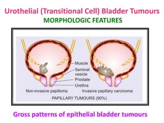
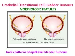
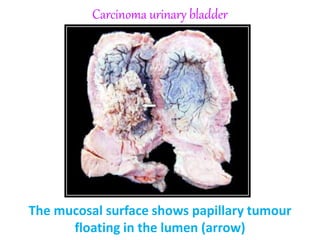
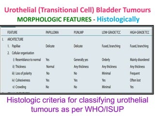
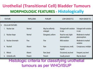
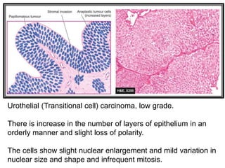
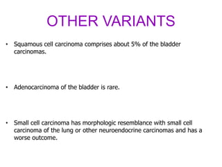
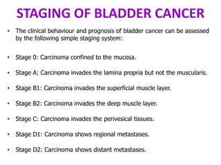
3. CONGENITAL ANOMALIE
DOUBLE URETER
• This is a condition in which the entire ureter or only the upper part is
duplicated. Double ureter is invariably associated with a double renal
pelvis, one in the upper part and the other in the lower part of the
kidney
4. CONGENITAL ANOMALIE
URETEROCELE
• Ureterocele is cystic dilatation of the terminal part of the ureter which
lies within the bladder wall.
• The cystic dilatation lies beneath the bladder mucosa and can be
visualised by cystoscopy
5. CONGENITAL ANOMALIE
ECTOPIA VESICAE (EXSTROPHY)
• rare condition
• congenital developmental deficiency of anterior wall of the bladder and
is associated with splitting of the overlying anterior abdominal wall.
• This results in exposed interior of the bladder.
• The condition in males is often associated with epispadias in which the
urethra opens on the dorsal aspect of penis.
• If the defect is not properly repaired, the exposed bladder mucosa gets
infected repeatedly and may undergo squamous metaplasia with
subsequent increased tendency to develop carcinoma of the bladder
7. URETERITIS
• Infection of the ureter is almost always secondary to pyelitis above, or
cystitis below.
• Ureteritis is usually mild but repeated and longstanding infection may
give rise to chronic ureteritis.
8. CYSTITIS
• Inflammation of the urinary bladder is called cystitis.
• Cystitis may occur by spread of infection from upper urinary tract as seen
following renal tuberculosis, or may spread from the urethra such as in
instrumentation.
• The most common pathogenic organism in UTI is E. coli, Enterobacter,
Klebsiella, Pseudomonas and Proteus.
• Infection with Candida albicans may occur in the bladder in immuno
suppressed patients.
• Besides bacterial and fungal organisms, parasitic infestations such as
with Schisto soma haematobium is common in the Middle-East
countries, particularly in Egypt.
9. CYSTITIS
• Chlamydia and Myco plasma may occasionally cause cystitis.
• In addition, radiation, direct exposure to chemical irritant, foreign
bodies and local trauma may all initiate cystitis.
• Cystitis, like UTI, is more common in females than in males.
• In males, prostatic obstruction is a frequent cause of cystitis.
• clinically characterised by a triad of symptoms—frequency (repeated
urination), dysuria (pain ful or burning micturition) & low
abdominal pain.
• There may, however, be systemic manifestations of bacteraemia such as
fever, chills and malaise
10. URETHRITIS
• Urethritis may be gonococcal or non-gonococcal.
• Gonococcal (gonorrhoeal) urethritis is an acute suppurative condition
caused by gonococci (Neisseria gonorrhoeae). Th e mucosa and
submucosa are eventually converted into granulation tissue which
becomes fi brosed and scarred resulting in urethral stricture.
• Non-gonococcal urethritis is more common and is most frequently caused
by E. coli. Th e infection of urethra often accompanies cystitis in females
and prostatitis in males. Urethritis is one of the components in the triad
of Reiter’s syndrome which comprises arthritis, conjunc tivitis and
urethritis. Th e pathologic changes are similar to infl ammation of the
lower urinary tract elsewhere but strictures are less common than
following gonococcal infection of the urethra
11. TUMOURS
• Majority of lower urinary tract tumours are epithelial.
• Both benign & malignant tumours occur; the latter being more common.
• About 90% of malignant tumours of the lower urinary tract occur in the
urinary bladder, 8% in the renal pelvis and remaining 2% are seen in the
urethra or ureters.
12. TUMOURS OF THE BLADDER
• The tumours of urinary bladder are divided into epithelial & non-
epithelial (uncommon).
• Thus, epithelial tumours are the main tumours, vast majority of which
are of transitional cell type (urothelial) tumours
14. Urothelial (Transitional Cell)
Bladder Tumours
• More than 90% of bladder tumours arise from transitional epithelial
(urothelium) lining of the bladder.
• Bladder cancer comprises about 3% of all cancers.
• Most of the cases appear beyond 5th decade of life
• 3-times higher preponderance in males than females
16. Urothelial (Transitional Cell) Bladder Tumours
ETIOPATHOGENESIS
• Several cytogenetic abnormalities have been seen in bladder cancer.
These include several mutations:
• fibroblast growth factor receptor 3,
• p53,
• RB gene
• p21 gene.
• These mutations are associated with higher rate of recurrences and
metastasis.
17. Urothelial (Transitional Cell) Bladder Tumours
MORPHOLOGIC FEATURES - Grossly
• Urothelial tumours may be single or multiple.
• About 90% of the tumours are papillary (non-invasive or invasive),
whereas the remaining 10% are flat indurated (non-invasive or invasive)
(Fig).
• Most common location in the bladder is lateral walls, followed by
posterior wall and region of trigone.
• The papillary tumours have free floating fern-like arrangement with a
broad or narrow pedicle.
• The non-papillary tumours are bulkier with ulcerated surface (Fig). More
common locations for either of the two types are the trigone, the region
of ureteral orifices and on the lateral walls
18. Urothelial (Transitional Cell) Bladder Tumours
MORPHOLOGIC FEATURES
• Th e WHO and ISUP (International Society of Urologic Pathology), in 1998
have proposed histologic criteria to categorise urothelial tumours into
• Papillomas (exophytic, inverted),
• Carcinoma in situ (CIS),
• Papillary urothelial neoplasms of low malignant potential
(PUNLMP)
• Urothelial carcinoma (low grade and high grade).
22. Urothelial (Transitional Cell) Bladder Tumours
MORPHOLOGIC FEATURES - Histologically
Histologic criteria for classifying urothelial
tumours as per WHO/ISUP
23. Urothelial (Transitional Cell) Bladder Tumours
MORPHOLOGIC FEATURES - Histologically
Histologic criteria for classifying urothelial
tumours as per WHO/ISUP
24. Urothelial (Transitional cell) carcinoma, low grade.
There is increase in the number of layers of epithelium in an
orderly manner and slight loss of polarity.
The cells show slight nuclear enlargement and mild variation in
nuclear size and shape and infrequent mitosis.
25. OTHER VARIANTS
• Squamous cell carcinoma comprises about 5% of the bladder
carcinomas.
• Adenocarcinoma of the bladder is rare.
• Small cell carcinoma has morphologic resemblance with small cell
carcinoma of the lung or other neuroendocrine carcinomas and has a
worse outcome.
26. STAGING OF BLADDER CANCER
• The clinical behaviour and prognosis of bladder cancer can be assessed
by the following simple staging system:
• Stage 0: Carcinoma confined to the mucosa.
• Stage A: Carcinoma invades the lamina propria but not the muscularis.
• Stage B1: Carcinoma invades the superficial muscle layer.
• Stage B2: Carcinoma invades the deep muscle layer.
• Stage C: Carcinoma invades the perivesical tissues.
• Stage D1: Carcinoma shows regional metastases.
• Stage D2: Carcinoma shows distant metastases.
28. Non-epithelial Bladder Tumours
BENIGN
• Benign mesenchymal tumour of the bladder is uncommon
• but most common is leiomyoma.
• Other less common examples are neurofibroma, haemangioma & granular
cell myoblastoma.
29. Non-epithelial Bladder Tumours
MALIGNANT
• Rhabdomyosarcoma is the most frequent malignant mesenchymal
tumour. It exists in 2 forms:
• Adult form
• occurring in adults over 40 years of age
• resembles the rhabdomyosarcoma of skeletal muscle.
• Childhood form
• occurring in infancy and childhood
• appears as large polypoid, soft, fleshy, grapelike mass and is also called
sarcoma botryoides or embryonal rhabdo myosarcoma.
• It is morphologically characterised by masses of embryonic mesenchyme
consisting of masses of highly pleomorphic stellate cells
30. TUMOURS OF RENAL PELVIS
AND URETERS
• Almost all the tumours of the renal pelvis and ureters are of epithelial
origin.
• They are of the same types as are seen in the urinary bladder. However,
tumours in the ureters are quite rare.