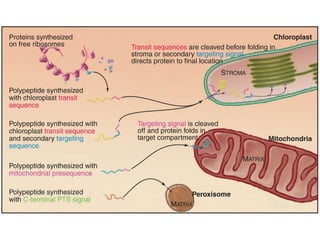
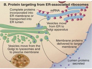
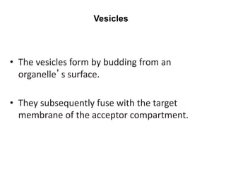
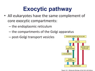
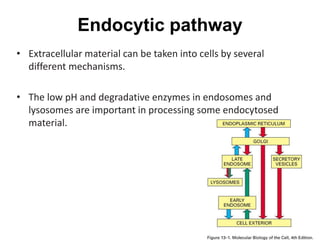
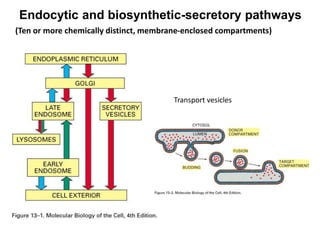
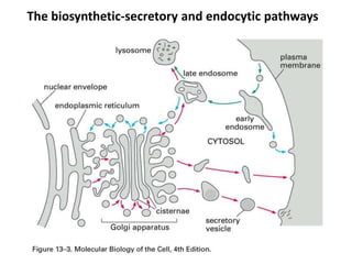
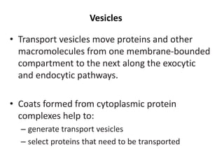
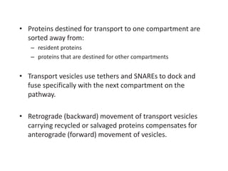
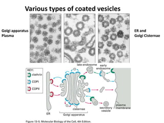
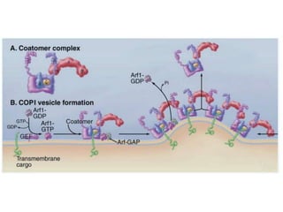
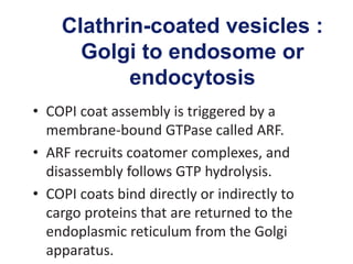
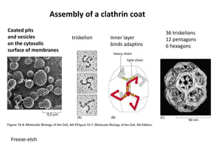
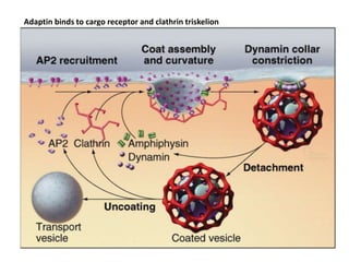
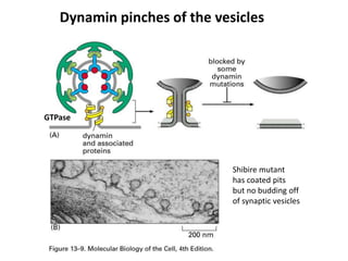
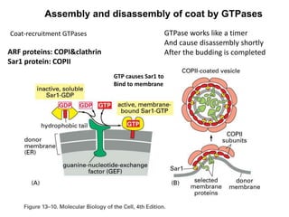
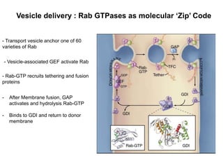
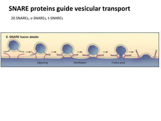
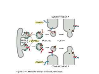
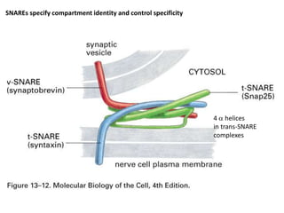
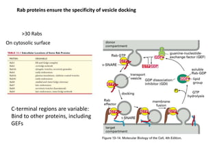
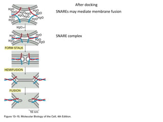
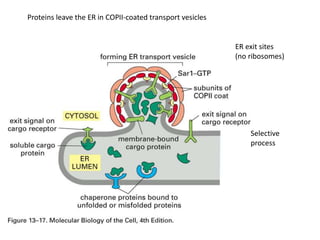
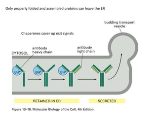
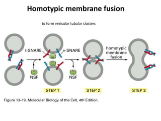
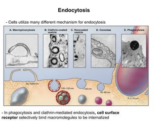
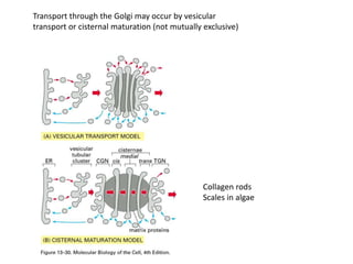
Advanced Cell Biology
2014 1nd Semester
Department of Animal Science
Chungbuk National University
5th Lecture
1st week : Introduction
3rd week :Research Strategies For Cell Biology
5nd week : Nucleus, Transcription and Splicing
7nd week : Membrane and Channel
9nd week : Membrane Trafficking
11nd week : Cell Signaling
13nd week : Cytoskeleton
15nd week : Cell Cycle
• Eukaryotic cells have an elaborate system of
internal membrane-bounded structures
called organelles.
• Each organelle:
– has a unique composition of (glyco)proteins and
(glyco)lipids
– carries out a particular set of functions
Why cell need Membrane Trafficking?
• An organelle comprises one or more
membrane-bounded compartments.
• Organelles may act autonomously or in
cooperation to accomplish a given function.
• In the endocytic and exocytic pathways,
cargo proteins are transferred between
compartments by transport vesicles.
Needs of Transport System..
• The vesicles form by budding from an
organelle’s surface.
• They subsequently fuse with the target
membrane of the acceptor compartment.
Vesicles
Exocytic pathway
• All eukaryotes have the same complement of
core exocytic compartments:
– the endoplasmic reticulum
– the compartments of the Golgi apparatus
– post-Golgi transport vesicles
Endocytic pathway
• Extracellular material can be taken into cells by several
different mechanisms.
• The low pH and degradative enzymes in endosomes and
lysosomes are important in processing some endocytosed
material.
Endocytic and biosynthetic-secretory pathways
Transport vesicles
(Ten or more chemically distinct, membrane-enclosed compartments)
The biosynthetic-secretory and endocytic pathways
March 30, 2006 Pabio552, Lecture 2 13
A 2. ER translocation is co-translational
March 30, 2006 Pabio552, Lecture 2 14
March 30, 2006 Pabio552, Lecture 2 15
Please review the signal hypothesis and mechanisms of co-translational translocation in
the Alberts’ textbook!
Vesicles
• Transport vesicles move proteins and other
macromolecules from one membrane-bounded
compartment to the next along the exocytic
and endocytic pathways.
• Coats formed from cytoplasmic protein
complexes help to:
– generate transport vesicles
– select proteins that need to be transported
• Proteins destined for transport to one compartment are
sorted away from:
– resident proteins
– proteins that are destined for other compartments
• Transport vesicles use tethers and SNAREs to dock and
fuse specifically with the next compartment on the
pathway.
• Retrograde (backward) movement of transport vesicles
carrying recycled or salvaged proteins compensates for
anterograde (forward) movement of vesicles.
Various types of coated vesicles
Golgi apparatus
Plasma
ER and
Golgi Cisternae
COPII-coated vesicles :
ER to Golgi
• ER membrane proteins activate Sar1p
GTPase.
• Activate Sar1p bring togerther a
transmembrane cargo receptor
• COPII coats deforms the membrane into a
bud
• After budding, coat component promotes
hydrolysis of GTP bound to Sar1p
COPI-coated vesicles :
Golgi to the ER
• COPI coat assembly is triggered by a
membrane-bound GTPase called ARF.
• ARF recruits coatomer complexes, and
disassembly follows GTP hydrolysis.
• COPI coats bind directly or indirectly to
cargo proteins that are returned to the
endoplasmic reticulum from the Golgi
apparatus.
Clathrin-coated vesicles :
Golgi to endosome or
endocytosis
• COPI coat assembly is triggered by a
membrane-bound GTPase called ARF.
• ARF recruits coatomer complexes, and
disassembly follows GTP hydrolysis.
• COPI coats bind directly or indirectly to
cargo proteins that are returned to the
endoplasmic reticulum from the Golgi
apparatus.
Assembly of a clathrin coat
triskelion
Coated pits
and vesicles
on the cytosolic
surface of membranes
Freeze-etch
36 triskelions
12 pentagons
6 hexagons
Inner layer
binds adaptins
Adaptin binds to cargo receptor and clathrin triskelion
Dynamin pinches of the vesicles
GTPase
Shibire mutant
has coated pits
but no budding off
of synaptic vesicles
ARF proteins: COPI&clathrin
Sar1 protein: COPII
GTP causes Sar1 to
Bind to membrane
Assembly and disassembly of coat by GTPases
Coat-recruitment GTPases GTPase works like a timer
And cause disassembly shortly
After the budding is completed
Vesicle delivery : Rab GTPases as molecular ‘Zip’ Code
- Transport vesicle anchor one of 60
varieties of Rab
- Vesicle-associated GEF activate Rab
- Rab-GTP recruits tethering and fusion
proteins
- After Membrane fusion, GAP
activates and hydrolysis Rab-GTP
- Binds to GDI and return to donor
membrane
SNARE proteins guide vesicular transport
20 SNAREs, v-SNAREs, t-SNAREs
SNAREs specify compartment identity and control specificity
4 a helices
in trans-SNARE
complexes
Rab proteins ensure the specificity of vesicle docking
>30 Rabs
On cytosolic surface
C-terminal regions are variable:
Bind to other proteins, including
GEFs
SNAREs may mediate membrane fusion
SNARE complex
After docking
The entry of enveloped viruses into cells
HIV
Similar to SNAREs
Proteins leave the ER in COPII-coated transport vesicles
ER exit sites
(no ribosomes)
Selective
process
Only properly folded and assembled proteins can leave the ER
Chaperones cover up exit signals
Homotypic membrane fusion
to form vesicular tubular clusters
The Golgi apparatus synthesizes sphingolipids, establishing a gradient of
sphingolipids and cholesterol and bilayer thickness from low in the ER to high
in the plasma membrane.
Transport through the Golgi apparatus: Membrane lipids, integral membrane
proteins, and soluble proteins in the lumen move from the cis-Golgi through the
stacks to the trans-Golgi.
An enzyme transfers the branched “core oligosaccharide” rich in mannose from dolichol
to the side chain of asparagine (single letter abbreviation N) as the protein enters the
lumen of the ER. An amide bond couples the first sugar to the side chain
Golgi apparatus glycosidases remove some sugars and
glycosyltransferases add sugars to remodel oligosaccharide side chains.
ER retrieval signals: KKXX in ER membrane proteins,
KDEL sequence in soluble ER resident proteins
Membrane proteins in Golgi and ER have shorter TM domains (15 aa)
Cholesterol
pH controls affinity of KDEL receptors
Targeting Protein and Membrane from TGN
The trans-Golgi network or TGN is a cluster of membrane-bounded tubules and vesicles
adjacent to the trans-most stack of the Golgi apparatus.
Central sorting and distribution point for membranes and cargo
coming through the secretory pathway and destined for lysosomes,
endosomes, and plasma membrane.
Roles of Trans-Golgi Networks
Lysosome and Endosome
Endosomes : sorting compartments between the plasma membrane and lysosomes
Lysosomes contain a variety of
hydrolytic enzymes that
degrade proteins, lipids,
polysaccharides, and nucleic
acids taken into the cell by
endocytosis (see next section)
as well as many cellular
molecules that turn over
normally.
Endocytosis
- Cells utilize many different mechanism for endocytosis
- In phagocytosis and clathrin-mediated endocytosis, cell surface
receptor selectively bind macromolegules to be internalized
Phagocytosis
Ingestion of large particles such as
bacteria, foreign bodies, and remnants of
dead cells
Four step in Phagocytosis
1. Attachments
2. Engulfment
- Formation of phagocuytic cup
- Growth of actin filaments
1. Fusion With Lysosome
2. Degradation
Phosphatidylinositol (PO) is important regulator of Endocytosis
Macropinocytosis
- Many Cell ingest extercellular fluid in large endocytic structure called macropinosomes
Caveolae mediated Endocytosis
Caveole : enriched in cholestrol
Stabillized by the protein called caveolin
Histochemical stains: biochemical
Compartmentalization of the Golgi
Functional
compartmentalization
Transport through the Golgi may occur by vesicular
transport or cisternal maturation (not mutually exclusive)
Collagen rods
Scales in algae
Summary
1. Vesicular transport, biosynthetic-secretory and
endocytic pathways;
2. Coated vesicles;
3. Coat assembly and disassembly, budding, dynamin,
coat-recruitment GTPases;
4. Targeting and fusion by Rab GTPases, SNAREs;
5. ER to Golgi: COPII, folding, fusion (cluster), retrograde;
6. Golgi apparatus structure and polarity;
7. Continuation of glycosylation;
8. Compartmentalization of Golgi cisternae;
9. By now we have introduced gated transport, transmembrane
transport and vesicular transport.

Cell biology Lecture 5

  • 1.
    Advanced Cell Biology 20141nd Semester Department of Animal Science Chungbuk National University 5th Lecture
  • 2.
    1st week :Introduction 3rd week :Research Strategies For Cell Biology 5nd week : Nucleus, Transcription and Splicing 7nd week : Membrane and Channel 9nd week : Membrane Trafficking 11nd week : Cell Signaling 13nd week : Cytoskeleton 15nd week : Cell Cycle
  • 3.
    • Eukaryotic cellshave an elaborate system of internal membrane-bounded structures called organelles. • Each organelle: – has a unique composition of (glyco)proteins and (glyco)lipids – carries out a particular set of functions Why cell need Membrane Trafficking?
  • 4.
    • An organellecomprises one or more membrane-bounded compartments. • Organelles may act autonomously or in cooperation to accomplish a given function. • In the endocytic and exocytic pathways, cargo proteins are transferred between compartments by transport vesicles.
  • 6.
  • 8.
    • The vesiclesform by budding from an organelle’s surface. • They subsequently fuse with the target membrane of the acceptor compartment. Vesicles
  • 9.
    Exocytic pathway • Alleukaryotes have the same complement of core exocytic compartments: – the endoplasmic reticulum – the compartments of the Golgi apparatus – post-Golgi transport vesicles
  • 10.
    Endocytic pathway • Extracellularmaterial can be taken into cells by several different mechanisms. • The low pH and degradative enzymes in endosomes and lysosomes are important in processing some endocytosed material.
  • 11.
    Endocytic and biosynthetic-secretorypathways Transport vesicles (Ten or more chemically distinct, membrane-enclosed compartments)
  • 12.
  • 13.
    March 30, 2006Pabio552, Lecture 2 13 A 2. ER translocation is co-translational
  • 14.
    March 30, 2006Pabio552, Lecture 2 14
  • 15.
    March 30, 2006Pabio552, Lecture 2 15 Please review the signal hypothesis and mechanisms of co-translational translocation in the Alberts’ textbook!
  • 16.
    Vesicles • Transport vesiclesmove proteins and other macromolecules from one membrane-bounded compartment to the next along the exocytic and endocytic pathways. • Coats formed from cytoplasmic protein complexes help to: – generate transport vesicles – select proteins that need to be transported
  • 17.
    • Proteins destinedfor transport to one compartment are sorted away from: – resident proteins – proteins that are destined for other compartments • Transport vesicles use tethers and SNAREs to dock and fuse specifically with the next compartment on the pathway. • Retrograde (backward) movement of transport vesicles carrying recycled or salvaged proteins compensates for anterograde (forward) movement of vesicles.
  • 18.
    Various types ofcoated vesicles Golgi apparatus Plasma ER and Golgi Cisternae
  • 19.
    COPII-coated vesicles : ERto Golgi • ER membrane proteins activate Sar1p GTPase. • Activate Sar1p bring togerther a transmembrane cargo receptor • COPII coats deforms the membrane into a bud • After budding, coat component promotes hydrolysis of GTP bound to Sar1p
  • 21.
    COPI-coated vesicles : Golgito the ER • COPI coat assembly is triggered by a membrane-bound GTPase called ARF. • ARF recruits coatomer complexes, and disassembly follows GTP hydrolysis. • COPI coats bind directly or indirectly to cargo proteins that are returned to the endoplasmic reticulum from the Golgi apparatus.
  • 23.
    Clathrin-coated vesicles : Golgito endosome or endocytosis • COPI coat assembly is triggered by a membrane-bound GTPase called ARF. • ARF recruits coatomer complexes, and disassembly follows GTP hydrolysis. • COPI coats bind directly or indirectly to cargo proteins that are returned to the endoplasmic reticulum from the Golgi apparatus.
  • 24.
    Assembly of aclathrin coat triskelion Coated pits and vesicles on the cytosolic surface of membranes Freeze-etch 36 triskelions 12 pentagons 6 hexagons Inner layer binds adaptins
  • 25.
    Adaptin binds tocargo receptor and clathrin triskelion
  • 26.
    Dynamin pinches ofthe vesicles GTPase Shibire mutant has coated pits but no budding off of synaptic vesicles
  • 27.
    ARF proteins: COPI&clathrin Sar1protein: COPII GTP causes Sar1 to Bind to membrane Assembly and disassembly of coat by GTPases Coat-recruitment GTPases GTPase works like a timer And cause disassembly shortly After the budding is completed
  • 28.
    Vesicle delivery :Rab GTPases as molecular ‘Zip’ Code - Transport vesicle anchor one of 60 varieties of Rab - Vesicle-associated GEF activate Rab - Rab-GTP recruits tethering and fusion proteins - After Membrane fusion, GAP activates and hydrolysis Rab-GTP - Binds to GDI and return to donor membrane
  • 29.
    SNARE proteins guidevesicular transport 20 SNAREs, v-SNAREs, t-SNAREs
  • 31.
    SNAREs specify compartmentidentity and control specificity 4 a helices in trans-SNARE complexes
  • 32.
    Rab proteins ensurethe specificity of vesicle docking >30 Rabs On cytosolic surface C-terminal regions are variable: Bind to other proteins, including GEFs
  • 33.
    SNAREs may mediatemembrane fusion SNARE complex After docking
  • 34.
    The entry ofenveloped viruses into cells HIV Similar to SNAREs
  • 35.
    Proteins leave theER in COPII-coated transport vesicles ER exit sites (no ribosomes) Selective process
  • 36.
    Only properly foldedand assembled proteins can leave the ER Chaperones cover up exit signals
  • 37.
    Homotypic membrane fusion toform vesicular tubular clusters
  • 39.
    The Golgi apparatussynthesizes sphingolipids, establishing a gradient of sphingolipids and cholesterol and bilayer thickness from low in the ER to high in the plasma membrane. Transport through the Golgi apparatus: Membrane lipids, integral membrane proteins, and soluble proteins in the lumen move from the cis-Golgi through the stacks to the trans-Golgi.
  • 40.
    An enzyme transfersthe branched “core oligosaccharide” rich in mannose from dolichol to the side chain of asparagine (single letter abbreviation N) as the protein enters the lumen of the ER. An amide bond couples the first sugar to the side chain
  • 41.
    Golgi apparatus glycosidasesremove some sugars and glycosyltransferases add sugars to remodel oligosaccharide side chains.
  • 42.
    ER retrieval signals:KKXX in ER membrane proteins, KDEL sequence in soluble ER resident proteins Membrane proteins in Golgi and ER have shorter TM domains (15 aa) Cholesterol pH controls affinity of KDEL receptors
  • 43.
    Targeting Protein andMembrane from TGN The trans-Golgi network or TGN is a cluster of membrane-bounded tubules and vesicles adjacent to the trans-most stack of the Golgi apparatus.
  • 44.
    Central sorting anddistribution point for membranes and cargo coming through the secretory pathway and destined for lysosomes, endosomes, and plasma membrane. Roles of Trans-Golgi Networks
  • 45.
    Lysosome and Endosome Endosomes: sorting compartments between the plasma membrane and lysosomes Lysosomes contain a variety of hydrolytic enzymes that degrade proteins, lipids, polysaccharides, and nucleic acids taken into the cell by endocytosis (see next section) as well as many cellular molecules that turn over normally.
  • 47.
    Endocytosis - Cells utilizemany different mechanism for endocytosis - In phagocytosis and clathrin-mediated endocytosis, cell surface receptor selectively bind macromolegules to be internalized
  • 48.
    Phagocytosis Ingestion of largeparticles such as bacteria, foreign bodies, and remnants of dead cells Four step in Phagocytosis 1. Attachments 2. Engulfment - Formation of phagocuytic cup - Growth of actin filaments 1. Fusion With Lysosome 2. Degradation
  • 49.
    Phosphatidylinositol (PO) isimportant regulator of Endocytosis
  • 50.
    Macropinocytosis - Many Cellingest extercellular fluid in large endocytic structure called macropinosomes
  • 51.
    Caveolae mediated Endocytosis Caveole: enriched in cholestrol Stabillized by the protein called caveolin
  • 52.
    Histochemical stains: biochemical Compartmentalizationof the Golgi Functional compartmentalization
  • 53.
    Transport through theGolgi may occur by vesicular transport or cisternal maturation (not mutually exclusive) Collagen rods Scales in algae
  • 54.
    Summary 1. Vesicular transport,biosynthetic-secretory and endocytic pathways; 2. Coated vesicles; 3. Coat assembly and disassembly, budding, dynamin, coat-recruitment GTPases; 4. Targeting and fusion by Rab GTPases, SNAREs; 5. ER to Golgi: COPII, folding, fusion (cluster), retrograde; 6. Golgi apparatus structure and polarity; 7. Continuation of glycosylation; 8. Compartmentalization of Golgi cisternae; 9. By now we have introduced gated transport, transmembrane transport and vesicular transport.