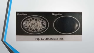
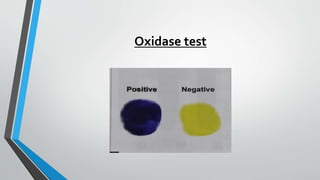
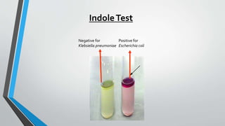
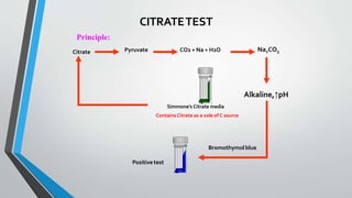
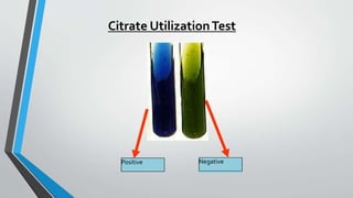
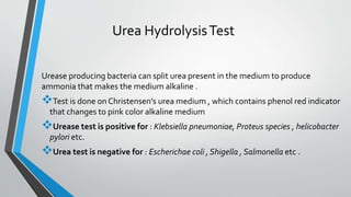
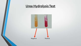
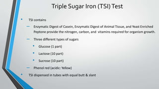
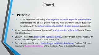
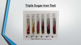
Biochemical Reaction
Dr Avinanda Biswas
Tutor of Microbiology
Biochemical Identification
Based on the type of colony morphology and Gram staining appearance observed in culture smear , the appropriate
biochemical tests are employed.
 Initially, catalase and oxidase tests are done on all types of colonies grown on media .
 For gram-negative bacilli: The following are the common biochemical tests done routinely , abbreviated as ‘ICUT’ :
 Indole test
 Citrate utilization test
 Urea hydrolysis test
 Triple sugar iron test
 For gram-positive cocci :The useful biochemical tests are as follows :
 Coagulase test ( for Staphylococcal aureus )
 CAMP test ( Christine – Atkins – Munch – Peterson) for group B Streptococcus .
 Bile esculin hydrolysis test (for Enterococcus)
 Inulin fermentation test and bile solubility test ( for pneumococcus)
 Antimicrobial susceptibility tests are done for bacterial identification are as follows :
 Optochin susceptibility test – done to differentiate pneumococcus (sensitive) from viridans streptococci.
 Bacitracin susceptibility test – done to differentiate group A ( sensitive) from group B (resistant) Streptococcus .
CatalaseTest
When a colony of any catalase producing bacteria is mixed with a drop of
hydrogen peroxide (3 % H2O2) placed on a slide , effervescence or bubble
appear due to breakdown of H2O2 by catalase to produce oxygen .
Catalase test is primarily used to differentiate between staphylococcus
(catalase positive) from Staphylococcus ( catalase negative) .
It is also positive for members of the families Enterobacteriaceae ,
Vibrionaceae , Pseudomonas etc .
OxidaseTest
It detects the presence of cytochrome oxidase enzyme in bacteria , which catalyzes
the oxidation of reduced cytochrome by atmospheric oxygen .
When a filter paper strip or disk , soaked in oxidase reagent is smeared with a
bacterial colony producing cytochrome oxidase enzyme , the smeared are turns
deep purple within 10 seconds due to oxidation of the dye to form a purple –
colored compound indophenol blue .
Examples :
 Oxidase positive (deep purple) : Pseudomonas ,Vibrio , Neisseria , Bacillus ,
Haemophillus etc .
 Oxidase negative ( no color change ) : members of family Enterobacteriaceae ,
Acinetobacter etc .
Oxidase test
IndoleTest
 Inoculate the test organism into tryptophane broth
 Incubate at 37°C for 24 hours
 After incubation interval, add 1 ml Kovacs reagent which contain 4 (p) –
dimethylamino
• benzaldehyde, shake the tube gently and read immediately
Method
Contd.
Result:
 A bright pink color in the top layer indicates the presence of indole
 The absence of color means that indole was not produced i.e. indole is negative .
Significance:
Indole Positive : A red-colored ring is formed near the surface of the broth . Example :
Escherichia coli , Proteus vulgaris , Vibrio cholerae etc .
Indole Negative : Yellow – colored ring is formed near the surface of the broth , e.g.,
Pseudomonas , Salmonella etc .
Positive for
Escherichia coli
Negative for
Klebsiella pneumoniae
IndoleTest
Citrate UtilizationTest
It detects the ability of a few bacteria to utilize citrate as the sole source of
carbon for their growth , with production of alkaline metabolic products .Test
is performed on Simmon’s citrate medium. Citrate utilizing bacteria produce
growth and a color change , i.e., original green color changes to blue .
Citrate test is positive for Klebsiella , Pneumonia , Citrobacter , Enterobacter
etc .
The test is negative for Escherichia coli , Shigella etc .
Principle:
Citrate Na2CO3
Alkaline,↑pH
Bromothymolblue
CO2 + Na + H2O
Pyruvate
Simmone’s Citrate media
ContainsCitrate as a sole of C source
Positivetest
CITRATETEST
Positive Negative
Citrate UtilizationTest
Urea HydrolysisTest
Urease producing bacteria can split urea present in the medium to produce
ammonia that makes the medium alkaline .
Test is done on Christensen’s urea medium , which contains phenol red indicator
that changes to pink color alkaline medium
Urease test is positive for : Klebsiella pneumoniae, Proteus species , helicobacter
pylori etc.
Urea test is negative for : Escherichae coli , Shigella , Salmonella etc .
Urea HydrolysisTest
Negative Positive
Triple Sugar Iron (TSI)Test
• TSI contains
– Enzymatic Digest of Casein, Enzymatic Digest of Animal Tissue, and Yeast Enriched
Peptone provide the nitrogen, carbon, and vitamins required for organism growth.
– Three different types of sugars
• Glucose (1 part)
• Lactose (10 part)
• Sucrose (10 part)
– Phenol red (acidic: Yellow)
• TSI dispensed in tubes with equal butt & slant
Contd.
• Principle
• – To determine the ability of an organism to attack a specific carbohydrate
incorporated into a basal growth medium, with or without the production of
gas, along with the determination of possible hydrogen sulphide production.
• When the carbohydrates are fermented, acid production is detected by the Phenol
Red pH indicator.
• SodiumThiosulfate is reduced to hydrogen sulfide, and hydrogen sulfide reacts with
an iron salt yielding the typical black iron sulfide.
• Ferric Ammonium Citrate is the hydrogen sulfide (H2S) indicator. Sodium Chloride
maintains the osmotic balance of the medium. Agar is the solidifying agent
Contd.
• Method:
– Inoculate TSI medium with an organism by inoculating
needle by stabbing the butt and streaking the slant
– Incubate at 37°C for 24 hours
Reaction on TSI
Result
Example
Butt
colo
r
Slan
t
colo
r
H2S
Red Red Negative
Alk/Alk/-
(Noaction on sugars)
Nonfermenter
e.g.
Pseudomonas
Yellow Red
Negative A/Alk/-
(Glucose
fermented
withoutH2S)
LNF
e.g. Shigella
Yellow Red
Positiv
e black
in butt
A/Alk/+
(Glucose
fermented
withH2S)
LNF
e.g. Salmonella &
Proteus
Yellow Yellow
Negative
A/A/-
(threesugars
are
fermented)
LF
e.g. E. coli,
Klebsiella,
Enterobacter
Result
Triple Sugar IronTest
Biochemical reaction

Biochemical reaction

  • 1.
    Biochemical Reaction Dr AvinandaBiswas Tutor of Microbiology
  • 2.
    Biochemical Identification Based onthe type of colony morphology and Gram staining appearance observed in culture smear , the appropriate biochemical tests are employed.  Initially, catalase and oxidase tests are done on all types of colonies grown on media .  For gram-negative bacilli: The following are the common biochemical tests done routinely , abbreviated as ‘ICUT’ :  Indole test  Citrate utilization test  Urea hydrolysis test  Triple sugar iron test  For gram-positive cocci :The useful biochemical tests are as follows :  Coagulase test ( for Staphylococcal aureus )  CAMP test ( Christine – Atkins – Munch – Peterson) for group B Streptococcus .  Bile esculin hydrolysis test (for Enterococcus)  Inulin fermentation test and bile solubility test ( for pneumococcus)  Antimicrobial susceptibility tests are done for bacterial identification are as follows :  Optochin susceptibility test – done to differentiate pneumococcus (sensitive) from viridans streptococci.  Bacitracin susceptibility test – done to differentiate group A ( sensitive) from group B (resistant) Streptococcus .
  • 3.
    CatalaseTest When a colonyof any catalase producing bacteria is mixed with a drop of hydrogen peroxide (3 % H2O2) placed on a slide , effervescence or bubble appear due to breakdown of H2O2 by catalase to produce oxygen . Catalase test is primarily used to differentiate between staphylococcus (catalase positive) from Staphylococcus ( catalase negative) . It is also positive for members of the families Enterobacteriaceae , Vibrionaceae , Pseudomonas etc .
  • 5.
    OxidaseTest It detects thepresence of cytochrome oxidase enzyme in bacteria , which catalyzes the oxidation of reduced cytochrome by atmospheric oxygen . When a filter paper strip or disk , soaked in oxidase reagent is smeared with a bacterial colony producing cytochrome oxidase enzyme , the smeared are turns deep purple within 10 seconds due to oxidation of the dye to form a purple – colored compound indophenol blue . Examples :  Oxidase positive (deep purple) : Pseudomonas ,Vibrio , Neisseria , Bacillus , Haemophillus etc .  Oxidase negative ( no color change ) : members of family Enterobacteriaceae , Acinetobacter etc .
  • 6.
  • 7.
    IndoleTest  Inoculate thetest organism into tryptophane broth  Incubate at 37°C for 24 hours  After incubation interval, add 1 ml Kovacs reagent which contain 4 (p) – dimethylamino • benzaldehyde, shake the tube gently and read immediately Method
  • 8.
    Contd. Result:  A brightpink color in the top layer indicates the presence of indole  The absence of color means that indole was not produced i.e. indole is negative . Significance: Indole Positive : A red-colored ring is formed near the surface of the broth . Example : Escherichia coli , Proteus vulgaris , Vibrio cholerae etc . Indole Negative : Yellow – colored ring is formed near the surface of the broth , e.g., Pseudomonas , Salmonella etc .
  • 9.
    Positive for Escherichia coli Negativefor Klebsiella pneumoniae IndoleTest
  • 10.
    Citrate UtilizationTest It detectsthe ability of a few bacteria to utilize citrate as the sole source of carbon for their growth , with production of alkaline metabolic products .Test is performed on Simmon’s citrate medium. Citrate utilizing bacteria produce growth and a color change , i.e., original green color changes to blue . Citrate test is positive for Klebsiella , Pneumonia , Citrobacter , Enterobacter etc . The test is negative for Escherichia coli , Shigella etc .
  • 11.
    Principle: Citrate Na2CO3 Alkaline,↑pH Bromothymolblue CO2 +Na + H2O Pyruvate Simmone’s Citrate media ContainsCitrate as a sole of C source Positivetest CITRATETEST
  • 12.
  • 13.
    Urea HydrolysisTest Urease producingbacteria can split urea present in the medium to produce ammonia that makes the medium alkaline . Test is done on Christensen’s urea medium , which contains phenol red indicator that changes to pink color alkaline medium Urease test is positive for : Klebsiella pneumoniae, Proteus species , helicobacter pylori etc. Urea test is negative for : Escherichae coli , Shigella , Salmonella etc .
  • 14.
  • 15.
    Triple Sugar Iron(TSI)Test • TSI contains – Enzymatic Digest of Casein, Enzymatic Digest of Animal Tissue, and Yeast Enriched Peptone provide the nitrogen, carbon, and vitamins required for organism growth. – Three different types of sugars • Glucose (1 part) • Lactose (10 part) • Sucrose (10 part) – Phenol red (acidic: Yellow) • TSI dispensed in tubes with equal butt & slant
  • 16.
    Contd. • Principle • –To determine the ability of an organism to attack a specific carbohydrate incorporated into a basal growth medium, with or without the production of gas, along with the determination of possible hydrogen sulphide production. • When the carbohydrates are fermented, acid production is detected by the Phenol Red pH indicator. • SodiumThiosulfate is reduced to hydrogen sulfide, and hydrogen sulfide reacts with an iron salt yielding the typical black iron sulfide. • Ferric Ammonium Citrate is the hydrogen sulfide (H2S) indicator. Sodium Chloride maintains the osmotic balance of the medium. Agar is the solidifying agent
  • 17.
    Contd. • Method: – InoculateTSI medium with an organism by inoculating needle by stabbing the butt and streaking the slant – Incubate at 37°C for 24 hours
  • 18.
    Reaction on TSI Result Example Butt colo r Slan t colo r H2S RedRed Negative Alk/Alk/- (Noaction on sugars) Nonfermenter e.g. Pseudomonas Yellow Red Negative A/Alk/- (Glucose fermented withoutH2S) LNF e.g. Shigella Yellow Red Positiv e black in butt A/Alk/+ (Glucose fermented withH2S) LNF e.g. Salmonella & Proteus Yellow Yellow Negative A/A/- (threesugars are fermented) LF e.g. E. coli, Klebsiella, Enterobacter Result
  • 19.