recent microbial techniques & advancement in identifying, cultivating,& handl...Karunanidhan3
I tried to include all techniques & diseases that are included in Pharm D 2nd year microbiology syllabus as per PCI. Do suggest if i have to improve my writing skills, on officialkarunanidhan@gmail.com
recent microbial techniques & advancement in identifying, cultivating,& handl...Karunanidhan3
I tried to include all techniques & diseases that are included in Pharm D 2nd year microbiology syllabus as per PCI. Do suggest if i have to improve my writing skills, on officialkarunanidhan@gmail.com
Each of the letters in “IMViC” stands for one of these tests. “I” is for indole; “M” is for methyl red; “V” is for Voges-Proskauer, and “C” is for citrate, lowercase “i” is added for the ease of pronunciation. IMViC is an acronym that stands for four different tests
Indole test
Methyl red test
Voges-Proskauer test
Citrate utilization test
Explore natural remedies for syphilis treatment in Singapore. Discover alternative therapies, herbal remedies, and lifestyle changes that may complement conventional treatments. Learn about holistic approaches to managing syphilis symptoms and supporting overall health.
Report Back from SGO 2024: What’s the Latest in Cervical Cancer?bkling
Are you curious about what’s new in cervical cancer research or unsure what the findings mean? Join Dr. Emily Ko, a gynecologic oncologist at Penn Medicine, to learn about the latest updates from the Society of Gynecologic Oncology (SGO) 2024 Annual Meeting on Women’s Cancer. Dr. Ko will discuss what the research presented at the conference means for you and answer your questions about the new developments.
Title: Sense of Smell
Presenter: Dr. Faiza, Assistant Professor of Physiology
Qualifications:
MBBS (Best Graduate, AIMC Lahore)
FCPS Physiology
ICMT, CHPE, DHPE (STMU)
MPH (GC University, Faisalabad)
MBA (Virtual University of Pakistan)
Learning Objectives:
Describe the primary categories of smells and the concept of odor blindness.
Explain the structure and location of the olfactory membrane and mucosa, including the types and roles of cells involved in olfaction.
Describe the pathway and mechanisms of olfactory signal transmission from the olfactory receptors to the brain.
Illustrate the biochemical cascade triggered by odorant binding to olfactory receptors, including the role of G-proteins and second messengers in generating an action potential.
Identify different types of olfactory disorders such as anosmia, hyposmia, hyperosmia, and dysosmia, including their potential causes.
Key Topics:
Olfactory Genes:
3% of the human genome accounts for olfactory genes.
400 genes for odorant receptors.
Olfactory Membrane:
Located in the superior part of the nasal cavity.
Medially: Folds downward along the superior septum.
Laterally: Folds over the superior turbinate and upper surface of the middle turbinate.
Total surface area: 5-10 square centimeters.
Olfactory Mucosa:
Olfactory Cells: Bipolar nerve cells derived from the CNS (100 million), with 4-25 olfactory cilia per cell.
Sustentacular Cells: Produce mucus and maintain ionic and molecular environment.
Basal Cells: Replace worn-out olfactory cells with an average lifespan of 1-2 months.
Bowman’s Gland: Secretes mucus.
Stimulation of Olfactory Cells:
Odorant dissolves in mucus and attaches to receptors on olfactory cilia.
Involves a cascade effect through G-proteins and second messengers, leading to depolarization and action potential generation in the olfactory nerve.
Quality of a Good Odorant:
Small (3-20 Carbon atoms), volatile, water-soluble, and lipid-soluble.
Facilitated by odorant-binding proteins in mucus.
Membrane Potential and Action Potential:
Resting membrane potential: -55mV.
Action potential frequency in the olfactory nerve increases with odorant strength.
Adaptation Towards the Sense of Smell:
Rapid adaptation within the first second, with further slow adaptation.
Psychological adaptation greater than receptor adaptation, involving feedback inhibition from the central nervous system.
Primary Sensations of Smell:
Camphoraceous, Musky, Floral, Pepperminty, Ethereal, Pungent, Putrid.
Odor Detection Threshold:
Examples: Hydrogen sulfide (0.0005 ppm), Methyl-mercaptan (0.002 ppm).
Some toxic substances are odorless at lethal concentrations.
Characteristics of Smell:
Odor blindness for single substances due to lack of appropriate receptor protein.
Behavioral and emotional influences of smell.
Transmission of Olfactory Signals:
From olfactory cells to glomeruli in the olfactory bulb, involving lateral inhibition.
Primitive, less old, and new olfactory systems with different path
These simplified slides by Dr. Sidra Arshad present an overview of the non-respiratory functions of the respiratory tract.
Learning objectives:
1. Enlist the non-respiratory functions of the respiratory tract
2. Briefly explain how these functions are carried out
3. Discuss the significance of dead space
4. Differentiate between minute ventilation and alveolar ventilation
5. Describe the cough and sneeze reflexes
Study Resources:
1. Chapter 39, Guyton and Hall Textbook of Medical Physiology, 14th edition
2. Chapter 34, Ganong’s Review of Medical Physiology, 26th edition
3. Chapter 17, Human Physiology by Lauralee Sherwood, 9th edition
4. Non-respiratory functions of the lungs https://academic.oup.com/bjaed/article/13/3/98/278874
Flu Vaccine Alert in Bangalore Karnatakaaddon Scans
As flu season approaches, health officials in Bangalore, Karnataka, are urging residents to get their flu vaccinations. The seasonal flu, while common, can lead to severe health complications, particularly for vulnerable populations such as young children, the elderly, and those with underlying health conditions.
Dr. Vidisha Kumari, a leading epidemiologist in Bangalore, emphasizes the importance of getting vaccinated. "The flu vaccine is our best defense against the influenza virus. It not only protects individuals but also helps prevent the spread of the virus in our communities," he says.
This year, the flu season is expected to coincide with a potential increase in other respiratory illnesses. The Karnataka Health Department has launched an awareness campaign highlighting the significance of flu vaccinations. They have set up multiple vaccination centers across Bangalore, making it convenient for residents to receive their shots.
To encourage widespread vaccination, the government is also collaborating with local schools, workplaces, and community centers to facilitate vaccination drives. Special attention is being given to ensuring that the vaccine is accessible to all, including marginalized communities who may have limited access to healthcare.
Residents are reminded that the flu vaccine is safe and effective. Common side effects are mild and may include soreness at the injection site, mild fever, or muscle aches. These side effects are generally short-lived and far less severe than the flu itself.
Healthcare providers are also stressing the importance of continuing COVID-19 precautions. Wearing masks, practicing good hand hygiene, and maintaining social distancing are still crucial, especially in crowded places.
Protect yourself and your loved ones by getting vaccinated. Together, we can help keep Bangalore healthy and safe this flu season. For more information on vaccination centers and schedules, residents can visit the Karnataka Health Department’s official website or follow their social media pages.
Stay informed, stay safe, and get your flu shot today!
Title: Sense of Taste
Presenter: Dr. Faiza, Assistant Professor of Physiology
Qualifications:
MBBS (Best Graduate, AIMC Lahore)
FCPS Physiology
ICMT, CHPE, DHPE (STMU)
MPH (GC University, Faisalabad)
MBA (Virtual University of Pakistan)
Learning Objectives:
Describe the structure and function of taste buds.
Describe the relationship between the taste threshold and taste index of common substances.
Explain the chemical basis and signal transduction of taste perception for each type of primary taste sensation.
Recognize different abnormalities of taste perception and their causes.
Key Topics:
Significance of Taste Sensation:
Differentiation between pleasant and harmful food
Influence on behavior
Selection of food based on metabolic needs
Receptors of Taste:
Taste buds on the tongue
Influence of sense of smell, texture of food, and pain stimulation (e.g., by pepper)
Primary and Secondary Taste Sensations:
Primary taste sensations: Sweet, Sour, Salty, Bitter, Umami
Chemical basis and signal transduction mechanisms for each taste
Taste Threshold and Index:
Taste threshold values for Sweet (sucrose), Salty (NaCl), Sour (HCl), and Bitter (Quinine)
Taste index relationship: Inversely proportional to taste threshold
Taste Blindness:
Inability to taste certain substances, particularly thiourea compounds
Example: Phenylthiocarbamide
Structure and Function of Taste Buds:
Composition: Epithelial cells, Sustentacular/Supporting cells, Taste cells, Basal cells
Features: Taste pores, Taste hairs/microvilli, and Taste nerve fibers
Location of Taste Buds:
Found in papillae of the tongue (Fungiform, Circumvallate, Foliate)
Also present on the palate, tonsillar pillars, epiglottis, and proximal esophagus
Mechanism of Taste Stimulation:
Interaction of taste substances with receptors on microvilli
Signal transduction pathways for Umami, Sweet, Bitter, Sour, and Salty tastes
Taste Sensitivity and Adaptation:
Decrease in sensitivity with age
Rapid adaptation of taste sensation
Role of Saliva in Taste:
Dissolution of tastants to reach receptors
Washing away the stimulus
Taste Preferences and Aversions:
Mechanisms behind taste preference and aversion
Influence of receptors and neural pathways
Impact of Sensory Nerve Damage:
Degeneration of taste buds if the sensory nerve fiber is cut
Abnormalities of Taste Detection:
Conditions: Ageusia, Hypogeusia, Dysgeusia (parageusia)
Causes: Nerve damage, neurological disorders, infections, poor oral hygiene, adverse drug effects, deficiencies, aging, tobacco use, altered neurotransmitter levels
Neurotransmitters and Taste Threshold:
Effects of serotonin (5-HT) and norepinephrine (NE) on taste sensitivity
Supertasters:
25% of the population with heightened sensitivity to taste, especially bitterness
Increased number of fungiform papillae
Tom Selleck Health: A Comprehensive Look at the Iconic Actor’s Wellness Journeygreendigital
Tom Selleck, an enduring figure in Hollywood. has captivated audiences for decades with his rugged charm, iconic moustache. and memorable roles in television and film. From his breakout role as Thomas Magnum in Magnum P.I. to his current portrayal of Frank Reagan in Blue Bloods. Selleck's career has spanned over 50 years. But beyond his professional achievements. fans have often been curious about Tom Selleck Health. especially as he has aged in the public eye.
Follow us on: Pinterest
Introduction
Many have been interested in Tom Selleck health. not only because of his enduring presence on screen but also because of the challenges. and lifestyle choices he has faced and made over the years. This article delves into the various aspects of Tom Selleck health. exploring his fitness regimen, diet, mental health. and the challenges he has encountered as he ages. We'll look at how he maintains his well-being. the health issues he has faced, and his approach to ageing .
Early Life and Career
Childhood and Athletic Beginnings
Tom Selleck was born on January 29, 1945, in Detroit, Michigan, and grew up in Sherman Oaks, California. From an early age, he was involved in sports, particularly basketball. which played a significant role in his physical development. His athletic pursuits continued into college. where he attended the University of Southern California (USC) on a basketball scholarship. This early involvement in sports laid a strong foundation for his physical health and disciplined lifestyle.
Transition to Acting
Selleck's transition from an athlete to an actor came with its physical demands. His first significant role in "Magnum P.I." required him to perform various stunts and maintain a fit appearance. This role, which he played from 1980 to 1988. necessitated a rigorous fitness routine to meet the show's demands. setting the stage for his long-term commitment to health and wellness.
Fitness Regimen
Workout Routine
Tom Selleck health and fitness regimen has evolved. adapting to his changing roles and age. During his "Magnum, P.I." days. Selleck's workouts were intense and focused on building and maintaining muscle mass. His routine included weightlifting, cardiovascular exercises. and specific training for the stunts he performed on the show.
Selleck adjusted his fitness routine as he aged to suit his body's needs. Today, his workouts focus on maintaining flexibility, strength, and cardiovascular health. He incorporates low-impact exercises such as swimming, walking, and light weightlifting. This balanced approach helps him stay fit without putting undue strain on his joints and muscles.
Importance of Flexibility and Mobility
In recent years, Selleck has emphasized the importance of flexibility and mobility in his fitness regimen. Understanding the natural decline in muscle mass and joint flexibility with age. he includes stretching and yoga in his routine. These practices help prevent injuries, improve posture, and maintain mobilit
Prix Galien International 2024 Forum ProgramLevi Shapiro
June 20, 2024, Prix Galien International and Jerusalem Ethics Forum in ROME. Detailed agenda including panels:
- ADVANCES IN CARDIOLOGY: A NEW PARADIGM IS COMING
- WOMEN’S HEALTH: FERTILITY PRESERVATION
- WHAT’S NEW IN THE TREATMENT OF INFECTIOUS,
ONCOLOGICAL AND INFLAMMATORY SKIN DISEASES?
- ARTIFICIAL INTELLIGENCE AND ETHICS
- GENE THERAPY
- BEYOND BORDERS: GLOBAL INITIATIVES FOR DEMOCRATIZING LIFE SCIENCE TECHNOLOGIES AND PROMOTING ACCESS TO HEALTHCARE
- ETHICAL CHALLENGES IN LIFE SCIENCES
- Prix Galien International Awards Ceremony
Couples presenting to the infertility clinic- Do they really have infertility...Sujoy Dasgupta
Dr Sujoy Dasgupta presented the study on "Couples presenting to the infertility clinic- Do they really have infertility? – The unexplored stories of non-consummation" in the 13th Congress of the Asia Pacific Initiative on Reproduction (ASPIRE 2024) at Manila on 24 May, 2024.
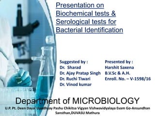
1. Department of MICROBIOLOGY
U.P. Pt. Deen Dayal Upadhyay Pashu Chikitsa Vigyan Vishwavidyalaya Evam Go-Ansundhan
Sansthan,DUVASU Mathura
Presentation on
Biochemical tests &
Serological tests for
Bacterial Identification
Suggested by :
Dr. Sharad
Dr. Ajay Pratap Singh
Dr. Ruchi Tiwari
Dr. Vinod kumar
Presented by :
Harshit Saxena
B.V.Sc & A.H.
Enroll. No. – V-1598/16
2. Diagnosis of a
infectious disease
A Clinician traces following path in diagnosing a disease:
1. History of Patient: By owner; Herd manager
↓
2.Signs & Symptoms: close external examination
↓
3.Isolation(if possible) & Identification of pathogen & its
reactions in body
4. 1. Urease Test
Purpose : Specifically- Differentiating between Proteus & Non Lactose
Fermenting enteric Bacteria (Salmonella & Sheigella)
Principle : Few bacteria undergo following reaction by Urease
Enzyme prodution
Urea + water → Ammonia + Carbondioxide + Water
Ammonia increase of pH → Colour of Phenol Red indicator changes
from orange red to deep pink or purplish red cerise
Materials : Cultures of Pseudomonas fluorescens & Proteus vulgaris
4 urea broth tubes
Inoculation loop
Marker, Bunsen burner
Urease
5. Method : Inoculate urea broth tubes with test bacteria
↓
Mix well & incubate at 37°C for 24-48 hrs
↓
Observe Change colour
Interpretation& Result: Tubes inoculated with Proteus vulgaris/
Urease Positive bacteria – Deep pink reaction (Positive Reaction)
Tubes inoculated with Pseudomonas fluorescence no change in
colour of broth(Negative Reaction)
6. 2.Nitrate Reduction Test
Purpose : Differentiation of members of Enterobacteriaceae from
other bacteria
Principle :
E. coli : Reduce nitrate to nitrite
Pseudomonas : Reduce completely to
molecular Nitrogen
S. aureus : Unable to reduce nitrate
Bacterial broth having 0.5% KNO3
↓
After incubation seen for gas CO2 & NO2
production(from nitrate reduction & citric acid cycle)
↓
By addition of 1. Sulfanilic acid
2. N,N di-CH3-1 naphthalamine
↓
Pink or Red colour produced due to nitrite in
medium
7. Materials: Culture of test bacteria, little soil, Nitrate broth tubes
Nitrate test reagent A
Nitrate test reagent B
Nitrate test reagent C or Zn powder
Test tubes
Procedure& Interpretation: Label bacterial tubes, last one is with Garden soil, 5th as control
↓
Inoculate respective bacterial tubes accordinly with different bacteria, fourth one with garden
soil
↓
Incubate all TT at 37°C for 24-48 hrs & observe after incubation for growth, control no growth
↓
In all TT add 0.5ml each of Nitrate Reagent A&B
↓
Development of RED colour – POSITIVE Reaction, no colour in control
↓
Confirm negative test by adding Nitrate C Reagent-5-10 drops or adding Zn powder
↓
Colour turn red in 5-10min if NO3 is present in medium i.e, Negative NO3 reduction test
9. Catalase Test
Purpose: Differentiate ‘Catalase’ enzyme producing(generally
AEROBIC) & Non producing bacteria
Differentiate Staphylococcus sp.(+test) from Stretococcus sp.(- test)
Principle: Catalase enzyme protect bacteria from destruction by H2O2
produced in Aerobic metabolism & release O2
2H2O2 2H2O + O2
Material: H2O2 , Inoculation loop, bacterial culture, Glass slides
Catalase
10. Procedure: Septically with sterile inoculation loop take bacterial
culture from petri plate to glass slide
↓
With pipette add 2-3 drops of H2 O2
Interpretation & Result:
Immediate appearance of Gas bubbles : POSITIVE Test
Absence of gas bubbles : NEGATIVE Test
Experimental observation
11. Lipid Hydrolysis Test
Purpose: Detect lipase producing ability of Bacteria detecting
pathogenicity of a bacteria
Enterobacteriaceae, Fusobacterium, Propionibacterium, Clostrid
ium,Pseudomonas, Mycoplasma, Corynebacterium,
and Staphylococcus give + test
Principle: Organism producing exoenzyme ‘lipase’ hydrolyses
Tributyrin oil in Tributyrin plates ( others oils too Corn oil,
soyabean oil, egg yolk etc)
Producing clear halo – Zone of hydrolysis in plates
Materials: Tributyrin agar plates
Culture of Salmonella typhimurium
Culture of Pseudomonas aeruginosa
Inoculation loop
12. Procedure:
Divide Tributyrin plate using marker in 2 halves
↓
Write P.aeruginosa on one and S.typhimurium on other side
↓
Streak respective bacterial culture on the plate
↓
Incubate at 30°C for 24-48 hours
Interpretation & Result:
Halo zone around streak of P.aeruginosa – POSITIVE Test
No zone of clearence - NEGATIVE Test
13. Oxidase Test
Purpose:To detect wheather a organism is Aerobe or not
Oxidase Positive Organisms: Pseudomonas, Neisseria, Alcaligens,
Aeromonas, Campylobacter, Vibrio, Brucella, Pasteurella, Moraxella,
Helicobacter pylori, Legionella pneumophila, etc.
Oxidase Negative Organisms: Enterobacteriaceae
Principle Detect whether the sample bacteria produces Cytochrome
c oxidase enzyme by:
N,N,N,N-tetramethyl-p-phenylenediamine(TMPD) a redox indicator
a. Appear blue when oxidised
b. Colourless when reduced
Materials: Culture of Pseudomonas aeruginosa,E.coli
Discs impregnated with TMPD solution
Inoculation loop
14. Procedure:
Take a impregnated disc over a glass slide
↓
Moisten it
↓
Take bacterial culture from petri plate or broth and place on the
disc
Interpretation & Result:
a. Disc turn dark purple : POSITIVE Test –Oxidising bacteria
(here P.aeruginosa
b. No change : NEGATIVE Test – Reducing bacteria
Experimental observation
15. DNase Production Test
Purpose: To detect DNase producing bacteria attributed to
Pathogenicity; Used when serum is not available to test
Principle : DNase culture contain nutrients for bacteria, DNA &methyl
green indicator
Methyl green a cation that binds → Negatively charged DNA
DNase breaks the DNA → No binding to indicator → Production of
clear halo
Materials :
DNase agar plate, Staph aureus/E.coli & Serratia. marcescens culture
1N HCl
Inoculation loop, Pipette
16. Method:
Divide DNase culture plate my marker to 2 halves mark the name
of 2 bacteria
↓
With sterile Inoculation loop streak heavily the respective bacterial
cultures ; Incubate at 37°C for 24-48 hrs
↓
Flood it with 1N HCl with pipette
Interpretation & Result:
a. Zone of clearence around streak of S. marcesens show DNase
activity & POSITIVE Test
b. No clearance: NEGATIVE Test
17. Starch hydrolysis or Amylase Production Test
Purpose: To see if the microbe can use starch as a source of
carbon and energy for growth & produce Amylase;Bacillus
subtilis and Escherichia coli, is compared on starch agar.
Principle: Exoenzyme Amylase produced by Bacteria hydrolyses
starch to Dextrins Maltose & Glucose
Addition of Iodine tells amount of satrch hydrolysed by bacteria
Starch Dextrins+Maltose+Glucose
Materials:
Test Bacteria (Bacillus subtilis & Escherichia coli)
Starch Agar media
Gram’s iodine soln.
Petri plates, Inoculation loop
Alpha- Amylase + H2O
18. Procedure:
Take Starch Agar plates mark according to bacterial culture to be
inoculated
↓
Using inoculation loop singly streak with respective bacteria;
incubate at 37°C for 24-48 hrs
↓
Flood the plate with I2 soln.
Interpretation & Result:
Clear Zone around bacterial colonies : POSITIVE Test
Dark blue colouration around bacterial colonies : NEGATIVE Test
Experimental observation
19. Gelatin hydrolysis(Gelatinase production)
Test
Purpose: Helpful in differentiating species of
Bacillus, Clostridium, Proteus, Pseudomonas, and Serratia;
Gelatinase-positive, pathogenic Staphylococcus aureus from the
Gelatinase-negative, nonpathogenic S. epidermidis .
Principle: Gelatin solidify at temperature below 28 °C this property
loses when gelatinase enzyme produced by bacteria act on gelatin
Materials: Bacterial culture of Bacillus subtilis, E.coli, Proteus vulgaris
12% Gelatin tubes
20. Method:
Take tubes containing gelatin with nutrient media
↓
Inoculate them heavily with test bacteria using Stab method
↓
Incubate for 48 hrs then place in refrigerator for 30 min.
Interpretation & Result:
Observe the tubes
a. Media is still intact –NEGATIVE Test
b. Media loses & gelatin hydrolyses to liquid media – POSITIVE
test
21. # Period of incubation can be extended for 2 weeks as some
bacteria produces Gelatin in very small quantities
22. IMVIC Tests
1.Indole Test
2.Methyl Red Test
3.Voges-Proskauer Test
4.Citrate Test
Purpose: For Identification of bacteria in
family ‘Enterobacteriaeae(Enterics)’
23. Indole Test
Purpose: To detect bacteria producing Tryptophanase enzyme
Principle: Some bacteria oxide EAA –
Tryptophane Indole+Pyruvic Acid +Ammonia
Indole + Kovac’s Reagent Rosindole (Cherry Red
(p-dimethylaminobenzaldehyde) Complex)
Materials:
E.coli & Enterobacter aerogens cultures in NB
1%tryptone broth in 3 test tubes
Kovac’s Reagent
Inoculation loop, Bunsenburner, Pipette
HCl Butanol
Tryptophanase
24. Procedure:
1% Peptone water in 3 test tubes 5ml each
↓
Inoculate the 2 test tube with 2 bacterial cultures separately
Incubate at 37°C for 24-48 hrs
↓
After 48 hours add 1ml kovac’s Reagent shake 15-20min with time gap
↓
Keep the tubes let reagent react to the surface
Interpretation & Result:
Development of Cherry red colour (E.coli) – POSITIVE Test
No colour production- NEGATIVE Test
25. Methyl Red & Voges Proskauer Test
•Clark and Lubs developed MR-VP Broth
•Both the MR and VP tests performed from the same inoculated
medium by aliquoting portions to different tubes
•Bacteria incubated in MR-VP broth having Glucose & Peptone
Methyl Red Test
Purpose: Originally used to distinguish between members of the
family Enterobacteriaceae
• But now also used to characterize other groups of bacteria including
Actinobacteria
Principe: Some bacteria have ability to :
•Glucose Lactic Acid +Acetic Acid+Formic Acid
Mixed Acid Pathway
26. •Acid so produced decreases the pH to 4.5 or below, which is
indicated by a change in the colour of methyl red from yellow to
red
Materials:
MRVP broth (pH 6.9)
Ingredients per liter of deionized water:
buffered peptone= 7.0 gm
glucose= 5.0 gm
dipotassium phosphate= 5.0 gm
Methyl red solution, 0.02%
a. Dissolve 0.1 g of methyl red in 300 ml of ethyl alcohol, 95%.
b. Add sufficient distilled water to make 500 ml.
c. Store at 4 to 8 degree C in a brown bottle. Solution is stable for
1 year.
27. Procedure:
Prior to inoculation, allow medium to equilibrate to room temperature
↓
Using organisms taken from an 18-24 hour pure culture, lightly inoculate
the medium
↓
Incubate aerobically at 37 degrees C. for 24 hours
↓
Following 24 hours of incubation, aliquot 1ml of the broth to a clean TT
↓
Reincubate the remaining broth for an additional 24 hours
↓
Add 2 to 3 drops of methyl red indicator to aliquot
Result & Interpretation:
Positive Reaction: A distinct red color (A)Examples: E. coli, Yersinia sps,
Negative Reaction: A yellow color (B)
Examples: Enterobacter aerogenes, Klebsiella pneumoniae, etc
29. Voges Proskauer Test
Principle: Used to determine if an organism produces acetylmethyl
carbinol from glucose fermentation
Acetylmethyl carbinol is converted to diacetyl in the presence of ∝-
naphthol, strong alkali (40% KOH), and atmospheric oxygen
The ∝-naphthol act as a color intensifier
The diacetyl and quanidine-containing compounds in the peptones of
the broth condense : pinkish red polymer
30. Materials:
Ingredients per liter of deionized water:
buffered peptone= 7.0 gm
glucose= 5.0 gm
dipotassium phosphate= 5.0 gm
Voges-Proskauer Reagent A: Barritt’s reagent AAlpha-Naphthol, 5%50
gmAbsolute Ethanol1000 ml
Voges-Proskauer Reagent B: Barritt’s reagent BPotassium Hydroxide400
gmDeionized Water1000 ml
Procedure:
Prior to inoculation, allow medium to equilibrate to room temperature
↓
Using organisms taken from an 18-24 hour pure culture, lightly inoculate
the medium
31. Incubate aerobically at 37 degrees C. for 24 hours
↓
Following 24 hours of incubation, aliquot 2 ml of the broth to a clean test
tube
↓
Re-incubate the remaining broth for an additional 24 hours
↓
Add 6 drops of 5% alpha-naphthol, and mix well to aerate
↓
Add 2 drops of 40% potassium hydroxide, and mix well to aerate
↓
Shake the tube vigorously during the 30-min period
Interpretation & Result:Positive Reaction: A pink-red color at the surface
Examples: Viridans group streptococci (except Streptococcus
vestibularis), Listeria, Enterobacter, Klebsiella, Serratia marcescens,
Hafnia alvei, Vibrio eltor, Vibrio alginolyticus, etc
32. Negative Reaction: A lack of a pink-red color
Examples: Streptococcus mitis, Citrobacter sp., Shigella, Yersinia,
Edwardsiella, Salmonella, Vibrio furnissii, Vibrio fluvialis, Vibrio
vulnificus, and Vibrio parahaemolyticus etc.
https://microbiologyinfo.com/voges-proskauer-vp-test-principle-reagents-procedure-and-
result/
33. Citrate Test
Purpose: Citrate utilising/fermenting bacteria are identified
List of Bacteria which gives positive citrate utilization test
Klebsiella pneumoniae.
Enterobacter species (minority of strains gives negative result)
Citrobacter freundii.
Salmonella other than Typhi and Paratyphi A.
Serratia marcescens.
Proteus mirabilis (minority of strains gives negative result)
Principle: Bacteria that can grow on Simmon’s citrate Agar(SCA)
produce an enzyme, citrate-permease, capable of converting
citrate to pyruvate
Bacteria metabolize citrate, the ammonium salts are broken down
to ammonia, which increases alkalinity
34. The shift in pH turns the bromthymol blue indicator in the medium
from green to blue above pH 7.6
Materials: E.coli & Enterobacter aerogens culture in NA
Simmon’s citrate agar plates
Inoculation loop & Needle
Method:
Inoculate SCA with bacterial cultures one kept as control
↓
Incubate all at 37°C for 24-48 hrs
Interpretation & Result:
Growth of bacteria=Prussian blue colour develops=POSITIVE Test
No growth=Medium remains green=Negative test
35.
36. H2S production & Motility
Purpose:Determines whether the microbe reduces :-
Sulfur-containing compounds to sulfides metabolism i.e,
Citrobacter spp; Clostridium perfringens
Principle: Reduction of compounds such as thiosulphate, sulphite,
sulphate by some bacteria produce H2 S
Materials:
•Plant cultures of Pseudomonas aerogenosa, Escherichia coli&
Proteus vulgaris
•Sulfide motility medium(SIM) tubes semi solid
•Inoculation loop & needle
37. Procedure:
Label all SIM agar tubes with name of bacteria to be tested
↓
Stab the tubes with respective bacteria incubate 37°C for 24-48 hrs
Interpretation & Result:
Proteus vulgaris tube=Blackening seen= POSITIVE Test
# Motility test = Zone of growth localised near stab(non motile) or
diffused in media(motile)
38. Carbohydrate fermentation test
Purpose:
Enterobacteriaceae family are glucose fermenters (they can
metabolize glucose anaerobically)
Maltose fermentation differentiates Proteus vulgaris (positive)
from Proteus mirabilis (negative)
Rapid carbohydrate utilization test can be performed to
identity Corynebacterium diphtheriae & other sp. Of this bacteria
Principle:
Tests for the presence of acid and/or gas produced from
carbohydrate fermentation
Basal medium containing a single carbohydrate source is used for
this purpose
39. •A pH indicator (such as Andrade’s solution) which will detect the
the lowering of the pH of the medium due to acid production
• Small inverted tubes called Durham tube immersed in the
medium to test production of the gas (hydrogen or CO2 )
Materials:
Composition of Phenol Red Carbohydrate Broth
Trypticase or proteose peptone No. 3: 10 g
Sodium Chloride (NaCl): 5 g
Beef extract (optional): 1 g
Phenol red (7.2 ml of 0.25% phenol red solution): 0.018 g
Carbohydrate source: 10 g
40. Procedure:
Prepare broth media
↓
Fill 4-5 ml of phenol red carbohydrate broth
↓
Insert a Durham tube to detect gas production.
Autoclave the prepared test media (at 121°C for 15 minutes) to
sterilize for 3 minute
The preferred carbohydrate concentration is 1%
↓
Aseptically inoculate each test tube with test microorganism
↓
Alternatively, inoculate each test tube with 1-2 drops of an 18- to
24-hour brain-heart infusion broth culture of the desired
organism
Incubate tubes at 35-37°C for 18-24 hours
41. Interpretation & Result:
Acid production:
Positive: Yellow colour
Negative: Red colour
Gas Production
Positive: Bubbles will be seen in the inverted Durham tube
Negative: No bubbles
42. Coagulase test
Purpose:To differentiate Staphylococcus aureus (positive) from
Coagulase negative sp. from Coagulase Negative (CONS)
Principle: Detects clumping factor (formerly referred as cell-
bound coagulase)
Clumping factor directly converts fibrinogen to fibrin causing
agglutination
Heavy suspension of organism is made on glass slide and mixed
with drop of plasma
Materials: Clean glass slide
Bacterial cuture to be tested
Serum, Inoculation loop
43. Procedure:
Emulsify a staphylococcal colony in a drop of water on a clean and
grease free glass slide
↓
Dip a inoculating wire into the undiluted plasma at room
temperature, withdraw, and stir the adhering traces of plasma (not
a loopful) into the suspension on the slide
↓
Flame the wire and repeat for the control suspensions
↓
Read clumping of cocci visible to the naked eye within 10 seconds
Interpretation & Result:
Coagulase positive: Macroscopic clumping in 10 second
Coagulase negative: No clumping in either drop
46. What is Serology & Serological Tests???
“Serology - is the science of identifying &measuring
antibody or antigen in body fluids”
Serological Tests refers to procedures applied in order to
scan either antibody of antigen in the body fluid of a
organism
Need???
•Diagnosis of a disease - 1° Use
•Checking competene
Eg: a)Transfusion
b)Feeding of new born with its Dam’s milk( IgG Ab testing)
47. Antigen Tests
Antibody Tests
•Isolation & Identification of a Pathogen
•Early diagnosis
•When Isolation & Identification of a Pathogen is not easy
•Transfusion Cases
•Early diagnosis
•Autoimmune diseases
•Checking effectivenes of treatment prescribed
49. Primary Serological Tests
Directly measure the binding of antigen and antibody
(i.e.; directly measure or visualize the immune
complex).
Most sensitive techniques
Eg:
1.Enzyme linked Immunosorbent assay (ELISA)
2. Radioimmunoassay (RIA)
3.Immnunoperoxidase Test
4.Flourescent Antibody Test
3. Western blotting
51. History:
1971 : two Swedish scientists : Eva Engvall and Peter
Perlman invented a test that revolutionized medicine
Why ELISA is one of the most celebrated diagnostic
technique???
56. Apply Antigen
1. Add 100 μl antigen diluted in coating solution to appropriate wells.
2. Incubate 1 hour.
3. Empty plate and tap out residual liquid.
Block Plate
1. Add 300 μl blocking solution to each well.
2. Incubate 15 minutes, empty plate and tap out residual liquid.
Add Secondary Antibody Solution
1. Add 100 μl secondary antibody solution to each well.
2. Incubate 1 hour.
3. Empty plate, tap out residual liquid.
Wash Plate
1. Fill each well with wash solution. 2. Empty plate, tap out residual liquid.
3. Repeat 3 - 5 times.
4. Give final 5 minute soak with wash solution; tap out residual liquid.
React Substrate
1. Dispense 100 μl substrate into each well.
2. If desired, after sufficient color development add 100 μl of the appropriate stop solution to
each well.
3. Read plate with plate reader. Recommended filters:ABTS: 405-415 nm TMB-based
substrates: unstopped 620-650 nm stopped 450 nm pNPP: 405-415 nm BluePhos: 595-650 nm
FirePhos: 460-505 nm
Direct ELISA(Protocol)
57. Indirect ELISA(Protocol)
Apply Antigen
1. Add 100 μl antigen diluted in coating solution to appropriate wells.
2. Incubate 1 hour.
3. Empty plate and tap out residual liquid.
Block Plate
1. Add 300 μl blocking solution to each well.
2. Incubate 15 minutes, empty plate and tap out residual liquid.
React Primary Antibody
1. Add 100 μl secondary antibody solution to each well.
2. Incubate 1 hour.
3. Empty plate, tap out residual liquid.
Wash Plate
1. Fill each well with wash solution.
2. Empty plate, tap out residual liquid.
3. Repeat 3 - 5 times.
Add Secondary Antibody Solution
1. Add 100 μl diluted secondary antibody to each well.incubate 1 hours
2.Empty plate, tap out residual liquid and wash as above.
3. Give final 5 minute soak with wash solution; tap out residual liquid.
React Substrate
1. Dispense 100 μl substrate into each well.
2. If desired, after sufficient color development add 100 ml appropriate stop solution to each well.
3. Read plate with plate reader
58. Advantages & Disadvantage
Direct ELISA• Quick, only one antibody and fewer steps are used.
• No cross-reactivity of secondary antibody
• Immune reactivity of the primary antibody might be adversely
affected by labeling.
• Minimal signal amplification
Indirect ELISA
• Versatile as many primary antibodies can be made in onespecies
and same labeled secondary antibody can be used for detection
• Maximum immune reactivity of the primary antibody is retained
because it is not labeled
• Sensitivity is increased because each primary antibody contains
several epitopes allowing for signal amplification.
59.
60. Sandwich ELISA-Protocol
Apply Capture Antibody :Same manner
Block Plate Same manner
React Sample Antigen
1. Add 100 μl secondary antibody solution to each well.
2. Incubate 1 hour or overnight.
3. Empty plate, tap out residual liquid.
Wash Plate
1. Fill each well with wash solution.
2. Empty plate, tap out residual liquid.
3. Repeat 3 - 5 times.
Add Secondary Antibody Solution
1. Add 100 μl diluted secondary antibody to each well.
2. Incubate 1 hour at room temperature.
3. Empty plate, tap out residual liquid and wash as above.
4. Give final 5 minute soak with wash solution; tap out residual liquid.
React Substrate
1. Dispense 100 μl substrate into each well.
2. If desired, after sufficient color development add 100 ml of the appropriate stop solution to
each well.
3. Read plate with plate reader
61. Immunoperoxidase Test
•Same as ELISA except it is used on tissue sections
•Also known as In situ ELISA
•Antibody conjugated with enzyme & when suitable
substrate added give colour seen by microscope
•Used for sections & smears that are formalin fixed thus
not use for pathogen isolation
•Detect Antigens & cellular changes caused by it
63. Fluorescent Antibody Test
•Same as ELISA except that here Ab are conjugated to Fluorescent dye
(say Fluorescein isothiocynateTetramethl, rhodamine, Alexafluor,
Auramine)in place of enzyme
•Dye after irradiation with UV light – 290&145nm emits green light
•Most suitable for Rabies Test
64. Procedure:
Infect coverslip with Goat pox virus of different dilutions
↓
Add maintainence media incubate at 37°C for 3 days
↓
Fix the monolayers & decant the medium from both test n control
↓
Dry monolayers stain with Hyperimmune serum leave for 1hour
↓
Wash with PBS
↓
Stain monolayers with FITC conjugate leave for a hour
↓
Wash molasses mount the coverslip on glass slide with 50%
glycerine saline
Examine under fluorescent microscope
65. Radioimmune Assay
•Developed by Rosalyn Sussman Yalow, Roger Guillemin, and Andrew
Schally
• This revolutionary development earned Dr. Yalow the Nobel Prize
for Medicine in 1977
Principle & Procedure:
66.
67. Secondary Serological Tests
Secondary binding tests are tests that detect and measure the
consequences (secondary effect) of antigen-antibody
Interaction
These consequences include:
• Precipitation of soluble antigens
• Clumping (agglutination) of particulate antigens
• Neutralization of bacteria, viruses, or toxins; and
•Activation of the complement system
•They are usually less sensitive than primary binding tests, but
may be easier to perform
68. Precipitation Tests
“Precipitation reactions are based on the interaction of two soluble
reactants antibodies and antigens that come together to make one
insoluble product”
•Antigen: Precipitinogen Antibody:Precipitin
•Presence of electrolyte & pH needed
•Produced by multivalent Ab & presence of Zonal Phenomenon
•Used for indentifying bacterial types; cross reactivity; Atg. relatedness
•Can be performed as :
Single dimension double diffusion: Oudin Test
Double dimension double diffusion: Ouchterlony Test
69. Lattice hypothesis
Multivalent Antigens react with Bivalent Antibodies - Varying
proportions-Depending on Ag-Ab ratio at zone of equivalence
Antigenic identity; Partial identity; Non Identity is also studied
70. A gel-diffusion, antibody-antigen precipitation test that depends on
simple vertical diffusion in one dimension
↓
Gel column is prepared containing a homogeneous distribution of
antibody molecules
↓
Above this is layered an aqueous suspension of antigen molecules is
poured
↓
As these formed diffuse into the gel, a moving zone of antigen-
antibody precipitates if the two are complementary : Positive Test
Oudin Test
71. Ouchterlony double immunodiffusion
a.Agar Gel Precipitation Test
Principle:
Antigen & Antibody both moves in solid agar to form insoluble
complexes & band formation
Visualization done against dark background with eluminated
object obliquely from bottom
Material used:-
1. Test sample as Antigen or Antibody
2. Known Antigen or Serum Containing known Antibodies
3. 1%Noble Agar(Difco) in PBS pH-7.4
72. 4. Staining soln. – Mixing n filtering
a. Coomsaie brilliant blue R-250
b. Glacial Acetic Acid
c. Methanol
d. Distilled Water
Procedure:
Pour 5ml molten agar in petri plates tranfer
to refrigerator at 4°C for 20minutes
↓
Punch well in Agar
↓
Remove agar from wells seal the base by 1.1% molten agar soln
↓
Fill the central well with Antigen & Periphera wells- (+)&(-) antisera with
pasteurpipettes
↓
Petri plate kept at humidified place for few hours- overnight
↓
Read pattern of precipitation line
73.
74. b.Countercurrent Immunoelectrophoresis
•In electric field:
Antigens: Negatively charge Migrate to anode
Antibody: Positively charge Migrate to Cathode
•Endosmotic flow & Double dimension double diffusion tests
•Same as AGPT with application of concept of Elecrophoresis
•Reduce duration in 30-60 minutes Antg-Atb complexes can be
visualized
•Electrophoresis use here 0.02M Barbitone buffer pH8.6
•Migaration of Antigens can be seen by adding Bromophenol
blue dye
75. Rocket Immunoelecrophoresis Test
Used in quantification of Antigen
•Length of Precipitation are formed when Antigen is
electrophresedd into an agar layer containing antibody
•In electric field:
Antigens: Negatively charge Migrate to anode
Antibody: Positively charge Migrate to Cathode
Antigens migrate- get dilute-Ag held back in complexes-Equivalence
point reached precipitate form
More antigen – precipitate redissolves move forwards
Antigens exhaust – Stable arc forms
Height of rockets directly proportional to concentration of antigen
76.
77. Agglutination Test
•Particulate antigen(Agglutinogen) mixed with a antibody(Agglutinin)
in presence of electrolyte(NaCl) at suitable temp & pH lead to clumping
& Agglutination
•More sensitive than precipitation
•Monovalent or incomplete antibodies not cause agglutination
•Most used test in identifying type specie
•Types
1.Slide Agglutination
2.Tube Agglutination
3.Heterophile Agglutination
4.Antiglobulin coombs test
78. Slide agglutination Test
• Suspension of unknown antigen is kept on slide and a drop of
standardized antiserum is added or vice versa
• A positive reaction is indicated by formation of visible clumps.
E.g. Widal test, RPR test.
79. Tube Agglutination Test
• Agglutination test performed in tube and standard quantitative
technique for determination of antibody titre
•In this method serum is diluted in a series of tubes and standard
antigen suspensions (specific for the suspected disease) are added
to it
•After incubation, antigen-antibody reaction is indicated visible
clumps of agglutination.
80. Heterophile Agglutination Test
This test depends on demonstration of heterophilic antibodies
in serum present in certain bacterial infection
Antiglobulin (Coombs) test:
Devised by Coombs, Mourant, and Race for detection of incomplete
anti-Rh antibodies that do not agglutinate Rh+ erythrocytes in saline
serum containing incomplete anti-Rh antibodies is mixed with Rh+
erythrocytes in saline
incomplete antibody antiglobulin coats the surface of erythrocytes but
does not cause any agglutination.
When such erythrocytes are treated with antiglobulin or Coombs
serum (rabbit antiserum against human gamma globulin), then the
cells are agglutinated.
81.
82. Complement Fixation Test
•Detection of the presence of either specific antibody or
specific antigen in a patient's serum, based on whether complement
fixation occurs
•Process
•Serum is separated from the patient
•Patients naturally have different levels of complement proteins in their
serum.
•To negate any effects this might have on the test, the complement
proteins in the patient's serum must be destroyed and replaced by a
known amount of standardized complement proteins
83. •A)The serum is heated in such a way that all of the complement
proteins—but none of the antibodies—within it are destroyed.
•B) known amount of standard complement proteins are added to
the serum. (guinea pig serum.)
•The antigen of interest is added to the serum
•Sheep red blood cells (sRBCs) [2] which have been pre-bound to anti-
sRBC antibodies are added to the serum
• The test is considered negative if the solution turns pink at this point
and positive otherwise
84.
85. Tertiary Binding Test
If an organism or antigen possesses biological activity, antibody can
be measured by their ability to neutralize this activity
The activities that may be neutralized include hemolysis of RBCs, lysis
of nucleated cells, and disease or death in animals
Reactions such as these are subject to high degree of variability
86. Neutrilization Test
1.In vivo –Schick’s Test
Schick test - used to determine whether or not a person is
susceptible to diphtheria (Corynebacterium diphtheria).
A small amount (0.1 ml) of diluted (1/50 MLD) diphtheria toxin
is injected intradermally into the arm of the person.
The skin around the injection will become red and swollen,
indicating a positive result.
This swelling disappears after a few days.
If the person has immunity, then little or no swelling and
redness will occur, indicating a negative result
2. In Vitro (Antistreptolysin “O” titration)
87. Test based on Type 4 Hypersensitivity
1.Tuberculin Test
2.Mallein Test
3.Johnin Test etcc..
Interadermal antigen inoculation is done and inflammatory
changes seen after 24-48 hours
Presence of inflammation is Positive Result
Absence of inflammation Negative result
# Tests based on Inoculation of test sample in live animals
also falls under the category of tertiary binding tests