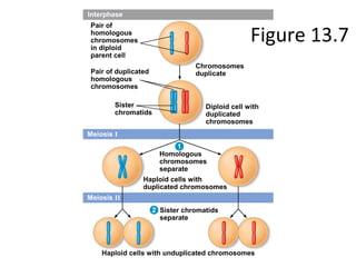
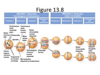
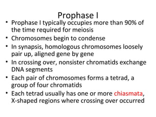
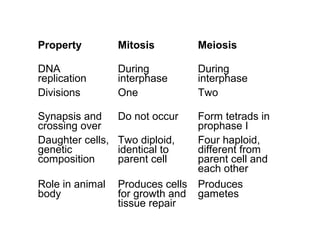
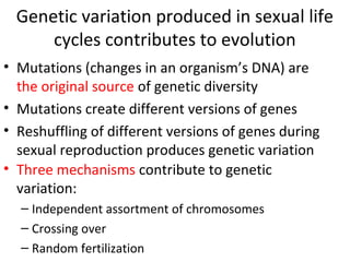
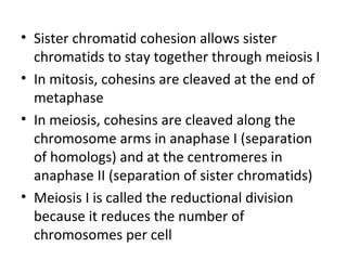
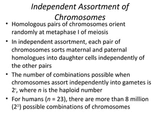
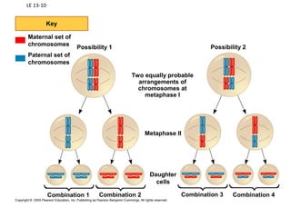
Chapter 13
Meiosis and Sexual
Life Cycles
Animation
Overview: Hereditary Similarity and
Variation
• Living organisms are distinguished by their
ability to reproduce their own kind
• Heredity is the transmission of traits from one
generation to the next
• Variation shows that offspring differ in
appearance from parents and siblings
• Genetics is the scientific study of heredity and
variation
Figure 13.1
Inheritance of Genes
• Genes are the units of heredity
• Genes are segments of DNA
• Each gene has a specific locus on a certain
chromosome
• One set of chromosomes is inherited from each
parent
• Reproductive cells called gametes (sperm and
eggs) unite, passing genes to the next
generation
3-17
The physical location of a gene on a
chromosome is called its locus.
Figure 3.3
• Gametes are typically haploid
– They contain a single set of chromosomes
• Gametes are 1n, while diploid cells are 2n
– A diploid human cell contains 46 chromosomes
– A human gamete only contains 23 chromosomes
• During meiosis, haploid cells are produced from
diploid cells
– Thus, the chromosomes must be correctly sorted and
distributed to reduce the chromosome number to
half its original value
• In humans, for example, a gamete must receive one
chromosome from each of the 23 pairs
Copyright ©The McGraw-Hill Companies, Inc. Permission required for reproduction or display
3-43
Sets of Chromosomes in Human Cells
• Each human somatic cell (any cell other than a
gamete) has 46 chromosomes arranged in pairs
• A karyotype is an ordered display of the pairs of
chromosomes from a cell
• The two chromosomes in each pair are called
homologous chromosomes, or homologues
• Both chromosomes in a pair carry genes
controlling the same inherited characteristics
LE 13-3
5 µmPair of homologous
chromosomes
Sister
chromatids
Centromere
• The sex chromosomes are called X and Y
• Human females have a homologous pair of X
chromosomes (XX)
• Human males have one X and one Y
chromosome
• The 22 pairs of chromosomes that do not
determine sex are called autosomes
Figure 13.4Key
Maternal set of
chromosomes (n = 3)
Paternal set of
chromosomes (n = 3)
2n = 6
Sister chromatids
of one duplicated
chromosome
Two nonsister
chromatids in
a homologous pair
Centromere
Pair of homologous
chromosomes
(one from each set)
1. Why is it more practical to prepare
karyotypes by viewing somatic diploid cells
rather than haploid gametes?
a. Somatic diploid cells do not contain organelles to
interfere with karyotyping.
b. Both sets of chromosomes, which are present in
somatic diploid cells, need to be examined.
c. DNA in haploid gametes will not stain.
d. The chromosomes are larger in a somatic diploid
cell.
e. Haploid gametes do not have sex chromosomes.
Behavior of Chromosome Sets in the Human
Life Cycle
• At sexual maturity, the ovaries and testes
produce haploid gametes
• Gametes are the only types of human cells
produced by meiosis, rather than mitosis
• Meiosis results in one set of chromosomes in
each gamete
• Fertilization, the fusing of gametes, restores the
diploid condition, forming a zygote
• The diploid zygote develops into an adult
Figure 13.5
Key
Haploid (n)
Diploid (2n)
Haploid gametes (n = 23)
Egg (n)
Sperm (n)
MEIOSIS FERTILIZATION
Testis
Diploid
zygote
(2n = 46)
Ovary
Mitosis and
development
Multicellular diploid
adults (2n = 46)
The Variety of Sexual Life Cycles
• The alternation of meiosis and fertilization is
common to all organisms that reproduce sexually
• The three main types of sexual life cycles differ in
the timing of meiosis and fertilization
Figure 13.6Key
Haploid (n)
Diploid (2n)
Gametes
Haploid multi-
cellular organism
(gametophyte)
MitosisMitosis Mitosis Mitosis
Haploid unicellular or
multicellular organism
Gametes
FERTILIZATIONMEIOSIS
n
2n
n
n
n
n
nn
n
nn
2n
2n
Zygote
MEIOSIS FERTILIZATION
Spores
Gametes
Mitosis
Diploid
multicellular
organism
(sporophyte)
Mitosis
Diploid
multicellular
organism
(a) Animals (b) Plants and some algae (c) Most fungi and
some protists
2n 2n
MEIOSIS FERTILIZATION
Zygote
n
Zygote
nn
• Plants and some algae exhibit an alternation of
generations
• This life cycle includes two multicellular
generations or stages: one diploid and one haploid
• The diploid organism, the sporophyte, makes
haploid spores by meiosis
• Each spore grows by mitosis into a haploid
organism called a gametophyte
• A gametophyte makes haploid gametes by mitosis
• In most fungi and some protists, the only diploid
stage is the single-celled zygote; there is no
multicellular diploid stage
• The zygote produces haploid cells by meiosis
• Each haploid cell grows by mitosis into a haploid
multicellular organism
• The haploid adult produces gametes by mitosis
• Depending on the type of life cycle, either haploid
or diploid cells can divide by mitosis
• However, only diploid cells can undergo meiosis
• In all three life cycles, chromosome halving and
doubling contribute to genetic variation in offspring
MEIOSIS
• Like mitosis, meiosis begins after a cell has progressed
through interphase of the cell cycle
• Unlike mitosis, meiosis involves two successive
divisions
– These are termed Meiosis I and II
– Each of these is subdivided into
• Prophase
• Prometaphase
• Metaphase
• Anaphase
• Telophase
• The two cell divisions result in four daughter cells, rather than the
two daughter cells in mitosis
• Each daughter cell has only half as many chromosomes as the parent
cell
The Stages of Meiosis
• In the first cell division (meiosis I), homologous
chromosomes separate
• Meiosis I results in two haploid daughter cells
with replicated chromosomes—reduction
division
• In the second cell division (meiosis II), sister
chromatids separate— equatorial division
• Meiosis II results in four haploid daughter cells
with unreplicated chromosomes
Figure 13.7
Interphase
Meiosis I
Meiosis II
Pair of
homologous
chromosomes
in diploid
parent cell
Pair of duplicated
homologous
chromosomes
Chromosomes
duplicate
Diploid cell with
duplicated
chromosomes
Sister
chromatids
Homologous
chromosomes
separate
Haploid cells with
duplicated chromosomes
Sister chromatids
separate
Haploid cells with unduplicated chromosomes
1
2
• Before Meiosis, chromosomes are replicated in
interphase
• The single centrosome replicates, forming two
centrosomes
Figure 13.8MEIOSIS I: Separates
homologous chromosomes
Prophase I Metaphase I Anaphase I
Telophase I
and
Cytokinesis
Prophase II Metaphase II Anaphase II
Telophase II
and
Cytokinesis
Centrosome
(with
centriole
pair)
Centromere
(with
kineto-
chore)
Sister
chromatids
remain
attachedMeta-
phase
plate
Sister
chroma-
tids
Chiasmata
Spindle
Homo-
logous
chromo-
somes Fragments
of nuclear
envelope
Microtubules
attached to kinetochore
Homologous
chromo-
somes
separate
Cleavage
furrow
Sister
chromatids
separate
Haploid
daughter
cells
forming
MEIOSIS II: Separates
sister chromatids
Prophase I
• Prophase I typically occupies more than 90% of
the time required for meiosis
• Chromosomes begin to condense
• In synapsis, homologous chromosomes loosely
pair up, aligned gene by gene
• In crossing over, nonsister chromatids exchange
DNA segments
• Each pair of chromosomes forms a tetrad, a
group of four chromatids
• Each tetrad usually has one or more chiasmata,
X-shaped regions where crossing over occurred
Crossing Over
• Crossing over produces recombinant
chromosomes, which combine genes inherited
from each parent
• Crossing over begins very early in prophase I, as
homologous chromosomes pair up gene by gene
• In crossing over, homologous portions of two
nonsister chromatids trade places
• Crossing over contributes to genetic variation by
combining DNA from two parents into a single
chromosome
LE 13-11
Prophase I
of meiosis
Tetrad
Nonsister
chromatids
Chiasma,
site of
crossing
over
Recombinant
chromosomes
Metaphase I
Metaphase II
Daughter
cells
Figure 13.9
DNA
breaks
Cohesins
Centromere
DNA
breaks
Pair of
homologous
chromosomes:
Paternal
sister
chromatids
Maternal
sister
chromatids
Synaptonemal
complex forming
Chiasmata
Crossover Crossover
1
2 4
3
Metaphase I
• At metaphase I, tetrads line up at the metaphase
plate, with one chromosome facing each pole
• Microtubules from one pole are attached to the
kinetochore of one chromosome of each tetrad
• Microtubules from the other pole are attached to
the kinetochore of the other chromosome
Anaphase I
• In anaphase I, pairs of homologous chromosomes
separate
• One chromosome moves toward each pole, guided
by the spindle apparatus
• Sister chromatids remain attached at the
centromere and move as one unit toward the pole
LE 13-8ab
Sister
chromatids
Chiasmata
Spindle
Centromere
(with kinetochore)
Metaphase
plate
Homologous
chromosomes
separate
Sister chromatids
remain attached
Microtubule
attached to
kinetochore
Tetrad
MEIOSIS I: Separates homologous chromosomes
PROPHASE I METAPHASE I ANAPHASE I
Homologous chromosomes
(red and blue) pair and
exchange segments; 2n = 6
in this example
Pairs of homologous
chromosomes split up
Tetrads line up
• At the end of meiosis, there are four daughter
cells, each with a haploid set of unreplicated
chromosomes
• Each daughter cell is genetically distinct from
the others and from the parent cell
2. How and at what stage do
chromosomes undergo independent
assortment?
a. meiosis I pairing of homologs
b. anaphase I separation of homologs
c. meiosis II separation of homologs
d. meiosis I metaphase alignment
e. meiosis I telophase separation
2. How and at what stage do
chromosomes undergo independent
assortment?
a. meiosis I pairing of homologs
b. anaphase I separation of homologs
c. meiosis II separation of homologs
d. meiosis I metaphase alignment
e. meiosis I telophase separation
A Comparison of Mitosis and Meiosis
• Mitosis conserves the number of chromosome
sets, producing cells that are genetically
identical to the parent cell
• Meiosis reduces the number of chromosomes
sets from two (diploid) to one (haploid),
producing cells that differ genetically from each
other and from the parent cell
• The mechanism for separating sister chromatids
is virtually identical in meiosis II and mitosis
• Three events are unique to meiosis, and all
three occur in meiosis l:
– Synapsis and crossing over in prophase I:
Homologous chromosomes physically connect and
exchange genetic information
– At the metaphase plate, there are paired
homologous chromosomes (tetrads), instead of
individual replicated chromosomes
– At anaphase I, it is homologous chromosomes,
instead of sister chromatids, that separate and are
carried to opposite poles of the cell
MITOSIS MEIOSIS
Prophase
Duplicated
chromosome
Metaphase
Anaphase
Telophase
2n 2n
Daughter cells
of mitosis
Sister
chromatids
separate.
Individual
chromosomes
line up.
Chromosome
duplication
Chromosome
duplication
2n = 6
Parent cell Chiasma MEIOSIS I
Prophase I
Homologous
chromosome
pair
Metaphase I
Anaphase I
Telophase I
MEIOSIS
II
Pairs of
homologous
chromosomes
line up.
Homologs
separate.
Sister
chroma-
tids
separate.
Daughter cells of meiosis II
Daughter
cells of
meiosis I
n n n n
Property Mitosis Meiosis
DNA
replication
During
interphase
During
interphase
Divisions One Two
Synapsis and
crossing over
Do not occur Form tetrads in
prophase I
Daughter cells,
genetic
composition
Two diploid,
identical to
parent cell
Four haploid,
different from
parent cell and
each other
Role in animal
body
Produces cells
for growth and
tissue repair
Produces
gametes
Genetic variation produced in sexual life
cycles contributes to evolution
• Mutations (changes in an organism’s DNA) are
the original source of genetic diversity
• Mutations create different versions of genes
• Reshuffling of different versions of genes during
sexual reproduction produces genetic variation
• Three mechanisms contribute to genetic
variation:
– Independent assortment of chromosomes
– Crossing over
– Random fertilization
• Sister chromatid cohesion allows sister
chromatids to stay together through meiosis I
• In mitosis, cohesins are cleaved at the end of
metaphase
• In meiosis, cohesins are cleaved along the
chromosome arms in anaphase I (separation
of homologs) and at the centromeres in
anaphase II (separation of sister chromatids)
• Meiosis I is called the reductional division
because it reduces the number of
chromosomes per cell
Independent Assortment of
Chromosomes
• Homologous pairs of chromosomes orient
randomly at metaphase I of meiosis
• In independent assortment, each pair of
chromosomes sorts maternal and paternal
homologues into daughter cells independently of
the other pairs
• The number of combinations possible when
chromosomes assort independently into gametes is
2n
, where n is the haploid number
• For humans (n = 23), there are more than 8 million
(223
) possible combinations of chromosomes
LE 13-10
Key
Maternal set of
chromosomes
Paternal set of
chromosomes
Possibility 1 Possibility 2
Combination 2Combination 1 Combination 3 Combination 4
Daughter
cells
Metaphase II
Two equally probable
arrangements of
chromosomes at
metaphase I
Random Fertilization
• Random fertilization adds to genetic variation
because any sperm can fuse with any ovum
(unfertilized egg)
• The fusion of gametes produces a zygote with
any of about 64 trillion diploid combinations
• Crossing over adds even more variation
• Each zygote has a unique genetic identity
Copyright © 2005 Pearson Education, Inc. publishing as Benjamin Cummings
Evolutionary Significance of Genetic Variation
Within Populations
• Natural selection results in accumulation of
genetic variations favored by the environment
• Sexual reproduction contributes to the genetic
variation in a population, which ultimately
results from mutations
Copyright © 2005 Pearson Education, Inc. publishing as Benjamin Cummings
3. Given the fact that 1 fg of DNA = 9.78 × 10⁵
base pairs (on
average), you can estimate the rate of DNA synthesis in
Saccharomyces cerevisiae.
Approximately how many base pairs per minute were
synthesized during the S phase of these yeast cells?
a. 0.19 (1.9 × 10⁻¹
) base pair
per minute
b. 100,000 (1.0 × 10⁵
) base
pairs per minute
c. 200,000 (2.0 × 10⁵
) base
pairs per minute
d. 11,000,000 (11.0 × 10⁶
)
base pairs per minute

Chapter 13: Meiosis

  • 1.
    Chapter 13 Meiosis andSexual Life Cycles Animation
  • 2.
    Overview: Hereditary Similarityand Variation • Living organisms are distinguished by their ability to reproduce their own kind • Heredity is the transmission of traits from one generation to the next • Variation shows that offspring differ in appearance from parents and siblings • Genetics is the scientific study of heredity and variation
  • 3.
  • 4.
    Inheritance of Genes •Genes are the units of heredity • Genes are segments of DNA • Each gene has a specific locus on a certain chromosome • One set of chromosomes is inherited from each parent • Reproductive cells called gametes (sperm and eggs) unite, passing genes to the next generation
  • 5.
    3-17 The physical locationof a gene on a chromosome is called its locus. Figure 3.3
  • 6.
    • Gametes aretypically haploid – They contain a single set of chromosomes • Gametes are 1n, while diploid cells are 2n – A diploid human cell contains 46 chromosomes – A human gamete only contains 23 chromosomes • During meiosis, haploid cells are produced from diploid cells – Thus, the chromosomes must be correctly sorted and distributed to reduce the chromosome number to half its original value • In humans, for example, a gamete must receive one chromosome from each of the 23 pairs Copyright ©The McGraw-Hill Companies, Inc. Permission required for reproduction or display 3-43
  • 7.
    Sets of Chromosomesin Human Cells • Each human somatic cell (any cell other than a gamete) has 46 chromosomes arranged in pairs • A karyotype is an ordered display of the pairs of chromosomes from a cell • The two chromosomes in each pair are called homologous chromosomes, or homologues • Both chromosomes in a pair carry genes controlling the same inherited characteristics
  • 8.
    LE 13-3 5 µmPairof homologous chromosomes Sister chromatids Centromere
  • 9.
    • The sexchromosomes are called X and Y • Human females have a homologous pair of X chromosomes (XX) • Human males have one X and one Y chromosome • The 22 pairs of chromosomes that do not determine sex are called autosomes
  • 10.
    Figure 13.4Key Maternal setof chromosomes (n = 3) Paternal set of chromosomes (n = 3) 2n = 6 Sister chromatids of one duplicated chromosome Two nonsister chromatids in a homologous pair Centromere Pair of homologous chromosomes (one from each set)
  • 11.
    1. Why isit more practical to prepare karyotypes by viewing somatic diploid cells rather than haploid gametes? a. Somatic diploid cells do not contain organelles to interfere with karyotyping. b. Both sets of chromosomes, which are present in somatic diploid cells, need to be examined. c. DNA in haploid gametes will not stain. d. The chromosomes are larger in a somatic diploid cell. e. Haploid gametes do not have sex chromosomes.
  • 12.
    1. Why isit more practical to prepare karyotypes by viewing somatic diploid cells rather than haploid gametes? a. Somatic diploid cells do not contain organelles to interfere with karyotyping. b. Both sets of chromosomes, which are present in somatic diploid cells, need to be examined. c. DNA in haploid gametes will not stain. d. The chromosomes are larger in a somatic diploid cell. e. Haploid gametes do not have sex chromosomes.
  • 13.
    Behavior of ChromosomeSets in the Human Life Cycle • At sexual maturity, the ovaries and testes produce haploid gametes • Gametes are the only types of human cells produced by meiosis, rather than mitosis • Meiosis results in one set of chromosomes in each gamete • Fertilization, the fusing of gametes, restores the diploid condition, forming a zygote • The diploid zygote develops into an adult
  • 14.
    Figure 13.5 Key Haploid (n) Diploid(2n) Haploid gametes (n = 23) Egg (n) Sperm (n) MEIOSIS FERTILIZATION Testis Diploid zygote (2n = 46) Ovary Mitosis and development Multicellular diploid adults (2n = 46)
  • 15.
    The Variety ofSexual Life Cycles • The alternation of meiosis and fertilization is common to all organisms that reproduce sexually • The three main types of sexual life cycles differ in the timing of meiosis and fertilization
  • 16.
    Figure 13.6Key Haploid (n) Diploid(2n) Gametes Haploid multi- cellular organism (gametophyte) MitosisMitosis Mitosis Mitosis Haploid unicellular or multicellular organism Gametes FERTILIZATIONMEIOSIS n 2n n n n n nn n nn 2n 2n Zygote MEIOSIS FERTILIZATION Spores Gametes Mitosis Diploid multicellular organism (sporophyte) Mitosis Diploid multicellular organism (a) Animals (b) Plants and some algae (c) Most fungi and some protists 2n 2n MEIOSIS FERTILIZATION Zygote n Zygote nn
  • 17.
    • Plants andsome algae exhibit an alternation of generations • This life cycle includes two multicellular generations or stages: one diploid and one haploid • The diploid organism, the sporophyte, makes haploid spores by meiosis • Each spore grows by mitosis into a haploid organism called a gametophyte • A gametophyte makes haploid gametes by mitosis
  • 18.
    • In mostfungi and some protists, the only diploid stage is the single-celled zygote; there is no multicellular diploid stage • The zygote produces haploid cells by meiosis • Each haploid cell grows by mitosis into a haploid multicellular organism • The haploid adult produces gametes by mitosis
  • 19.
    • Depending onthe type of life cycle, either haploid or diploid cells can divide by mitosis • However, only diploid cells can undergo meiosis • In all three life cycles, chromosome halving and doubling contribute to genetic variation in offspring
  • 20.
    MEIOSIS • Like mitosis,meiosis begins after a cell has progressed through interphase of the cell cycle • Unlike mitosis, meiosis involves two successive divisions – These are termed Meiosis I and II – Each of these is subdivided into • Prophase • Prometaphase • Metaphase • Anaphase • Telophase • The two cell divisions result in four daughter cells, rather than the two daughter cells in mitosis • Each daughter cell has only half as many chromosomes as the parent cell
  • 21.
    The Stages ofMeiosis • In the first cell division (meiosis I), homologous chromosomes separate • Meiosis I results in two haploid daughter cells with replicated chromosomes—reduction division • In the second cell division (meiosis II), sister chromatids separate— equatorial division • Meiosis II results in four haploid daughter cells with unreplicated chromosomes
  • 22.
    Figure 13.7 Interphase Meiosis I MeiosisII Pair of homologous chromosomes in diploid parent cell Pair of duplicated homologous chromosomes Chromosomes duplicate Diploid cell with duplicated chromosomes Sister chromatids Homologous chromosomes separate Haploid cells with duplicated chromosomes Sister chromatids separate Haploid cells with unduplicated chromosomes 1 2
  • 23.
    • Before Meiosis,chromosomes are replicated in interphase • The single centrosome replicates, forming two centrosomes
  • 24.
    Figure 13.8MEIOSIS I:Separates homologous chromosomes Prophase I Metaphase I Anaphase I Telophase I and Cytokinesis Prophase II Metaphase II Anaphase II Telophase II and Cytokinesis Centrosome (with centriole pair) Centromere (with kineto- chore) Sister chromatids remain attachedMeta- phase plate Sister chroma- tids Chiasmata Spindle Homo- logous chromo- somes Fragments of nuclear envelope Microtubules attached to kinetochore Homologous chromo- somes separate Cleavage furrow Sister chromatids separate Haploid daughter cells forming MEIOSIS II: Separates sister chromatids
  • 25.
    Prophase I • ProphaseI typically occupies more than 90% of the time required for meiosis • Chromosomes begin to condense • In synapsis, homologous chromosomes loosely pair up, aligned gene by gene • In crossing over, nonsister chromatids exchange DNA segments • Each pair of chromosomes forms a tetrad, a group of four chromatids • Each tetrad usually has one or more chiasmata, X-shaped regions where crossing over occurred
  • 26.
    Crossing Over • Crossingover produces recombinant chromosomes, which combine genes inherited from each parent • Crossing over begins very early in prophase I, as homologous chromosomes pair up gene by gene • In crossing over, homologous portions of two nonsister chromatids trade places • Crossing over contributes to genetic variation by combining DNA from two parents into a single chromosome
  • 27.
    LE 13-11 Prophase I ofmeiosis Tetrad Nonsister chromatids Chiasma, site of crossing over Recombinant chromosomes Metaphase I Metaphase II Daughter cells
  • 28.
  • 29.
    Metaphase I • Atmetaphase I, tetrads line up at the metaphase plate, with one chromosome facing each pole • Microtubules from one pole are attached to the kinetochore of one chromosome of each tetrad • Microtubules from the other pole are attached to the kinetochore of the other chromosome
  • 30.
    Anaphase I • Inanaphase I, pairs of homologous chromosomes separate • One chromosome moves toward each pole, guided by the spindle apparatus • Sister chromatids remain attached at the centromere and move as one unit toward the pole
  • 31.
    LE 13-8ab Sister chromatids Chiasmata Spindle Centromere (with kinetochore) Metaphase plate Homologous chromosomes separate Sisterchromatids remain attached Microtubule attached to kinetochore Tetrad MEIOSIS I: Separates homologous chromosomes PROPHASE I METAPHASE I ANAPHASE I Homologous chromosomes (red and blue) pair and exchange segments; 2n = 6 in this example Pairs of homologous chromosomes split up Tetrads line up
  • 32.
    • At theend of meiosis, there are four daughter cells, each with a haploid set of unreplicated chromosomes • Each daughter cell is genetically distinct from the others and from the parent cell
  • 33.
    2. How andat what stage do chromosomes undergo independent assortment? a. meiosis I pairing of homologs b. anaphase I separation of homologs c. meiosis II separation of homologs d. meiosis I metaphase alignment e. meiosis I telophase separation
  • 34.
    2. How andat what stage do chromosomes undergo independent assortment? a. meiosis I pairing of homologs b. anaphase I separation of homologs c. meiosis II separation of homologs d. meiosis I metaphase alignment e. meiosis I telophase separation
  • 35.
    A Comparison ofMitosis and Meiosis • Mitosis conserves the number of chromosome sets, producing cells that are genetically identical to the parent cell • Meiosis reduces the number of chromosomes sets from two (diploid) to one (haploid), producing cells that differ genetically from each other and from the parent cell • The mechanism for separating sister chromatids is virtually identical in meiosis II and mitosis
  • 36.
    • Three eventsare unique to meiosis, and all three occur in meiosis l: – Synapsis and crossing over in prophase I: Homologous chromosomes physically connect and exchange genetic information – At the metaphase plate, there are paired homologous chromosomes (tetrads), instead of individual replicated chromosomes – At anaphase I, it is homologous chromosomes, instead of sister chromatids, that separate and are carried to opposite poles of the cell
  • 37.
    MITOSIS MEIOSIS Prophase Duplicated chromosome Metaphase Anaphase Telophase 2n 2n Daughtercells of mitosis Sister chromatids separate. Individual chromosomes line up. Chromosome duplication Chromosome duplication 2n = 6 Parent cell Chiasma MEIOSIS I Prophase I Homologous chromosome pair Metaphase I Anaphase I Telophase I MEIOSIS II Pairs of homologous chromosomes line up. Homologs separate. Sister chroma- tids separate. Daughter cells of meiosis II Daughter cells of meiosis I n n n n
  • 38.
    Property Mitosis Meiosis DNA replication During interphase During interphase DivisionsOne Two Synapsis and crossing over Do not occur Form tetrads in prophase I Daughter cells, genetic composition Two diploid, identical to parent cell Four haploid, different from parent cell and each other Role in animal body Produces cells for growth and tissue repair Produces gametes
  • 39.
    Genetic variation producedin sexual life cycles contributes to evolution • Mutations (changes in an organism’s DNA) are the original source of genetic diversity • Mutations create different versions of genes • Reshuffling of different versions of genes during sexual reproduction produces genetic variation • Three mechanisms contribute to genetic variation: – Independent assortment of chromosomes – Crossing over – Random fertilization
  • 40.
    • Sister chromatidcohesion allows sister chromatids to stay together through meiosis I • In mitosis, cohesins are cleaved at the end of metaphase • In meiosis, cohesins are cleaved along the chromosome arms in anaphase I (separation of homologs) and at the centromeres in anaphase II (separation of sister chromatids) • Meiosis I is called the reductional division because it reduces the number of chromosomes per cell
  • 41.
    Independent Assortment of Chromosomes •Homologous pairs of chromosomes orient randomly at metaphase I of meiosis • In independent assortment, each pair of chromosomes sorts maternal and paternal homologues into daughter cells independently of the other pairs • The number of combinations possible when chromosomes assort independently into gametes is 2n , where n is the haploid number • For humans (n = 23), there are more than 8 million (223 ) possible combinations of chromosomes
  • 42.
    LE 13-10 Key Maternal setof chromosomes Paternal set of chromosomes Possibility 1 Possibility 2 Combination 2Combination 1 Combination 3 Combination 4 Daughter cells Metaphase II Two equally probable arrangements of chromosomes at metaphase I
  • 43.
    Random Fertilization • Randomfertilization adds to genetic variation because any sperm can fuse with any ovum (unfertilized egg) • The fusion of gametes produces a zygote with any of about 64 trillion diploid combinations • Crossing over adds even more variation • Each zygote has a unique genetic identity
  • 44.
    Copyright © 2005Pearson Education, Inc. publishing as Benjamin Cummings Evolutionary Significance of Genetic Variation Within Populations • Natural selection results in accumulation of genetic variations favored by the environment • Sexual reproduction contributes to the genetic variation in a population, which ultimately results from mutations
  • 45.
    Copyright © 2005Pearson Education, Inc. publishing as Benjamin Cummings 3. Given the fact that 1 fg of DNA = 9.78 × 10⁵ base pairs (on average), you can estimate the rate of DNA synthesis in Saccharomyces cerevisiae. Approximately how many base pairs per minute were synthesized during the S phase of these yeast cells? a. 0.19 (1.9 × 10⁻¹ ) base pair per minute b. 100,000 (1.0 × 10⁵ ) base pairs per minute c. 200,000 (2.0 × 10⁵ ) base pairs per minute d. 11,000,000 (11.0 × 10⁶ ) base pairs per minute
  • 46.
    Copyright © 2005Pearson Education, Inc. publishing as Benjamin Cummings 3. Given the fact that 1 fg of DNA = 9.78 × 10⁵ base pairs (on average), you can estimate the rate of DNA synthesis in Saccharomyces cerevisiae. Approximately how many base pairs per minute were synthesized during the S phase of these yeast cells? a. 0.19 (1.9 × 10⁻¹ ) base pair per minute b. 100,000 (1.0 × 10⁵ ) base pairs per minute c. 200,000 (2.0 × 10⁵ ) base pairs per minute d. 11,000,000 (11.0 × 10⁶ ) base pairs per minute

Editor's Notes

  • #4 Figure 13.1 What accounts for family resemblance?
  • #11 Figure 13.4 Describing chromosomes
  • #12 Answer: B
  • #13 Answer: B
  • #15 Figure 13.5 The human life cycle
  • #17 Figure 13.6 Three types of sexual life cycles
  • #23 Figure 13.7 Overview of meiosis: how meiosis reduces chromosome number
  • #25 Figure 13.8 Exploring meiosis in an animal cell
  • #29 Figure 13.9 Crossing over and synapsis in prophase I: a closer look
  • #34 Answer: D
  • #35 Answer: D
  • #38 Figure 13.10a A comparison of mitosis and meiosis (part 1: mitosis vs. meiosis art)
  • #46 Answer: C Because the S phase took place from approximately the 1-hour mark to the 3-hour mark, start by finding the difference between the amount of DNA at 3 hours and the amount at 1 hour: 47.0 fg – 24.0 fg = 23.0 fg. Now calculate the rate of fg synthesized per minute by dividing that amount by the number of minutes in two hours: 23.0 fg/ 120 minutes = 0.192 fg/min. Finally, you need to convert from fg/min to base pairs/min: 0.192 fg/min × (9.78 × 105 base pairs/fg) = 187,800, or approximately 200,000 base pairs/min.
  • #47 Answer: C Because the S phase took place from approximately the 1-hour mark to the 3-hour mark, start by finding the difference between the amount of DNA at 3 hours and the amount at 1 hour: 47.0 fg – 24.0 fg = 23.0 fg. Now calculate the rate of fg synthesized per minute by dividing that amount by the number of minutes in two hours: 23.0 fg/ 120 minutes = 0.192 fg/min. Finally, you need to convert from fg/min to base pairs/min: 0.192 fg/min × (9.78 × 105 base pairs/fg) = 187,800, or approximately 200,000 base pairs/min.