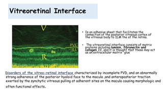
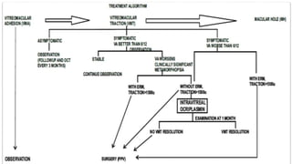
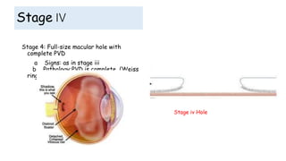
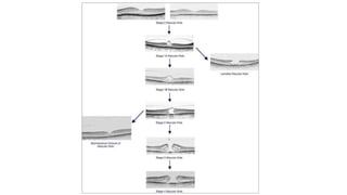
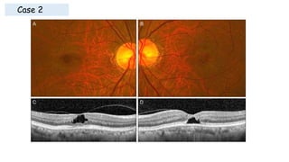
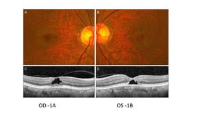
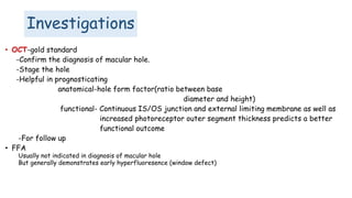
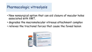
Vitreoretinal Interface Disorders
Eranda Wannigama
Overview
• Anatomy.
• Posterior Vitreous Detachment (PVD).
• Types Vitreoretinal Interface Disorders.
• Management of Vitreoretinal Interface Disorders.
• Summary.
Anatomy
• The vitreous gel is a transparent
extracellular matrix that fills the
cavity behind the lens of the eye.
• It occupies an average volume of
4.4 ml in adulthood
• The outer cortex of vitreous which
is made up of dense collagen (Type
II) is firmly attached to internal
limiting membrane (Type IV
collagen).
Vitreous Attachments
• Vitreous Base-
a ring shaped area
encircling the ora serrata
(2mm anterior and 4mm
posterior to it).
• Peripheral margin of the
Optic nerve head
• Margin of the fovea
• Main retinal vessels
Abnormal areas
– Lattice degeneration
– Cystic retinal tufts
– Chorioretinal scars
• Posterior Vitreous Detachment (PVD)
• Is a separation between the posterior vitreous cortex and the neurosensory retina, with
the vitreous collapsing anteriorly towards the vitreous base.
• PVD affects most eyes by the eighth decade of life. Age at onset is generally in sixth to
seventh decade and men and women appear to be equally affected.
• PVD occurs earlier in myopic eyes, in eyes with inflammatory disease and following blunt
trauma or cataract surgery (especially when there is surgical vitreous loss).
Incomplete posterior vitreous detachment. Posterior hyaloid is detached from fovea and remains
partially attached to optic disc.
Pathophysiology of PVD
• The initial event is liquefaction (Synchisis)
• Then the collapse (Syneresis) of the central vitreous.
Synchysis Syneresis
A rupture develops in the posterior hyaloid (or vitreous cortex) through
which liquefied vitreous flows into the retrovitreous space, separating
the posterior hyaloid from the retina.
• It typically starts as a partial PVD in the perifoveal region and is usually asymptomatic
until it progresses to the optic disc, when separation of the peripapillary glial tissue
from the optic nerve head occurs, usually with formation of a Weiss ring and
accompanying symptoms
PVD ctd
Vitreoretinal Interface
• Is an adhesive sheet that facilitates the
connection of the posterior vitreous cortex of
the vitreous body to ILM the of the retina.
• The vitreoretinal interface consists of matrix
proteins including laminin, fibronectin and
collagen IV, and it is thought that these may act
as an extracellular matrix 'glue'
Disorders of the vitreo-retinal interface characterized by incomplete PVD, and an abnormally
strong adherence of the posterior hyaloid face to the macula ,and anteroposterior traction
exerted by the synchytic vitreous pulling at adherent sites on the macula causing morphologic and
often functional effects.
Vitreoretinal Interface Disorders
• Vitreomacular Adhesion: focal adhesion of the vitreous face within macular
region.
• Vitreomacular Traction: VMA causing focal tractional distortion of macula.
• Macular Hole
• Macular oedema with Posterior hyaloid traction(PHT)
Vitreoretinal Interface imaging with Optical coherence tomography (OCT)
has contributed significantly to the diagnosis and management of
Vitreoretinal Interface diseases
Case 1
66year old Female retired teacher from Kolonnawa
OD-BCVA 6/6
OS-BCVA 6/9
3D OCT -OD 3D OCT -OS
VMT• VMA
Vitreomacular Adhesion
In VMAThe following must be present on at least one OCT image
(i) Partial vitreous detachment as indicated by elevation of cortical vitreous above the retinal surface in the
perifoveal area
(ii) Persistent vitreous attachment to the macula within a 3-mm radius from the center of the fovea
(iii) Acute angle between posterior hyaloid and inner retinal surface
(iv) Absence of changes in foveal contour or retinal morphology.
Focal vitreomacular adhesion with normal foveal contour and retina morphology.
VMA-OCT-based classification
(i) Focal: Width of attachment ≤1500 μm
(ii) Broad: Width of attachment >1500 μm
(i) Concurrent: Associated with other macular abnormalities (e.g.
age-related macular degeneration, retinal vein occlusion, diabetic
macular edema)
(ii) Isolated: Not associated with other macular abnormalities
Vitreomacular traction (VMT)
The following must be present on at least one OCT image:
(i) Partial PVD as indicated by elevation of cortical vitreous above the retinal surface in the perifoveal area
(ii) Persistent vitreous attachment to the macula within a 3-mm radius from the center of the fovea
(iii) Acute angle between posterior hyaloid and inner retinal surface
(iv) Presence of changes in foveal contour or retinal morphology (distortion of foveal surface, intraretinal
structural changes such as pseudocyst formation, elevation of fovea from the RPE, or a combination of any of
these three features)
(v) Absence of full thickness interruption of all retinal layers
VMT-OCT-based classification
(i) Focal: Width of attachment ≤1500 μm
(ii) Broad: Width of attachment >1500μm
(i) V shaped - the vitreous cortex is detached from the retina both temporal and nasal to the
fovea, and attached only to the fovea,
(ii) J shaped or arc shaped - the vitreous cortex is detached from the retina temporal to the
fovea, but remains attached to the retina nasal to the fovea and to the fovea itself.
675um
1752um
VMT & VMA
Symptoms
• VMA: Asymptomatic
VMT: Symptomatic with blurred or reduced vision, metamorphopsia, micropsia, scotoma,
and difficulties with daily vision-related tasks such as reading. Onset and progression of
symptoms are usually gradual, except in a few cases of sudden onset of vision
loss/scotoma due to severe traction causing foveal detachment.
Complications can include:
1. Full thickness macular hole
2. Tractional macular schisis
3. Tractional foveal/macular detachment
Management
1. Observation
2. Surgery
Release anteroposterior traction by means of pars plana vitrectomy (PPV) and
tangential traction with ILM peeling is a common treatment for VMT The
reconstitution of retinal architecture is expected to occur with time, after
removal of the tractional forces.
Symptomatic and visual improvements are expected to follow. Outcomes are variable depending
on morphology and duration
3. Medical therapyThe medical therapy of VMT consists of
pharmacologic vitreolysis.-Ocriplasmin (formerly, microplasmin) is a recombinant
truncated form of human plasmin that has proteolytic activity against these proteins
anchoring the vitreoretinal interface.
4 pneumatic Vitreolysis ;risk of Glaucoma and Inducing tears
Macular hole
• Incidence 0.3% in 3in 1000
• 12% Bilateral
• Common in women 3:1 aged 70 years
• In other eye if PVD present less
than 1% chance to have a macular
hole. If PVD not present risk
increase by 20%.
Etiology
• Common causes:
• Idiopathic -90%
• Other causes:
• Trauma
• high myopia
• ERM
• cystoid macular edema
• proliferative diabetic retinopathy
• severe hypertensive retinopathy
• Choroidal neovasculatrisation
• Solar retinopathy
Classification
• Primary macular hole: is commonly an idiopathic
macular hole
• Caused by vitreous traction on the fovea from an
abnormal vitreous separation
• Secondary Macular hole: caused by other pathologies
not associated with vitereomacular traction
• blunt trauma, high myopia, macular
telangiectasia,ERM
Stages of Macular hole
• Gass first described a series of
stages of formation of idiopathic
macular hole 1988.
Stage 1 a
• Anteroposterior traction by prefoveolar cortical
vitreous split the foveolar retina
Stage 1a: ‘Impending’ macular hole
a Signs: yellow spot
b Pathology: Müller cell cone detach from the
underlying photoreceptor layer, with the formation
of a schisis cavity (pseudocyst)
pseudocyst
Stage 1 a
(A)showing a cyst in the inner part of the fovea (asterisk) due to the traction exerted by the incompletely
detached posterior hyaloid (arrows).
(B)(B) Magnification of (A) showing that the central cyst is divided into several cystic spaces (asterisks) by
septa. The inner segment/outer segment (IS/OS) line is intact, but the cone outer segment tips (COST)
line is elevated at the foveal center.
Stage 1 b
Stage 1b: Occult macular
hole
a Signs: a yellow ring
(donut-shaped) 200-300μm in
diameter
b Pathology: photoreceptor
layer undergo centrifugal
displacement
The posterior hyaloid (PH) is still attached to the roof
of the cyst. The cystic space extends posteriorly and
there is a break in the photoreceptor layer. Note also
the cystic cavities around the central defect. Stage 1B
is an occult macular hole.
Stage II
Stage 2: Small full-thickness hole
a Signs: < 400 µm , central, slightly
eccentric or crescent-shaped.
b Pathology: dehiscence centrally or
eccentrically seen in the roof of the
schitic cavity, pseudo-operculum
The posterior hyaloid (PH) is still attached to the operculum
(aarrow). The operculum is only partially detached from the hole
edge.
Stage III
Stage 3: Full-size macular hole
a Signs:
- > 400 µm
- red base with yellow-white dots
- surrounding grey cuff of
subretinal fluid
b Pathology: avulsion of the roof
of the cyst with an operculum and
persistent peripapillary
attachment of the vitreous
cortex.
The posterior hyaloid (PH) is detached from the
macular surface and contains the operculum (blue
arrow). The edge of the hole has been thickened by
cystic spaces and the photoreceptors are elevated
(black arrows mark the end of the outer segments of
the elevated photoreceptors).
Stage IV
Stage 4: Full-size macular hole with
complete PVD
a Signs: as in stage iii
b Pathology:PVD is complete (Weiss
ring)
Stage iv Hole
Case 2
OD -1A OS -1B
Clinical features
• Visual acuity the first indicator but sometimes
misleading
• Mild loss of central vision (Stage 1a & 1b)
• Metamorphopsia
Amsler Grid
• Non specific distortion rather than scotomas.But Small absolute
scotomas can be detected in 30 -40 % of patients
Watzke RC, Allen L. Subjective slit-
beam sign for macular disease.
• FTMH is diagnosed on slit lamp biomicroscopy
• By slowly sweeping a vertical or horizontal narrow slit beam across the
eye we can study the contour of hole and vitreous interface
differentiates FTMH from other lesions
Positive & Negative Watzke - Allen Sign
Investigations
• OCT-gold standard
-Confirm the diagnosis of macular hole.
-Stage the hole
-Helpful in prognosticating
anatomical-hole form factor(ratio between base
diameter and height)
functional- Continuous IS/OS junction and external limiting membrane as well as
increased photoreceptor outer segment thickness predicts a better
functional outcome
-For follow up
• FFA
Usually not indicated in diagnosis of macular hole
But generally demonstrates early hyperfluoresence (window defect)
Differential Diagnosis
• Epiretinal membrane with pseudomacular hole
• Lamellar macular hole
• Chronic cystoid macular edema
Lamellar macular hole
• Sharply circumscribed
• Partial-thickness defects of the macula
• Represents either as an aborted full-
thickness lesions or a complication of chronic
cystoid macular edema.
• Characterized by a flat, reddish hue-type
lesion with intact outer retinal tissue
• Careful evaluation will reveal retinal tissue in
the base of the lesion
• No evidence of subretinal fluid
• Do not progress to full-thickness lesions
Management
• Depending on the Stage
Stage i > 50% resolve spontaneously -Observation
Stage ii,iii,iv- Surgery is benefitial for both Functional and Anatomical
closure.
Surgery
TPPV +ILM peeling and Tamponade
Release anteroposterior traction by means of pars plana vitrectomy (PPV) and
tangential traction with ILM peeling is a common treatment for MH The
reconstitution of retinal architecture is expected to occur with time, after
removal of the tractional forces
• Surgical and Medical
Pharmacologic vitreolysis
• New nonsurgical option that can aid closure of macular holes
associated with VMT.
• degrades the macromolecular vitreous attachment complex
• relieves the tractional forces that cause the foveal lesion
• On the basis of post operative OCT findings closed macular
holes are:
• Type 1 & type 2 closure
Types of closure
• Indicates that macular hole is closed without foveal defect of the
neurosensory retina
• U shape-perfect closure
• V shape-closure with central thinning
• .
Type 1 closure
Type 2 closure
• Indicates a foveal defect of neurosensory retina persists
postoperatively(w shape closure)
• Although the whole rim of macular hole is attached to the
underlying RPE with flattening of the cuff
Post operative parameters
• OCT parameters: Type 1 closure of MH without
neurosensory defect) has a better visual outcome
compared to Type 2 closure (with neurosensory defect)
• Continuous IS/OS junction and external limiting
membrane as well as increased photoreceptor outer
segment thickness predicts a better functional
outcome
Pre operative parameters
Hole form factor > 0.9 and Macular Hole
index > 0.5 also have a better prognosis
a = base diameter, b = minimum diameter
c = left arm length, d = right arm length
Case 3
73 year old female house wife came
with reduced visual acuity in right
eye
OD-BCVA 2/60
OS-BCVA 6/9
Advantages of ILM peeling
• Removes the scaffold for proliferation of cellular components Like
myofibroblasts, fibrocytes,RPE cells, fibrous astrocyts.
• Eleminate tangential traction around fovea.
• The benefit of ILM peeling is that it ensures complete removal of the
posterior hyaloid or any overlying epiretinal membranes.
• ILM peeling may reduce the duration of face-down positioning
required for macular hole closure.
Case 4
Macular oedema with Posterior
hyaloid traction(PHT)
• Vitrectomy including removal of the ILM leads to expedited
resolution of diffuse diabetic macular edema and
improvement of visual acuity without subsequent epiretinal
membrane formation.
• Complete release of tractional forces and inhibition of
reproliferation of fibrous astrocytes seem to be prudent in
the eyes of patients with diabetes and advanced
vitreoretinal interface disease of the macula.
Vitreoretinal interface disorders

Vitreoretinal interface disorders

  • 1.
  • 2.
    Overview • Anatomy. • PosteriorVitreous Detachment (PVD). • Types Vitreoretinal Interface Disorders. • Management of Vitreoretinal Interface Disorders. • Summary.
  • 3.
    Anatomy • The vitreousgel is a transparent extracellular matrix that fills the cavity behind the lens of the eye. • It occupies an average volume of 4.4 ml in adulthood • The outer cortex of vitreous which is made up of dense collagen (Type II) is firmly attached to internal limiting membrane (Type IV collagen).
  • 4.
    Vitreous Attachments • VitreousBase- a ring shaped area encircling the ora serrata (2mm anterior and 4mm posterior to it). • Peripheral margin of the Optic nerve head • Margin of the fovea • Main retinal vessels Abnormal areas – Lattice degeneration – Cystic retinal tufts – Chorioretinal scars
  • 5.
    • Posterior VitreousDetachment (PVD) • Is a separation between the posterior vitreous cortex and the neurosensory retina, with the vitreous collapsing anteriorly towards the vitreous base. • PVD affects most eyes by the eighth decade of life. Age at onset is generally in sixth to seventh decade and men and women appear to be equally affected. • PVD occurs earlier in myopic eyes, in eyes with inflammatory disease and following blunt trauma or cataract surgery (especially when there is surgical vitreous loss). Incomplete posterior vitreous detachment. Posterior hyaloid is detached from fovea and remains partially attached to optic disc.
  • 6.
    Pathophysiology of PVD •The initial event is liquefaction (Synchisis) • Then the collapse (Syneresis) of the central vitreous. Synchysis Syneresis A rupture develops in the posterior hyaloid (or vitreous cortex) through which liquefied vitreous flows into the retrovitreous space, separating the posterior hyaloid from the retina.
  • 7.
    • It typicallystarts as a partial PVD in the perifoveal region and is usually asymptomatic until it progresses to the optic disc, when separation of the peripapillary glial tissue from the optic nerve head occurs, usually with formation of a Weiss ring and accompanying symptoms PVD ctd
  • 8.
    Vitreoretinal Interface • Isan adhesive sheet that facilitates the connection of the posterior vitreous cortex of the vitreous body to ILM the of the retina. • The vitreoretinal interface consists of matrix proteins including laminin, fibronectin and collagen IV, and it is thought that these may act as an extracellular matrix 'glue' Disorders of the vitreo-retinal interface characterized by incomplete PVD, and an abnormally strong adherence of the posterior hyaloid face to the macula ,and anteroposterior traction exerted by the synchytic vitreous pulling at adherent sites on the macula causing morphologic and often functional effects.
  • 9.
    Vitreoretinal Interface Disorders •Vitreomacular Adhesion: focal adhesion of the vitreous face within macular region. • Vitreomacular Traction: VMA causing focal tractional distortion of macula. • Macular Hole • Macular oedema with Posterior hyaloid traction(PHT) Vitreoretinal Interface imaging with Optical coherence tomography (OCT) has contributed significantly to the diagnosis and management of Vitreoretinal Interface diseases
  • 10.
    Case 1 66year oldFemale retired teacher from Kolonnawa OD-BCVA 6/6 OS-BCVA 6/9 3D OCT -OD 3D OCT -OS
  • 11.
  • 12.
    Vitreomacular Adhesion In VMAThefollowing must be present on at least one OCT image (i) Partial vitreous detachment as indicated by elevation of cortical vitreous above the retinal surface in the perifoveal area (ii) Persistent vitreous attachment to the macula within a 3-mm radius from the center of the fovea (iii) Acute angle between posterior hyaloid and inner retinal surface (iv) Absence of changes in foveal contour or retinal morphology. Focal vitreomacular adhesion with normal foveal contour and retina morphology.
  • 13.
    VMA-OCT-based classification (i) Focal:Width of attachment ≤1500 μm (ii) Broad: Width of attachment >1500 μm (i) Concurrent: Associated with other macular abnormalities (e.g. age-related macular degeneration, retinal vein occlusion, diabetic macular edema) (ii) Isolated: Not associated with other macular abnormalities
  • 14.
    Vitreomacular traction (VMT) Thefollowing must be present on at least one OCT image: (i) Partial PVD as indicated by elevation of cortical vitreous above the retinal surface in the perifoveal area (ii) Persistent vitreous attachment to the macula within a 3-mm radius from the center of the fovea (iii) Acute angle between posterior hyaloid and inner retinal surface (iv) Presence of changes in foveal contour or retinal morphology (distortion of foveal surface, intraretinal structural changes such as pseudocyst formation, elevation of fovea from the RPE, or a combination of any of these three features) (v) Absence of full thickness interruption of all retinal layers
  • 15.
    VMT-OCT-based classification (i) Focal:Width of attachment ≤1500 μm (ii) Broad: Width of attachment >1500μm (i) V shaped - the vitreous cortex is detached from the retina both temporal and nasal to the fovea, and attached only to the fovea, (ii) J shaped or arc shaped - the vitreous cortex is detached from the retina temporal to the fovea, but remains attached to the retina nasal to the fovea and to the fovea itself. 675um 1752um
  • 17.
    VMT & VMA Symptoms •VMA: Asymptomatic VMT: Symptomatic with blurred or reduced vision, metamorphopsia, micropsia, scotoma, and difficulties with daily vision-related tasks such as reading. Onset and progression of symptoms are usually gradual, except in a few cases of sudden onset of vision loss/scotoma due to severe traction causing foveal detachment. Complications can include: 1. Full thickness macular hole 2. Tractional macular schisis 3. Tractional foveal/macular detachment
  • 18.
    Management 1. Observation 2. Surgery Releaseanteroposterior traction by means of pars plana vitrectomy (PPV) and tangential traction with ILM peeling is a common treatment for VMT The reconstitution of retinal architecture is expected to occur with time, after removal of the tractional forces. Symptomatic and visual improvements are expected to follow. Outcomes are variable depending on morphology and duration 3. Medical therapyThe medical therapy of VMT consists of pharmacologic vitreolysis.-Ocriplasmin (formerly, microplasmin) is a recombinant truncated form of human plasmin that has proteolytic activity against these proteins anchoring the vitreoretinal interface. 4 pneumatic Vitreolysis ;risk of Glaucoma and Inducing tears
  • 20.
    Macular hole • Incidence0.3% in 3in 1000 • 12% Bilateral • Common in women 3:1 aged 70 years • In other eye if PVD present less than 1% chance to have a macular hole. If PVD not present risk increase by 20%.
  • 21.
    Etiology • Common causes: •Idiopathic -90% • Other causes: • Trauma • high myopia • ERM • cystoid macular edema • proliferative diabetic retinopathy • severe hypertensive retinopathy • Choroidal neovasculatrisation • Solar retinopathy
  • 22.
    Classification • Primary macularhole: is commonly an idiopathic macular hole • Caused by vitreous traction on the fovea from an abnormal vitreous separation • Secondary Macular hole: caused by other pathologies not associated with vitereomacular traction • blunt trauma, high myopia, macular telangiectasia,ERM
  • 23.
    Stages of Macularhole • Gass first described a series of stages of formation of idiopathic macular hole 1988.
  • 24.
    Stage 1 a •Anteroposterior traction by prefoveolar cortical vitreous split the foveolar retina Stage 1a: ‘Impending’ macular hole a Signs: yellow spot b Pathology: Müller cell cone detach from the underlying photoreceptor layer, with the formation of a schisis cavity (pseudocyst) pseudocyst
  • 25.
    Stage 1 a (A)showinga cyst in the inner part of the fovea (asterisk) due to the traction exerted by the incompletely detached posterior hyaloid (arrows). (B)(B) Magnification of (A) showing that the central cyst is divided into several cystic spaces (asterisks) by septa. The inner segment/outer segment (IS/OS) line is intact, but the cone outer segment tips (COST) line is elevated at the foveal center.
  • 26.
    Stage 1 b Stage1b: Occult macular hole a Signs: a yellow ring (donut-shaped) 200-300μm in diameter b Pathology: photoreceptor layer undergo centrifugal displacement The posterior hyaloid (PH) is still attached to the roof of the cyst. The cystic space extends posteriorly and there is a break in the photoreceptor layer. Note also the cystic cavities around the central defect. Stage 1B is an occult macular hole.
  • 27.
    Stage II Stage 2:Small full-thickness hole a Signs: < 400 µm , central, slightly eccentric or crescent-shaped. b Pathology: dehiscence centrally or eccentrically seen in the roof of the schitic cavity, pseudo-operculum The posterior hyaloid (PH) is still attached to the operculum (aarrow). The operculum is only partially detached from the hole edge.
  • 28.
    Stage III Stage 3:Full-size macular hole a Signs: - > 400 µm - red base with yellow-white dots - surrounding grey cuff of subretinal fluid b Pathology: avulsion of the roof of the cyst with an operculum and persistent peripapillary attachment of the vitreous cortex. The posterior hyaloid (PH) is detached from the macular surface and contains the operculum (blue arrow). The edge of the hole has been thickened by cystic spaces and the photoreceptors are elevated (black arrows mark the end of the outer segments of the elevated photoreceptors).
  • 29.
    Stage IV Stage 4:Full-size macular hole with complete PVD a Signs: as in stage iii b Pathology:PVD is complete (Weiss ring) Stage iv Hole
  • 31.
  • 32.
  • 33.
    Clinical features • Visualacuity the first indicator but sometimes misleading • Mild loss of central vision (Stage 1a & 1b) • Metamorphopsia Amsler Grid • Non specific distortion rather than scotomas.But Small absolute scotomas can be detected in 30 -40 % of patients
  • 34.
    Watzke RC, AllenL. Subjective slit- beam sign for macular disease. • FTMH is diagnosed on slit lamp biomicroscopy • By slowly sweeping a vertical or horizontal narrow slit beam across the eye we can study the contour of hole and vitreous interface differentiates FTMH from other lesions Positive & Negative Watzke - Allen Sign
  • 35.
    Investigations • OCT-gold standard -Confirmthe diagnosis of macular hole. -Stage the hole -Helpful in prognosticating anatomical-hole form factor(ratio between base diameter and height) functional- Continuous IS/OS junction and external limiting membrane as well as increased photoreceptor outer segment thickness predicts a better functional outcome -For follow up • FFA Usually not indicated in diagnosis of macular hole But generally demonstrates early hyperfluoresence (window defect)
  • 36.
    Differential Diagnosis • Epiretinalmembrane with pseudomacular hole • Lamellar macular hole • Chronic cystoid macular edema
  • 37.
    Lamellar macular hole •Sharply circumscribed • Partial-thickness defects of the macula • Represents either as an aborted full- thickness lesions or a complication of chronic cystoid macular edema. • Characterized by a flat, reddish hue-type lesion with intact outer retinal tissue • Careful evaluation will reveal retinal tissue in the base of the lesion • No evidence of subretinal fluid • Do not progress to full-thickness lesions
  • 38.
    Management • Depending onthe Stage Stage i > 50% resolve spontaneously -Observation Stage ii,iii,iv- Surgery is benefitial for both Functional and Anatomical closure. Surgery TPPV +ILM peeling and Tamponade Release anteroposterior traction by means of pars plana vitrectomy (PPV) and tangential traction with ILM peeling is a common treatment for MH The reconstitution of retinal architecture is expected to occur with time, after removal of the tractional forces • Surgical and Medical
  • 39.
    Pharmacologic vitreolysis • Newnonsurgical option that can aid closure of macular holes associated with VMT. • degrades the macromolecular vitreous attachment complex • relieves the tractional forces that cause the foveal lesion
  • 40.
    • On thebasis of post operative OCT findings closed macular holes are: • Type 1 & type 2 closure Types of closure
  • 41.
    • Indicates thatmacular hole is closed without foveal defect of the neurosensory retina • U shape-perfect closure • V shape-closure with central thinning • . Type 1 closure
  • 42.
    Type 2 closure •Indicates a foveal defect of neurosensory retina persists postoperatively(w shape closure) • Although the whole rim of macular hole is attached to the underlying RPE with flattening of the cuff
  • 43.
    Post operative parameters •OCT parameters: Type 1 closure of MH without neurosensory defect) has a better visual outcome compared to Type 2 closure (with neurosensory defect) • Continuous IS/OS junction and external limiting membrane as well as increased photoreceptor outer segment thickness predicts a better functional outcome
  • 44.
    Pre operative parameters Holeform factor > 0.9 and Macular Hole index > 0.5 also have a better prognosis a = base diameter, b = minimum diameter c = left arm length, d = right arm length
  • 45.
    Case 3 73 yearold female house wife came with reduced visual acuity in right eye OD-BCVA 2/60 OS-BCVA 6/9
  • 46.
    Advantages of ILMpeeling • Removes the scaffold for proliferation of cellular components Like myofibroblasts, fibrocytes,RPE cells, fibrous astrocyts. • Eleminate tangential traction around fovea. • The benefit of ILM peeling is that it ensures complete removal of the posterior hyaloid or any overlying epiretinal membranes. • ILM peeling may reduce the duration of face-down positioning required for macular hole closure.
  • 47.
  • 49.
    Macular oedema withPosterior hyaloid traction(PHT) • Vitrectomy including removal of the ILM leads to expedited resolution of diffuse diabetic macular edema and improvement of visual acuity without subsequent epiretinal membrane formation. • Complete release of tractional forces and inhibition of reproliferation of fibrous astrocytes seem to be prudent in the eyes of patients with diabetes and advanced vitreoretinal interface disease of the macula.

Editor's Notes

  • #30 Peripapillary vitreous condensation ring
  • #35 With the ring use of a thin slit beam during biomicroscopy, an absolute scotoma may appear to the patient as a break in the beam when it is centered over larger holes
  • #42 Color fundus photograph and vertical spectral-domain optical coherence tomography scan of case 1 at presentation showing stage IV macular hole (MH). (c, d) One month following primary surgery, the MH was open with a cuff of subretinal fl uid. (e, f) Following repeat fluid-gas exchange, type 1 closure occurred at 1-month follow-up
  • #43  pre-operative (a, c) and post-operative (b, d) result at most recent exam for cases of type 2 (c, d) closure. Type 1 closure demonstrates relative restoration of the outer retina compared with persistence of a central neurosensory retinal defect observed with type 2 closure
  • #44 preoperative (a), postoperative at 6 weeks (b), and 6 months (c). Foveal disruption of the photoreceptor layer is seen at 6 weeks after the surgery, whereas the ELM is continuous above the defect in IS–OS (BCVA 20 out of 60). At 6 months, the IS–OS layer has been completely restored (BCVA 20 out of 30). SD-OCT, spectral domain optical coherence tomography; ELM, external limiting membrane; IS–OS, inner segment–outer segment junction layer; ELMc/IS–OSd, ELM continuous and IS–OS discontinuous; BCVA, best-corrected visual acuity.