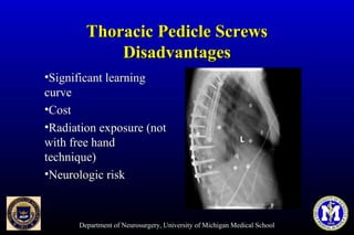
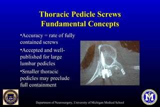
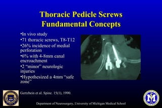
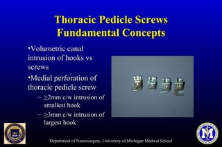
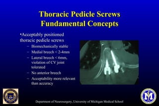
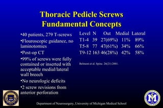
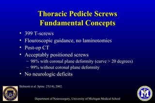
SlideShare a Scribd company logo
1 of 25
Department of Neurosurgery, University of Michigan Medical School
Thoracic Pedicle ScrewsThoracic Pedicle Screws
Anatomy and TechniqueAnatomy and Technique
Frank La Marca M. D.Frank La Marca M. D.
Department of NeurosurgeryDepartment of Neurosurgery
University of MichiganUniversity of Michigan
Department of Neurosurgery, University of Michigan Medical School
Relevant AnatomyRelevant Anatomy
• a = chord lengtha = chord length
• b = transverse diameterb = transverse diameter
• c = angle of insertionc = angle of insertion
• d = pedicle lengthd = pedicle length
Vaccaro et al. JBJS 77(8), 1995.Vaccaro et al. JBJS 77(8), 1995.
Department of Neurosurgery, University of Michigan Medical School
Basic TechniqueBasic Technique
•Starting pointStarting point
– Vertical landmarksVertical landmarks
– Horizontal landmarksHorizontal landmarks
•Medio-lateralMedio-lateral
inclinationinclination
•Cephalo-caudadCephalo-caudad
inclinationinclination
•Pedicle widthPedicle width
•Chord lengthChord length
Department of Neurosurgery, University of Michigan Medical School
Basic TechniqueBasic Technique
HistoryHistory
•Starting pointStarting point
– Mid-articular lineMid-articular line
(vertically)(vertically)
– Bisection of transverseBisection of transverse
process (horizontally)process (horizontally)
•Screw directedScrew directed
perpendicular to plane of theperpendicular to plane of the
posterior elementsposterior elements
•Accuracy not documentedAccuracy not documented
Roy-Camille et al. Clin Orth. 203, 1986.Roy-Camille et al. Clin Orth. 203, 1986.
Department of Neurosurgery, University of Michigan Medical School
Basic TechniqueBasic Technique
HistoryHistory
•Modified starting pointModified starting point
– Mid-articular lineMid-articular line
(vertically)(vertically)
– Superior border ofSuperior border of
transverse process attransverse process at
juncture with the laminajuncture with the lamina
(horizontally)(horizontally)
Xu et al, Spine. 24(2), 1999.Xu et al, Spine. 24(2), 1999.
Vaccaro et al. JBJS 77(8), 1995.Vaccaro et al. JBJS 77(8), 1995.
Department of Neurosurgery, University of Michigan Medical School
Pedicle MorphometryPedicle Morphometry
•Chord lengthChord length
– T4 (39mm)T4 (39mm)
– T12 (47mm)T12 (47mm)
•Transverse diameterTransverse diameter
– T4 (4.5mm)T4 (4.5mm)
– T12 (7.8mm)T12 (7.8mm)
Vaccaro et al. JBJS 77(8), 1995.Vaccaro et al. JBJS 77(8), 1995.
Department of Neurosurgery, University of Michigan Medical School
Pedicle MorphometryPedicle Morphometry
•Sagittal diameterSagittal diameter
– T4(10mm)T4(10mm)
– T12 (14.7mm)T12 (14.7mm)
•Angle of insertionAngle of insertion
– T4 (14 degrees)T4 (14 degrees)
– T12 (0.3 degrees)T12 (0.3 degrees)
Vaccaro et al. JBJS 77(8), 1995.Vaccaro et al. JBJS 77(8), 1995.
Department of Neurosurgery, University of Michigan Medical School
Pedicle MorphometryPedicle Morphometry
•Chord lengthChord length
– Constant betweenConstant between
individuals and vertebralindividuals and vertebral
levelslevels
•Insertion angleInsertion angle
– 15 degree angle increases15 degree angle increases
chord length 1 cmchord length 1 cm
Krag et al. Spine 13(1), 1988.Krag et al. Spine 13(1), 1988.
Department of Neurosurgery, University of Michigan Medical School
Pedicle MorphometryPedicle Morphometry
ScoliosisScoliosis
• Prospective analysis of 337Prospective analysis of 337
pedicles in 29 scoliotic ptspedicles in 29 scoliotic pts
•T4 to L4, all standardT4 to L4, all standard
measurementsmeasurements
•Concave vs convexConcave vs convex
– Endosteal width of concaveEndosteal width of concave
pedicle is significantlypedicle is significantly
smallersmaller
Liljenqvist et al. Spine. 25(10), 2000.Liljenqvist et al. Spine. 25(10), 2000.
Department of Neurosurgery, University of Michigan Medical School
Pedicle MorphometryPedicle Morphometry
• Medio-lateralMedio-lateral
inclinationinclination
– TranspedicularTranspedicular
techniquetechnique
– In-out-in techniqueIn-out-in technique
•Cephalo-caudadCephalo-caudad
inclinationinclination
Department of Neurosurgery, University of Michigan Medical School
Surrounding StructuresSurrounding Structures
•MedialMedial
– Spinal cordSpinal cord
•LateralLateral
– PleuraPleura
•Cephalo/caudadCephalo/caudad
– Nerve rootNerve root
•AnteriorAnterior
– Vascular/visceral structuresVascular/visceral structures
(5mm)(5mm)
Vaccaro et al. JBJS 77(8), 1995.Vaccaro et al. JBJS 77(8), 1995.
Department of Neurosurgery, University of Michigan Medical School
Thoracic Pedicle ScrewsThoracic Pedicle Screws
Basic Surgical StepsBasic Surgical Steps
1.1. Identify anatomic landmarksIdentify anatomic landmarks
2.2. Access pedicle entry zone … soundAccess pedicle entry zone … sound
pediclepedicle
3.3. TapTap
4.4. Screw placementScrew placement
upper thoracic – 4.5mmupper thoracic – 4.5mm
middle thoracic – 4.5 to 5.5mmmiddle thoracic – 4.5 to 5.5mm
lower thoracic – 5.5 to 6.5mmlower thoracic – 5.5 to 6.5mm
Department of Neurosurgery, University of Michigan Medical School
Lenke – 12-Step “Free Hand”Lenke – 12-Step “Free Hand”
Technique of Thoracic PedicleTechnique of Thoracic Pedicle
Screw (TPS) PlacementScrew (TPS) Placement
1.1. ExposureExposure
2.2. Starting pointStarting point
3.3. Cortical burrCortical burr
4.4. Pedicle gearshift – lateralPedicle gearshift – lateral
5.5. Pedicle gearshift – medialPedicle gearshift – medial
6.6. Pedicle palpationPedicle palpation
7.7. Pedicle length measurementPedicle length measurement
8.8. Pedicle tappingPedicle tapping
9.9. Repeat pedicle palpationRepeat pedicle palpation
10.10. Screw placementScrew placement
11.11. Intraoperative x-raysIntraoperative x-rays
12.12. Screw EMG stimulationScrew EMG stimulation
Department of Neurosurgery, University of Michigan Medical School
Pedicle Screw EMGPedicle Screw EMG
StimulationStimulation
Screw levelScrew level Recording MuscleRecording Muscle
T6-T12T6-T12 Rectus abdominusRectus abdominus
L1-L2L1-L2 AdductorsAdductors
L3-L4L3-L4 QuadricepsQuadriceps
L5L5 Tibialis AnteriorTibialis Anterior
S1S1 GastrocnemiusGastrocnemius
Department of Neurosurgery, University of Michigan Medical School
Thoracic Pedicle ScrewsThoracic Pedicle Screws
AdvantagesAdvantages
•BiomechanicallyBiomechanically
superior anchor pointsuperior anchor point
•Can be performed safelyCan be performed safely
•Enhance curveEnhance curve
correctioncorrection
•Save fusion levelsSave fusion levels
•Address all curve typesAddress all curve types
Department of Neurosurgery, University of Michigan Medical School
Thoracic Pedicle ScrewsThoracic Pedicle Screws
DisadvantagesDisadvantages
•Significant learningSignificant learning
curvecurve
•CostCost
•Radiation exposure (notRadiation exposure (not
with free handwith free hand
technique)technique)
•Neurologic riskNeurologic risk
Department of Neurosurgery, University of Michigan Medical School
Thoracic Pedicle ScrewsThoracic Pedicle Screws
Fundamental ConceptsFundamental Concepts
•Accuracy = rate of fullyAccuracy = rate of fully
contained screwscontained screws
•Accepted and well-Accepted and well-
published for largepublished for large
lumbar pedicleslumbar pedicles
•Smaller thoracicSmaller thoracic
pedicles may precludepedicles may preclude
full containmentfull containment
Department of Neurosurgery, University of Michigan Medical School
Thoracic Pedicle ScrewsThoracic Pedicle Screws
Fundamental ConceptsFundamental Concepts
•In vivo studyIn vivo study
•71 thoracic screws, T8-T1271 thoracic screws, T8-T12
•26% incidence of medial26% incidence of medial
perforationperforation
•6% with 4-8mm canal6% with 4-8mm canal
encroachmentencroachment
•2 “minor” neurologic2 “minor” neurologic
injuriesinjuries
•Hypothesized a 4mm “safeHypothesized a 4mm “safe
zone”zone”
Gertzbein et al. Spine. 15(1), 1990.Gertzbein et al. Spine. 15(1), 1990.
Department of Neurosurgery, University of Michigan Medical School
Thoracic Pedicle ScrewsThoracic Pedicle Screws
Fundamental ConceptsFundamental Concepts
•Volumetric canalVolumetric canal
intrusion of hooks vsintrusion of hooks vs
screwsscrews
•Medial perforation ofMedial perforation of
thoracic pedicle screwthoracic pedicle screw
– >>2mm c/w intrusion of2mm c/w intrusion of
smallest hooksmallest hook
– >>3mm c/w intrusion of3mm c/w intrusion of
largest hooklargest hook
Department of Neurosurgery, University of Michigan Medical School
Thoracic Pedicle ScrewsThoracic Pedicle Screws
Fundamental ConceptsFundamental Concepts
•Acceptably positionedAcceptably positioned
thoracic pedicle screwsthoracic pedicle screws
– Biomechanically stableBiomechanically stable
– Medial breech < 2-4mmMedial breech < 2-4mm
– Lateral breech < 6mm,Lateral breech < 6mm,
violation of CV jointviolation of CV joint
toleratedtolerated
– No anterior breechNo anterior breech
– Acceptability more relevantAcceptability more relevant
than accuracythan accuracy
Department of Neurosurgery, University of Michigan Medical School
Thoracic Pedicle ScrewsThoracic Pedicle Screws
Fundamental ConceptsFundamental Concepts
•40 patients, 279 T-screws40 patients, 279 T-screws
•Flouroscopic guidance, noFlouroscopic guidance, no
laminotomieslaminotomies
•Post-op CTPost-op CT
•99% of screws were fully99% of screws were fully
contained or inserted withcontained or inserted with
acceptable medial/lateralacceptable medial/lateral
wall breechwall breech
•No neurologic deficitsNo neurologic deficits
•2 screw revisions from2 screw revisions from
anterior perforationanterior perforation
Level N Out Medial LateralLevel N Out Medial Lateral
T1-4 39 27(69%) 11% 89%T1-4 39 27(69%) 11% 89%
T5-8 77 47(61%) 34% 66%T5-8 77 47(61%) 34% 66%
T9-12 163 46(28%) 42% 58%T9-12 163 46(28%) 42% 58%
Belmont et al. Spine. 26(21) 2001.Belmont et al. Spine. 26(21) 2001.
Department of Neurosurgery, University of Michigan Medical School
Thoracic Pedicle ScrewsThoracic Pedicle Screws
Fundamental ConceptsFundamental Concepts
• 399 T-screws399 T-screws
• Flouroscopic guidance, no laminotomiesFlouroscopic guidance, no laminotomies
• Post-op CTPost-op CT
• Acceptably positioned screwsAcceptably positioned screws
– 98% with coronal plane deformity (curve > 20 degrees)98% with coronal plane deformity (curve > 20 degrees)
– 99% without coronal plane deformity99% without coronal plane deformity
• No neurologic deficitsNo neurologic deficits
Belmont et al. Spine. 27(14), 2002.Belmont et al. Spine. 27(14), 2002.
Department of Neurosurgery, University of Michigan Medical School
ConclusionConclusion
•Understanding of pedicle morphometry is essentialUnderstanding of pedicle morphometry is essential
•Free-hand technique safe and effectiveFree-hand technique safe and effective
•Acceptable vs accuracyAcceptable vs accuracy

More Related Content

What's hot

Free hand screw
Free hand screwFree hand screw
Free hand screwSumit2018
 
Posterior Spine Fixation
Posterior Spine FixationPosterior Spine Fixation
Posterior Spine FixationGhazwan Bayaty
 
Endoscopic spinal surgery
Endoscopic spinal surgeryEndoscopic spinal surgery
Endoscopic spinal surgeryLocal Konnect
 
Minimal invasive techniques in lumbar degenerative diseases
Minimal invasive techniques in lumbar degenerative diseasesMinimal invasive techniques in lumbar degenerative diseases
Minimal invasive techniques in lumbar degenerative diseasesProf. Dr. Mohamed Mohi Eldin
 
Biomechanics and biology of relative stability
Biomechanics and biology of relative stabilityBiomechanics and biology of relative stability
Biomechanics and biology of relative stabilityOrthosurg2016
 
Posterior lumbar fusion vs Lumbar interbody fusion Evidence based.pptx
Posterior lumbar fusion vs Lumbar interbody fusion Evidence based.pptxPosterior lumbar fusion vs Lumbar interbody fusion Evidence based.pptx
Posterior lumbar fusion vs Lumbar interbody fusion Evidence based.pptxsuresh Bishokarma
 
Lumbar interbody fusion indications techniques and complications
Lumbar interbody fusion indications techniques and complicationsLumbar interbody fusion indications techniques and complications
Lumbar interbody fusion indications techniques and complicationsDr Praveen kumar tripathi
 
Poller screw Tibia.pptx
Poller screw Tibia.pptxPoller screw Tibia.pptx
Poller screw Tibia.pptxSureshPandey32
 
Cervical Arthritis / Cervical Spondylotic Myelopathy / Cervical Stenosis by P...
Cervical Arthritis / Cervical Spondylotic Myelopathy / Cervical Stenosis by P...Cervical Arthritis / Cervical Spondylotic Myelopathy / Cervical Stenosis by P...
Cervical Arthritis / Cervical Spondylotic Myelopathy / Cervical Stenosis by P...Pablo Pazmino
 
Surgical approach for thoracic and lumbosacral spine
Surgical approach for thoracic and lumbosacral spineSurgical approach for thoracic and lumbosacral spine
Surgical approach for thoracic and lumbosacral spineDr Ajay Shah IOM TUTH Nepal
 

What's hot (20)

Free hand screw
Free hand screwFree hand screw
Free hand screw
 
Posterior Spine Fixation
Posterior Spine FixationPosterior Spine Fixation
Posterior Spine Fixation
 
Ossified Posterior Longitudinal Ligament (OPLL)
Ossified Posterior Longitudinal Ligament (OPLL)Ossified Posterior Longitudinal Ligament (OPLL)
Ossified Posterior Longitudinal Ligament (OPLL)
 
Endoscopic spinal surgery
Endoscopic spinal surgeryEndoscopic spinal surgery
Endoscopic spinal surgery
 
Minimal invasive techniques in lumbar degenerative diseases
Minimal invasive techniques in lumbar degenerative diseasesMinimal invasive techniques in lumbar degenerative diseases
Minimal invasive techniques in lumbar degenerative diseases
 
Fusion techniques spine
Fusion techniques spineFusion techniques spine
Fusion techniques spine
 
Ao philosophy
Ao philosophyAo philosophy
Ao philosophy
 
Lateral mass screws
Lateral mass screwsLateral mass screws
Lateral mass screws
 
Approaches to spine
Approaches to spineApproaches to spine
Approaches to spine
 
Subaxial cervical fixation techniques
Subaxial cervical fixation techniquesSubaxial cervical fixation techniques
Subaxial cervical fixation techniques
 
Gap nonunion
Gap nonunionGap nonunion
Gap nonunion
 
Biomechanics and biology of relative stability
Biomechanics and biology of relative stabilityBiomechanics and biology of relative stability
Biomechanics and biology of relative stability
 
Spinal balance
Spinal balanceSpinal balance
Spinal balance
 
Posterior lumbar fusion vs Lumbar interbody fusion Evidence based.pptx
Posterior lumbar fusion vs Lumbar interbody fusion Evidence based.pptxPosterior lumbar fusion vs Lumbar interbody fusion Evidence based.pptx
Posterior lumbar fusion vs Lumbar interbody fusion Evidence based.pptx
 
Lumbar interbody fusion indications techniques and complications
Lumbar interbody fusion indications techniques and complicationsLumbar interbody fusion indications techniques and complications
Lumbar interbody fusion indications techniques and complications
 
Poller screw Tibia.pptx
Poller screw Tibia.pptxPoller screw Tibia.pptx
Poller screw Tibia.pptx
 
Cervical Arthritis / Cervical Spondylotic Myelopathy / Cervical Stenosis by P...
Cervical Arthritis / Cervical Spondylotic Myelopathy / Cervical Stenosis by P...Cervical Arthritis / Cervical Spondylotic Myelopathy / Cervical Stenosis by P...
Cervical Arthritis / Cervical Spondylotic Myelopathy / Cervical Stenosis by P...
 
Scoliosis
Scoliosis Scoliosis
Scoliosis
 
Lumbar interbody fusion.pptx
Lumbar interbody fusion.pptxLumbar interbody fusion.pptx
Lumbar interbody fusion.pptx
 
Surgical approach for thoracic and lumbosacral spine
Surgical approach for thoracic and lumbosacral spineSurgical approach for thoracic and lumbosacral spine
Surgical approach for thoracic and lumbosacral spine
 

Viewers also liked

Spinal - Implants - Pedicular screws
Spinal - Implants - Pedicular screwsSpinal - Implants - Pedicular screws
Spinal - Implants - Pedicular screwsperfect-spine
 
TLIF by Pablo Pazmino MD
TLIF by Pablo Pazmino MDTLIF by Pablo Pazmino MD
TLIF by Pablo Pazmino MDPablo Pazmino
 
OLIF-oblique lumbar interbody fusion
OLIF-oblique lumbar interbody fusionOLIF-oblique lumbar interbody fusion
OLIF-oblique lumbar interbody fusionMano Ranjitha Kumari
 
Spinal anatomy and biomechanics
Spinal anatomy and biomechanicsSpinal anatomy and biomechanics
Spinal anatomy and biomechanicsAmbrish Verma
 
Medtronic cd horizonadvanceddeformityst
Medtronic cd horizonadvanceddeformitystMedtronic cd horizonadvanceddeformityst
Medtronic cd horizonadvanceddeformitystDan Melendez
 
XLIF by Pablo Pazmino MD
XLIF by Pablo Pazmino MDXLIF by Pablo Pazmino MD
XLIF by Pablo Pazmino MDPablo Pazmino
 
Approach side during anterior cervical discectomy
Approach side during anterior cervical discectomyApproach side during anterior cervical discectomy
Approach side during anterior cervical discectomyMQ_Library
 
Vertebral Canal Lecture Notes
Vertebral Canal Lecture NotesVertebral Canal Lecture Notes
Vertebral Canal Lecture NotesAnan
 
Referat – Knochentumore
Referat – KnochentumoreReferat – Knochentumore
Referat – KnochentumoreKerstin Hoppe
 
Matthias Honl Habilitation
Matthias Honl HabilitationMatthias Honl Habilitation
Matthias Honl HabilitationMatthias Honl
 
Cervical Compressive Myelopathy
Cervical Compressive MyelopathyCervical Compressive Myelopathy
Cervical Compressive MyelopathySivaraj Sadhasivam
 
Anterior Cervical Discectomy with Fusion
Anterior Cervical Discectomy with FusionAnterior Cervical Discectomy with Fusion
Anterior Cervical Discectomy with Fusionbsni
 
REOPERATIONS AFTER MINIMALLY INVASIVE LUMBAR SPINE SURGERY
REOPERATIONS AFTER MINIMALLY INVASIVE LUMBAR SPINE SURGERYREOPERATIONS AFTER MINIMALLY INVASIVE LUMBAR SPINE SURGERY
REOPERATIONS AFTER MINIMALLY INVASIVE LUMBAR SPINE SURGERYYunus Aydın
 
Symptoms of Cervical Myelopathy
Symptoms of Cervical MyelopathySymptoms of Cervical Myelopathy
Symptoms of Cervical MyelopathyRa'Kerry Rahman
 

Viewers also liked (18)

Spinal - Implants - Pedicular screws
Spinal - Implants - Pedicular screwsSpinal - Implants - Pedicular screws
Spinal - Implants - Pedicular screws
 
TLIF by Pablo Pazmino MD
TLIF by Pablo Pazmino MDTLIF by Pablo Pazmino MD
TLIF by Pablo Pazmino MD
 
OLIF-oblique lumbar interbody fusion
OLIF-oblique lumbar interbody fusionOLIF-oblique lumbar interbody fusion
OLIF-oblique lumbar interbody fusion
 
Spinetrauma 2
Spinetrauma 2Spinetrauma 2
Spinetrauma 2
 
Spinal anatomy and biomechanics
Spinal anatomy and biomechanicsSpinal anatomy and biomechanics
Spinal anatomy and biomechanics
 
Anatomy of spine
Anatomy of spineAnatomy of spine
Anatomy of spine
 
Medtronic cd horizonadvanceddeformityst
Medtronic cd horizonadvanceddeformitystMedtronic cd horizonadvanceddeformityst
Medtronic cd horizonadvanceddeformityst
 
XLIF by Pablo Pazmino MD
XLIF by Pablo Pazmino MDXLIF by Pablo Pazmino MD
XLIF by Pablo Pazmino MD
 
MIA in Thoraco-lumbar Trauma (SPINE 2009)
MIA in Thoraco-lumbar Trauma (SPINE 2009)MIA in Thoraco-lumbar Trauma (SPINE 2009)
MIA in Thoraco-lumbar Trauma (SPINE 2009)
 
Approach side during anterior cervical discectomy
Approach side during anterior cervical discectomyApproach side during anterior cervical discectomy
Approach side during anterior cervical discectomy
 
Vertebral Canal Lecture Notes
Vertebral Canal Lecture NotesVertebral Canal Lecture Notes
Vertebral Canal Lecture Notes
 
The cervical myelopathy.
The cervical myelopathy.The cervical myelopathy.
The cervical myelopathy.
 
Referat – Knochentumore
Referat – KnochentumoreReferat – Knochentumore
Referat – Knochentumore
 
Matthias Honl Habilitation
Matthias Honl HabilitationMatthias Honl Habilitation
Matthias Honl Habilitation
 
Cervical Compressive Myelopathy
Cervical Compressive MyelopathyCervical Compressive Myelopathy
Cervical Compressive Myelopathy
 
Anterior Cervical Discectomy with Fusion
Anterior Cervical Discectomy with FusionAnterior Cervical Discectomy with Fusion
Anterior Cervical Discectomy with Fusion
 
REOPERATIONS AFTER MINIMALLY INVASIVE LUMBAR SPINE SURGERY
REOPERATIONS AFTER MINIMALLY INVASIVE LUMBAR SPINE SURGERYREOPERATIONS AFTER MINIMALLY INVASIVE LUMBAR SPINE SURGERY
REOPERATIONS AFTER MINIMALLY INVASIVE LUMBAR SPINE SURGERY
 
Symptoms of Cervical Myelopathy
Symptoms of Cervical MyelopathySymptoms of Cervical Myelopathy
Symptoms of Cervical Myelopathy
 

Similar to Thoracic pedicle screws

Steps total knee replacement
Steps total knee replacement Steps total knee replacement
Steps total knee replacement AdityaApte11
 
Knee arthroplasty for FRCS Orth course Newcastle
Knee arthroplasty for FRCS Orth course NewcastleKnee arthroplasty for FRCS Orth course Newcastle
Knee arthroplasty for FRCS Orth course NewcastleProfessor Deiary Kader
 
The Future is Now with Robotic Spine Surgery
The Future is Now with Robotic Spine Surgery The Future is Now with Robotic Spine Surgery
The Future is Now with Robotic Spine Surgery Atlantic Brain & Spine
 
Arthroscopi Bankart's Repair-Dr. Sunit hazra
Arthroscopi Bankart's Repair-Dr. Sunit hazraArthroscopi Bankart's Repair-Dr. Sunit hazra
Arthroscopi Bankart's Repair-Dr. Sunit hazraTheRightDoctors
 
Hip Joint anatomy, surgical approches & AVN review
Hip Joint anatomy, surgical approches & AVN reviewHip Joint anatomy, surgical approches & AVN review
Hip Joint anatomy, surgical approches & AVN reviewdocortho Patel
 
pediatric fracture around elbow.pptx
pediatric fracture around elbow.pptxpediatric fracture around elbow.pptx
pediatric fracture around elbow.pptxbishwabandhuniraula
 
Swine Spine Presentation Patricia 10_28_16
Swine Spine Presentation Patricia 10_28_16Swine Spine Presentation Patricia 10_28_16
Swine Spine Presentation Patricia 10_28_16Patricia Stan
 
Percutaneous drilling tibial osteotomy for correction of genu varum in children
Percutaneous drilling tibial osteotomy for correction of genu varum in childrenPercutaneous drilling tibial osteotomy for correction of genu varum in children
Percutaneous drilling tibial osteotomy for correction of genu varum in childrenAhmed Abd El Razek
 
ThesisprotocolPresentation14262635363.pptx
ThesisprotocolPresentation14262635363.pptxThesisprotocolPresentation14262635363.pptx
ThesisprotocolPresentation14262635363.pptx2rgscwvdvb
 
AHSS Registrar Review Course. Scaphoid and carpal fractures
AHSS Registrar Review Course. Scaphoid and carpal fracturesAHSS Registrar Review Course. Scaphoid and carpal fractures
AHSS Registrar Review Course. Scaphoid and carpal fracturesAvanthiMandaleson
 
Clavicle fractures
Clavicle fracturesClavicle fractures
Clavicle fracturesSICOTEduDay
 
Advancement in back pain treatment
Advancement in back pain treatmentAdvancement in back pain treatment
Advancement in back pain treatmentManish Raj
 
NCCT OR CECT PELVIS BONE.pptx, INDICATIONS OF PELVIS BONE
NCCT OR CECT PELVIS BONE.pptx, INDICATIONS OF PELVIS BONENCCT OR CECT PELVIS BONE.pptx, INDICATIONS OF PELVIS BONE
NCCT OR CECT PELVIS BONE.pptx, INDICATIONS OF PELVIS BONERukamaneeYadav
 
The Elbow - Anatomy, Injury, and Rehabilitation
The Elbow - Anatomy, Injury, and RehabilitationThe Elbow - Anatomy, Injury, and Rehabilitation
The Elbow - Anatomy, Injury, and RehabilitationC Noel Henley MD
 
Regional anaesthesia strategies for upper limb surgeries
Regional anaesthesia strategies for upper limb surgeriesRegional anaesthesia strategies for upper limb surgeries
Regional anaesthesia strategies for upper limb surgeriesGopan Gopalakrisna Pillai
 

Similar to Thoracic pedicle screws (20)

Steps total knee replacement
Steps total knee replacement Steps total knee replacement
Steps total knee replacement
 
Knee arthroplasty for FRCS Orth course Newcastle
Knee arthroplasty for FRCS Orth course NewcastleKnee arthroplasty for FRCS Orth course Newcastle
Knee arthroplasty for FRCS Orth course Newcastle
 
The Future is Now with Robotic Spine Surgery
The Future is Now with Robotic Spine Surgery The Future is Now with Robotic Spine Surgery
The Future is Now with Robotic Spine Surgery
 
Arthroscopi Bankart's Repair-Dr. Sunit hazra
Arthroscopi Bankart's Repair-Dr. Sunit hazraArthroscopi Bankart's Repair-Dr. Sunit hazra
Arthroscopi Bankart's Repair-Dr. Sunit hazra
 
Hip Joint anatomy, surgical approches & AVN review
Hip Joint anatomy, surgical approches & AVN reviewHip Joint anatomy, surgical approches & AVN review
Hip Joint anatomy, surgical approches & AVN review
 
pediatric fracture around elbow.pptx
pediatric fracture around elbow.pptxpediatric fracture around elbow.pptx
pediatric fracture around elbow.pptx
 
Swine Spine Presentation Patricia 10_28_16
Swine Spine Presentation Patricia 10_28_16Swine Spine Presentation Patricia 10_28_16
Swine Spine Presentation Patricia 10_28_16
 
Osteoarthritis of the hand
Osteoarthritis of the handOsteoarthritis of the hand
Osteoarthritis of the hand
 
Percutaneous drilling tibial osteotomy for correction of genu varum in children
Percutaneous drilling tibial osteotomy for correction of genu varum in childrenPercutaneous drilling tibial osteotomy for correction of genu varum in children
Percutaneous drilling tibial osteotomy for correction of genu varum in children
 
ThesisprotocolPresentation14262635363.pptx
ThesisprotocolPresentation14262635363.pptxThesisprotocolPresentation14262635363.pptx
ThesisprotocolPresentation14262635363.pptx
 
Hip anatomy, approaches & AVN
Hip anatomy, approaches & AVN Hip anatomy, approaches & AVN
Hip anatomy, approaches & AVN
 
AHSS Registrar Review Course. Scaphoid and carpal fractures
AHSS Registrar Review Course. Scaphoid and carpal fracturesAHSS Registrar Review Course. Scaphoid and carpal fractures
AHSS Registrar Review Course. Scaphoid and carpal fractures
 
SPINE SBRT for beginners
SPINE SBRT for beginnersSPINE SBRT for beginners
SPINE SBRT for beginners
 
Clavicle fractures
Clavicle fracturesClavicle fractures
Clavicle fractures
 
Terrible triad of the elbow
Terrible triad of the elbowTerrible triad of the elbow
Terrible triad of the elbow
 
Advancement in back pain treatment
Advancement in back pain treatmentAdvancement in back pain treatment
Advancement in back pain treatment
 
NCCT OR CECT PELVIS BONE.pptx, INDICATIONS OF PELVIS BONE
NCCT OR CECT PELVIS BONE.pptx, INDICATIONS OF PELVIS BONENCCT OR CECT PELVIS BONE.pptx, INDICATIONS OF PELVIS BONE
NCCT OR CECT PELVIS BONE.pptx, INDICATIONS OF PELVIS BONE
 
The Elbow - Anatomy, Injury, and Rehabilitation
The Elbow - Anatomy, Injury, and RehabilitationThe Elbow - Anatomy, Injury, and Rehabilitation
The Elbow - Anatomy, Injury, and Rehabilitation
 
eidelman2016.pdf
eidelman2016.pdfeidelman2016.pdf
eidelman2016.pdf
 
Regional anaesthesia strategies for upper limb surgeries
Regional anaesthesia strategies for upper limb surgeriesRegional anaesthesia strategies for upper limb surgeries
Regional anaesthesia strategies for upper limb surgeries
 

Thoracic pedicle screws

  • 1. Department of Neurosurgery, University of Michigan Medical School Thoracic Pedicle ScrewsThoracic Pedicle Screws Anatomy and TechniqueAnatomy and Technique Frank La Marca M. D.Frank La Marca M. D. Department of NeurosurgeryDepartment of Neurosurgery University of MichiganUniversity of Michigan
  • 2. Department of Neurosurgery, University of Michigan Medical School Relevant AnatomyRelevant Anatomy • a = chord lengtha = chord length • b = transverse diameterb = transverse diameter • c = angle of insertionc = angle of insertion • d = pedicle lengthd = pedicle length Vaccaro et al. JBJS 77(8), 1995.Vaccaro et al. JBJS 77(8), 1995.
  • 3. Department of Neurosurgery, University of Michigan Medical School Basic TechniqueBasic Technique •Starting pointStarting point – Vertical landmarksVertical landmarks – Horizontal landmarksHorizontal landmarks •Medio-lateralMedio-lateral inclinationinclination •Cephalo-caudadCephalo-caudad inclinationinclination •Pedicle widthPedicle width •Chord lengthChord length
  • 4. Department of Neurosurgery, University of Michigan Medical School Basic TechniqueBasic Technique HistoryHistory •Starting pointStarting point – Mid-articular lineMid-articular line (vertically)(vertically) – Bisection of transverseBisection of transverse process (horizontally)process (horizontally) •Screw directedScrew directed perpendicular to plane of theperpendicular to plane of the posterior elementsposterior elements •Accuracy not documentedAccuracy not documented Roy-Camille et al. Clin Orth. 203, 1986.Roy-Camille et al. Clin Orth. 203, 1986.
  • 5. Department of Neurosurgery, University of Michigan Medical School Basic TechniqueBasic Technique HistoryHistory •Modified starting pointModified starting point – Mid-articular lineMid-articular line (vertically)(vertically) – Superior border ofSuperior border of transverse process attransverse process at juncture with the laminajuncture with the lamina (horizontally)(horizontally) Xu et al, Spine. 24(2), 1999.Xu et al, Spine. 24(2), 1999. Vaccaro et al. JBJS 77(8), 1995.Vaccaro et al. JBJS 77(8), 1995.
  • 6. Department of Neurosurgery, University of Michigan Medical School Pedicle MorphometryPedicle Morphometry •Chord lengthChord length – T4 (39mm)T4 (39mm) – T12 (47mm)T12 (47mm) •Transverse diameterTransverse diameter – T4 (4.5mm)T4 (4.5mm) – T12 (7.8mm)T12 (7.8mm) Vaccaro et al. JBJS 77(8), 1995.Vaccaro et al. JBJS 77(8), 1995.
  • 7. Department of Neurosurgery, University of Michigan Medical School Pedicle MorphometryPedicle Morphometry •Sagittal diameterSagittal diameter – T4(10mm)T4(10mm) – T12 (14.7mm)T12 (14.7mm) •Angle of insertionAngle of insertion – T4 (14 degrees)T4 (14 degrees) – T12 (0.3 degrees)T12 (0.3 degrees) Vaccaro et al. JBJS 77(8), 1995.Vaccaro et al. JBJS 77(8), 1995.
  • 8. Department of Neurosurgery, University of Michigan Medical School Pedicle MorphometryPedicle Morphometry •Chord lengthChord length – Constant betweenConstant between individuals and vertebralindividuals and vertebral levelslevels •Insertion angleInsertion angle – 15 degree angle increases15 degree angle increases chord length 1 cmchord length 1 cm Krag et al. Spine 13(1), 1988.Krag et al. Spine 13(1), 1988.
  • 9. Department of Neurosurgery, University of Michigan Medical School Pedicle MorphometryPedicle Morphometry ScoliosisScoliosis • Prospective analysis of 337Prospective analysis of 337 pedicles in 29 scoliotic ptspedicles in 29 scoliotic pts •T4 to L4, all standardT4 to L4, all standard measurementsmeasurements •Concave vs convexConcave vs convex – Endosteal width of concaveEndosteal width of concave pedicle is significantlypedicle is significantly smallersmaller Liljenqvist et al. Spine. 25(10), 2000.Liljenqvist et al. Spine. 25(10), 2000.
  • 10. Department of Neurosurgery, University of Michigan Medical School Pedicle MorphometryPedicle Morphometry • Medio-lateralMedio-lateral inclinationinclination – TranspedicularTranspedicular techniquetechnique – In-out-in techniqueIn-out-in technique •Cephalo-caudadCephalo-caudad inclinationinclination
  • 11. Department of Neurosurgery, University of Michigan Medical School Surrounding StructuresSurrounding Structures •MedialMedial – Spinal cordSpinal cord •LateralLateral – PleuraPleura •Cephalo/caudadCephalo/caudad – Nerve rootNerve root •AnteriorAnterior – Vascular/visceral structuresVascular/visceral structures (5mm)(5mm) Vaccaro et al. JBJS 77(8), 1995.Vaccaro et al. JBJS 77(8), 1995.
  • 12. Department of Neurosurgery, University of Michigan Medical School Thoracic Pedicle ScrewsThoracic Pedicle Screws Basic Surgical StepsBasic Surgical Steps 1.1. Identify anatomic landmarksIdentify anatomic landmarks 2.2. Access pedicle entry zone … soundAccess pedicle entry zone … sound pediclepedicle 3.3. TapTap 4.4. Screw placementScrew placement upper thoracic – 4.5mmupper thoracic – 4.5mm middle thoracic – 4.5 to 5.5mmmiddle thoracic – 4.5 to 5.5mm lower thoracic – 5.5 to 6.5mmlower thoracic – 5.5 to 6.5mm
  • 13. Department of Neurosurgery, University of Michigan Medical School Lenke – 12-Step “Free Hand”Lenke – 12-Step “Free Hand” Technique of Thoracic PedicleTechnique of Thoracic Pedicle Screw (TPS) PlacementScrew (TPS) Placement 1.1. ExposureExposure 2.2. Starting pointStarting point 3.3. Cortical burrCortical burr 4.4. Pedicle gearshift – lateralPedicle gearshift – lateral 5.5. Pedicle gearshift – medialPedicle gearshift – medial 6.6. Pedicle palpationPedicle palpation 7.7. Pedicle length measurementPedicle length measurement 8.8. Pedicle tappingPedicle tapping 9.9. Repeat pedicle palpationRepeat pedicle palpation 10.10. Screw placementScrew placement 11.11. Intraoperative x-raysIntraoperative x-rays 12.12. Screw EMG stimulationScrew EMG stimulation
  • 14. Department of Neurosurgery, University of Michigan Medical School Lenke – 12-Step “Free Hand”Lenke – 12-Step “Free Hand” Technique of Thoracic PedicleTechnique of Thoracic Pedicle Screw (TPS) PlacementScrew (TPS) Placement 1.1. ExposureExposure 2.2. Starting pointStarting point 3.3. Cortical burrCortical burr 4.4. Pedicle gearshift – lateralPedicle gearshift – lateral 5.5. Pedicle gearshift – medialPedicle gearshift – medial 6.6. Pedicle palpationPedicle palpation 7.7. Pedicle length measurementPedicle length measurement 8.8. Pedicle tappingPedicle tapping 9.9. Repeat pedicle palpationRepeat pedicle palpation 10.10. Screw placementScrew placement 11.11. Intraoperative x-raysIntraoperative x-rays 12.12. Screw EMG stimulationScrew EMG stimulation
  • 15. Department of Neurosurgery, University of Michigan Medical School Lenke – 12-Step “Free Hand”Lenke – 12-Step “Free Hand” Technique of Thoracic PedicleTechnique of Thoracic Pedicle Screw (TPS) PlacementScrew (TPS) Placement 1.1. ExposureExposure 2.2. Starting pointStarting point 3.3. Cortical burrCortical burr 4.4. Pedicle gearshift – lateralPedicle gearshift – lateral 5.5. Pedicle gearshift – medialPedicle gearshift – medial 6.6. Pedicle palpationPedicle palpation 7.7. Pedicle length measurementPedicle length measurement 8.8. Pedicle tappingPedicle tapping 9.9. Repeat pedicle palpationRepeat pedicle palpation 10.10. Screw placementScrew placement 11.11. Intraoperative x-raysIntraoperative x-rays 12.12. Screw EMG stimulationScrew EMG stimulation
  • 16. Department of Neurosurgery, University of Michigan Medical School Pedicle Screw EMGPedicle Screw EMG StimulationStimulation Screw levelScrew level Recording MuscleRecording Muscle T6-T12T6-T12 Rectus abdominusRectus abdominus L1-L2L1-L2 AdductorsAdductors L3-L4L3-L4 QuadricepsQuadriceps L5L5 Tibialis AnteriorTibialis Anterior S1S1 GastrocnemiusGastrocnemius
  • 17. Department of Neurosurgery, University of Michigan Medical School Thoracic Pedicle ScrewsThoracic Pedicle Screws AdvantagesAdvantages •BiomechanicallyBiomechanically superior anchor pointsuperior anchor point •Can be performed safelyCan be performed safely •Enhance curveEnhance curve correctioncorrection •Save fusion levelsSave fusion levels •Address all curve typesAddress all curve types
  • 18. Department of Neurosurgery, University of Michigan Medical School Thoracic Pedicle ScrewsThoracic Pedicle Screws DisadvantagesDisadvantages •Significant learningSignificant learning curvecurve •CostCost •Radiation exposure (notRadiation exposure (not with free handwith free hand technique)technique) •Neurologic riskNeurologic risk
  • 19. Department of Neurosurgery, University of Michigan Medical School Thoracic Pedicle ScrewsThoracic Pedicle Screws Fundamental ConceptsFundamental Concepts •Accuracy = rate of fullyAccuracy = rate of fully contained screwscontained screws •Accepted and well-Accepted and well- published for largepublished for large lumbar pedicleslumbar pedicles •Smaller thoracicSmaller thoracic pedicles may precludepedicles may preclude full containmentfull containment
  • 20. Department of Neurosurgery, University of Michigan Medical School Thoracic Pedicle ScrewsThoracic Pedicle Screws Fundamental ConceptsFundamental Concepts •In vivo studyIn vivo study •71 thoracic screws, T8-T1271 thoracic screws, T8-T12 •26% incidence of medial26% incidence of medial perforationperforation •6% with 4-8mm canal6% with 4-8mm canal encroachmentencroachment •2 “minor” neurologic2 “minor” neurologic injuriesinjuries •Hypothesized a 4mm “safeHypothesized a 4mm “safe zone”zone” Gertzbein et al. Spine. 15(1), 1990.Gertzbein et al. Spine. 15(1), 1990.
  • 21. Department of Neurosurgery, University of Michigan Medical School Thoracic Pedicle ScrewsThoracic Pedicle Screws Fundamental ConceptsFundamental Concepts •Volumetric canalVolumetric canal intrusion of hooks vsintrusion of hooks vs screwsscrews •Medial perforation ofMedial perforation of thoracic pedicle screwthoracic pedicle screw – >>2mm c/w intrusion of2mm c/w intrusion of smallest hooksmallest hook – >>3mm c/w intrusion of3mm c/w intrusion of largest hooklargest hook
  • 22. Department of Neurosurgery, University of Michigan Medical School Thoracic Pedicle ScrewsThoracic Pedicle Screws Fundamental ConceptsFundamental Concepts •Acceptably positionedAcceptably positioned thoracic pedicle screwsthoracic pedicle screws – Biomechanically stableBiomechanically stable – Medial breech < 2-4mmMedial breech < 2-4mm – Lateral breech < 6mm,Lateral breech < 6mm, violation of CV jointviolation of CV joint toleratedtolerated – No anterior breechNo anterior breech – Acceptability more relevantAcceptability more relevant than accuracythan accuracy
  • 23. Department of Neurosurgery, University of Michigan Medical School Thoracic Pedicle ScrewsThoracic Pedicle Screws Fundamental ConceptsFundamental Concepts •40 patients, 279 T-screws40 patients, 279 T-screws •Flouroscopic guidance, noFlouroscopic guidance, no laminotomieslaminotomies •Post-op CTPost-op CT •99% of screws were fully99% of screws were fully contained or inserted withcontained or inserted with acceptable medial/lateralacceptable medial/lateral wall breechwall breech •No neurologic deficitsNo neurologic deficits •2 screw revisions from2 screw revisions from anterior perforationanterior perforation Level N Out Medial LateralLevel N Out Medial Lateral T1-4 39 27(69%) 11% 89%T1-4 39 27(69%) 11% 89% T5-8 77 47(61%) 34% 66%T5-8 77 47(61%) 34% 66% T9-12 163 46(28%) 42% 58%T9-12 163 46(28%) 42% 58% Belmont et al. Spine. 26(21) 2001.Belmont et al. Spine. 26(21) 2001.
  • 24. Department of Neurosurgery, University of Michigan Medical School Thoracic Pedicle ScrewsThoracic Pedicle Screws Fundamental ConceptsFundamental Concepts • 399 T-screws399 T-screws • Flouroscopic guidance, no laminotomiesFlouroscopic guidance, no laminotomies • Post-op CTPost-op CT • Acceptably positioned screwsAcceptably positioned screws – 98% with coronal plane deformity (curve > 20 degrees)98% with coronal plane deformity (curve > 20 degrees) – 99% without coronal plane deformity99% without coronal plane deformity • No neurologic deficitsNo neurologic deficits Belmont et al. Spine. 27(14), 2002.Belmont et al. Spine. 27(14), 2002.
  • 25. Department of Neurosurgery, University of Michigan Medical School ConclusionConclusion •Understanding of pedicle morphometry is essentialUnderstanding of pedicle morphometry is essential •Free-hand technique safe and effectiveFree-hand technique safe and effective •Acceptable vs accuracyAcceptable vs accuracy