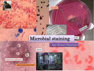
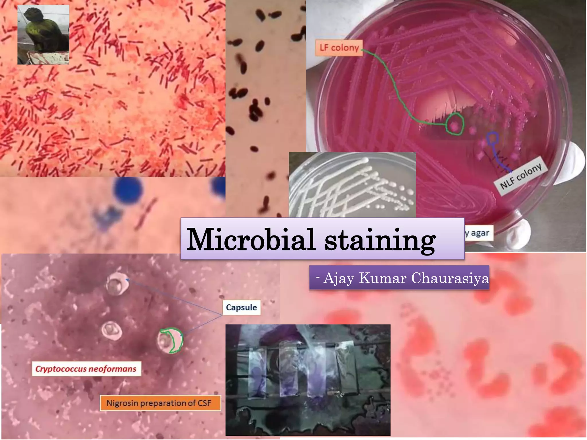
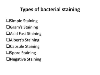
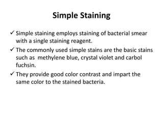
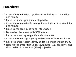
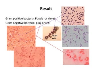
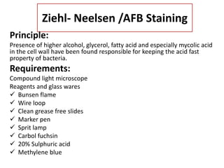
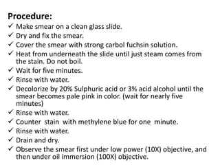
Microbial staining
- Ajay Kumar Chaurasiya
Staining
Stain: to leave a mark on something that is difficult
to remove.
Dye : A dye is a colored substance that has an affinity
to the substrate to which it is being applied.
Staining: Staining is an auxiliary technique used in
microscopy to enhance contrast in the microscopic
image.
Types of bacterial staining
Simple Staining
Gram’s Staining
Acid Fast Staining
Albert’s Staining
Capsule Staining
Spore Staining
Negative Staining
Simple Staining
 Simple staining employs staining of bacterial smear
with a single staining reagent.
 The commonly used simple stains are the basic stains
such as methylene blue, crystal violet and carbol
fuchsin.
 They provide good color contrast and impart the
same color to the stained bacteria.
Gram’s Staining
 It is a differential stain and thus used to differentiate
Gram positive and Gram negative bacteria.
 It was originally devised by a Danish bacteriologist,
Hans Christian Joachim Gram (1884) as a method of
staining bacteria.
Principle:
The reaction is dependent on permeability of the
bacterial cell wall and cytoplasmic membrane, to the dye
–iodine complex.
In Gram positive bacteria, the crystal violet dye iodine
complex combines to form a larger molecule which
precipitates within the cell.
Also the alcohol /acetone mixture which acts as
decolorizing agent, cause dehydration of the multi-layered
peptidoglycan of the cell wall. This causes decreasing of
the space between the molecules causing the cell wall to
trap the crystal violet iodine complex within the cell.
Hence the Gram positive bacteria do not get decolorized
and retain primary dye appearing violet. Also, Gram
positive bacteria have more acidic protoplasm and hence
bind to the basic dye more firmly.
In the case of Gram negative bacteria, the alcohol ,being
a lipid solvent, dissolves the outer lipopolysaccharide
membrane the of cell wall and also damages the
cytoplasmic membrane to which the peptidoglycan is
attached. As a result , the dye-iodine complex is not
retained within the cell and permeates out of it during
the process of decolonization. Hence when a counter stain
is added, they take up the color of the stain and appear
pink.
Requirements:
a) Compound light microscope
b) Reagents and glass wares
Bunsen flame
Wire loop
Clean grease free slides
Marker pen
Crystal violet (Basic dye)
Gram’s iodine(mordant)
95% ethanol (decolorizing agent)
1% safranine or dilute carbol fuchsin or neutral
red
c) Specimen
Preparation of bacterial smear: from liquid culture
 Take a clean, and grease free slide for making smear.
 Take one or two loopful of the bacterial cell suspension
and place on the slide with a bacteriological loop.
 Then with circular movement of the loop, spread the cell
suspension into a thin area.
 Allow the smear to air dry.
 Heat fix the smear while holding the slide at one end,
and by quickly passing the smear over the flame of
Bunsen burner two to three times.
Preparation of bacterial smear: from the solid
medium
 Take a clean, and grease free slide for making smear.
 Take a loopful of 0.85% saline i. e. physiological saline
and place it on the Centre of the slide.
 With a straight wire touch the surface of a well isolated
colony from the solid media and emulsify in the saline
drop forming a thin film.
 Allow the smear to air dry.
 Heat fix the smear while holding the slide at one end,
and by quickly passing the smear over the flame of
Bunsen burner two to three times.
Procedure:
 Cover the smear with crystal violet and allow it to stand for
one minute.
 Rinse the smear gently under tap water.
 Cover the smear with Gram’s iodine and allow it to stand for
one minute.
 Rinse smear again gently under tap water.
 Decolorize the smear with 95% alcohol.
 Rinse the smear again gently under tap water.
 Cover the smear again gently with safranine for one minute.
 Rinse the smear again gently under tap water and air dry it.
 Observe the smear first under low power (10X) objective, and
then under oil immersion (100X) objective.
Result
Gram positive bacteria: Purple or violet
Gram negative bacteria: pink or red
Yeast
cells
Ziehl- Neelsen /AFB Staining
Principle:
Presence of higher alcohol, glycerol, fatty acid and especially mycolic acid
in the cell wall have been found responsible for keeping the acid fast
property of bacteria.
Requirements:
Compound light microscope
Reagents and glass wares
 Bunsen flame
 Wire loop
 Clean grease free slides
 Marker pen
 Sprit lamp
 Carbol fuchsin
 20% Sulphuric acid
 Methylene blue
Procedure:
 Make smear on a clean glass slide.
 Dry and fix the smear.
 Cover the smear with strong carbol fuchsin solution.
 Heat from underneath the slide until just steam comes from
the stain. Do not boil.
 Wait for five minutes.
 Rinse with water.
 Decolorize by 20% Sulphuric acid or 3% acid alcohol until the
smear becomes pale pink in color. (wait for nearly five
minutes)
 Rinse with water.
 Counter stain with methylene blue for one minute.
 Rinse with water.
 Drain and dry.
 Observe the smear first under low power (10X) objective, and
then under oil immersion (100X) objective.
Result:
AFB: pink or red bacillus
Back ground: Blue
Reporting:
International Union Against Tuberculosis and Lung
Disease (IUATLD):
No organism seen-Negative
1-9/100 OIF: Exact number
10-99/100 OIF :+
1-10/OIF :++
>10/OIF :+++
Albert’s Staining
It is used to stain Corynebacterium
diphtheriae.
Capsule Staining
 It is used to stain capsule.
Spore staining
 It is used to stain spore.
Negative staining
 Negative staining is so called because the
background gets stained and the organism remains
colorless.
 India ink , nigrosin or eosin are the stains used in
negative staining for demonstration of capsule.
Bibliography
1. Forbes BA, Sahm DF and Weissfeld AS. Bailey and Scott’s
Diagnostic Microbiology . 11th ed.2002
2. Koneman EW, Allen SD, Janda WM, Schreckenbergu PC and
Winn Jr. WC . Color Atlas and Textbook of Diagnostic
Microbiology. 5th ed.
3. Evans EGV, Killington RA , Heritage J. Introductory
Microbiology, 1996.
4. WHO . Guidelines on Standard Operating Procedures for
Microbiology . Chapter 4: Staining techniques.
Bacterial staining methods

Bacterial staining methods

  • 1.
    Microbial staining - AjayKumar Chaurasiya
  • 2.
    Staining Stain: to leavea mark on something that is difficult to remove. Dye : A dye is a colored substance that has an affinity to the substrate to which it is being applied. Staining: Staining is an auxiliary technique used in microscopy to enhance contrast in the microscopic image.
  • 3.
    Types of bacterialstaining Simple Staining Gram’s Staining Acid Fast Staining Albert’s Staining Capsule Staining Spore Staining Negative Staining
  • 4.
    Simple Staining  Simplestaining employs staining of bacterial smear with a single staining reagent.  The commonly used simple stains are the basic stains such as methylene blue, crystal violet and carbol fuchsin.  They provide good color contrast and impart the same color to the stained bacteria.
  • 5.
    Gram’s Staining  Itis a differential stain and thus used to differentiate Gram positive and Gram negative bacteria.  It was originally devised by a Danish bacteriologist, Hans Christian Joachim Gram (1884) as a method of staining bacteria. Principle: The reaction is dependent on permeability of the bacterial cell wall and cytoplasmic membrane, to the dye –iodine complex. In Gram positive bacteria, the crystal violet dye iodine complex combines to form a larger molecule which precipitates within the cell.
  • 6.
    Also the alcohol/acetone mixture which acts as decolorizing agent, cause dehydration of the multi-layered peptidoglycan of the cell wall. This causes decreasing of the space between the molecules causing the cell wall to trap the crystal violet iodine complex within the cell. Hence the Gram positive bacteria do not get decolorized and retain primary dye appearing violet. Also, Gram positive bacteria have more acidic protoplasm and hence bind to the basic dye more firmly. In the case of Gram negative bacteria, the alcohol ,being a lipid solvent, dissolves the outer lipopolysaccharide membrane the of cell wall and also damages the cytoplasmic membrane to which the peptidoglycan is attached. As a result , the dye-iodine complex is not retained within the cell and permeates out of it during the process of decolonization. Hence when a counter stain is added, they take up the color of the stain and appear pink.
  • 7.
    Requirements: a) Compound lightmicroscope b) Reagents and glass wares Bunsen flame Wire loop Clean grease free slides Marker pen Crystal violet (Basic dye) Gram’s iodine(mordant) 95% ethanol (decolorizing agent) 1% safranine or dilute carbol fuchsin or neutral red
  • 8.
    c) Specimen Preparation ofbacterial smear: from liquid culture  Take a clean, and grease free slide for making smear.  Take one or two loopful of the bacterial cell suspension and place on the slide with a bacteriological loop.  Then with circular movement of the loop, spread the cell suspension into a thin area.  Allow the smear to air dry.  Heat fix the smear while holding the slide at one end, and by quickly passing the smear over the flame of Bunsen burner two to three times.
  • 9.
    Preparation of bacterialsmear: from the solid medium  Take a clean, and grease free slide for making smear.  Take a loopful of 0.85% saline i. e. physiological saline and place it on the Centre of the slide.  With a straight wire touch the surface of a well isolated colony from the solid media and emulsify in the saline drop forming a thin film.  Allow the smear to air dry.  Heat fix the smear while holding the slide at one end, and by quickly passing the smear over the flame of Bunsen burner two to three times.
  • 10.
    Procedure:  Cover thesmear with crystal violet and allow it to stand for one minute.  Rinse the smear gently under tap water.  Cover the smear with Gram’s iodine and allow it to stand for one minute.  Rinse smear again gently under tap water.  Decolorize the smear with 95% alcohol.  Rinse the smear again gently under tap water.  Cover the smear again gently with safranine for one minute.  Rinse the smear again gently under tap water and air dry it.  Observe the smear first under low power (10X) objective, and then under oil immersion (100X) objective.
  • 11.
    Result Gram positive bacteria:Purple or violet Gram negative bacteria: pink or red Yeast cells
  • 12.
    Ziehl- Neelsen /AFBStaining Principle: Presence of higher alcohol, glycerol, fatty acid and especially mycolic acid in the cell wall have been found responsible for keeping the acid fast property of bacteria. Requirements: Compound light microscope Reagents and glass wares  Bunsen flame  Wire loop  Clean grease free slides  Marker pen  Sprit lamp  Carbol fuchsin  20% Sulphuric acid  Methylene blue
  • 13.
    Procedure:  Make smearon a clean glass slide.  Dry and fix the smear.  Cover the smear with strong carbol fuchsin solution.  Heat from underneath the slide until just steam comes from the stain. Do not boil.  Wait for five minutes.  Rinse with water.  Decolorize by 20% Sulphuric acid or 3% acid alcohol until the smear becomes pale pink in color. (wait for nearly five minutes)  Rinse with water.  Counter stain with methylene blue for one minute.  Rinse with water.  Drain and dry.  Observe the smear first under low power (10X) objective, and then under oil immersion (100X) objective.
  • 14.
    Result: AFB: pink orred bacillus Back ground: Blue Reporting: International Union Against Tuberculosis and Lung Disease (IUATLD): No organism seen-Negative 1-9/100 OIF: Exact number 10-99/100 OIF :+ 1-10/OIF :++ >10/OIF :+++
  • 15.
    Albert’s Staining It isused to stain Corynebacterium diphtheriae. Capsule Staining  It is used to stain capsule. Spore staining  It is used to stain spore.
  • 16.
    Negative staining  Negativestaining is so called because the background gets stained and the organism remains colorless.  India ink , nigrosin or eosin are the stains used in negative staining for demonstration of capsule.
  • 17.
    Bibliography 1. Forbes BA,Sahm DF and Weissfeld AS. Bailey and Scott’s Diagnostic Microbiology . 11th ed.2002 2. Koneman EW, Allen SD, Janda WM, Schreckenbergu PC and Winn Jr. WC . Color Atlas and Textbook of Diagnostic Microbiology. 5th ed. 3. Evans EGV, Killington RA , Heritage J. Introductory Microbiology, 1996. 4. WHO . Guidelines on Standard Operating Procedures for Microbiology . Chapter 4: Staining techniques.