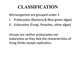
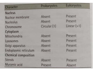
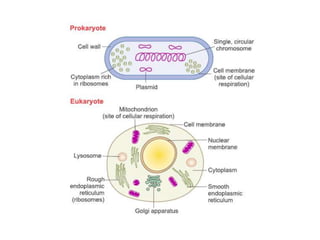
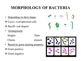
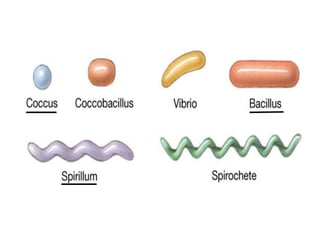
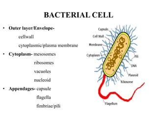
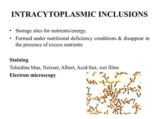
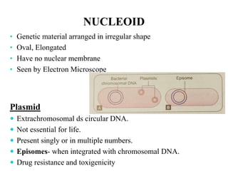
BACTERIAL ANATOMY
Dr. Sumesh Kumar Dash
Department of Microbiology
IMS & SUM Hospital
CLASSIFICATION
Microorganism are grouped under 2
I. Prokaryotes (Bacteria & Blue-green algae)
II. Eukaryotes (Fungi, Parasites, other algae)
Viruses are neither prokaryotes nor
eukaryotes as they lack the characteristics of
living thinks except replication.
SIZE
• Bacteria size in micrometer and virus size in
nanometer
• Bacteria observed by light microscope and
Viruses observed by electron microscope
• To see microorganisms – staining is used
MORPHOLOGY OF BACTERIA
• Depending on their shape;
 Cocci- oval/spherical cells
 Bacilli- rod shaped
• Arrangement;
Singles Pairs
Chains clusters
• Based on gram staining property;
 Gram positive
 Gram negative
BACTERIAL CELL
• Outer layer/Envelope-
cellwall
cytoplasmic/plasma membrane
• Cytoplasm- mesosomes
ribosomes
vacuoles
nucleoid
• Appendages- capsule
flagella
fimbriae/pili
CELL WALL
• Chemically composed of peptidoglycan
GRAM POSITIVE CELL WALL
 Peptidoglycan layer
50-100 layers thick
16-80nm
 Teichoic acids
2 types:
Cell wall teichoic acid
Lipoteichoic acid
GRAM NEGATIVE CELL WALL
 Peptidoglycan layer
1-2 layer thick
2-3nm
Outer membrane
1. Porin proteins
2. Lipopolysaccharide (LPS)
• Lipid A
• Core polysaccharide
• O side chain /O antigen
DIFFERENCE BETWEEN GRAM POSITIVE &
GRAM NEGATIVE CELL WALL
Characters GRAM + GRAM -
peptidoglycan layer Thick Thin
Aromatic and sulphur
containing amino acids
Absent Present
Teichoic acids Present Absent
Gram stain Violet Red
FUNCTIONS OF CELL WALL
Maintaining the cells characteristic shape
Providing a rigid platform for surface appendages- flagella,
fimbriae/pili all emanate from the wall and extend beyond it.
Providing attachment sites for bacteriophage
Be the sites of major antigenic determinant of cell surface-
endotoxcity
Site of action of many antibiotics & resistance
CYTOPLASMIC/CELL MEMBRANE
5-10nm thick
Phospholipid bilayer
With integral and peripheral proteins
Selective permeability i:e -
semipermeable
Site of ATP production
Viability
CYTOPLASM
• Viscous watery solution containing organic & inorganic
solutes, biosynthetic components which required for the
growth & cell division, together with genetic material.
• Contains RIBOSOMES,
MESOSOMES
INTRACYTOPLASMIC INCLUSION BODIES
VACUOLES
NUCLEOUS
RIBOSOMES
• Sites for protein
synthesis.
• 10-20 nm in size.
MESOSOMES / Chondroids
• Vesicular/convoluted/multilaminated
• More prominent in G+.
• LOCATION:
Often found next to septa, in dividing
bacteria or seen attached to nuclear body.
• FUNCTION:
Site of bacterial respiration
INTRACYTOPLASMIC INCLUSIONS
• Storage sites for nutrients/energy.
• Formed under nutritional deficiency conditions & disappear in
the presence of excess nutrients
Staining
Toluidine blue, Neisser, Albert, Acid-fast, wet films
Electron microscopy
VACUOLES
• Vacuoles are fluid containing cavities
• Separated from cytoplasm by a membrane
• Function - uncertain
NUCLEOID
• Genetic material arranged in irregular shape
• Oval, Elongated
• Have no nuclear membrane
• Seen by Electron Microscope
Plasmid
 Extrachromosomal ds circular DNA.
 Not essential for life.
 Present singly or in multiple numbers.
 Episomes- when integrated with chromosomal DNA.
 Drug resistance and toxigenicity
THANK
YOU

Bacterial anatomy

  • 1.
    BACTERIAL ANATOMY Dr. SumeshKumar Dash Department of Microbiology IMS & SUM Hospital
  • 2.
    CLASSIFICATION Microorganism are groupedunder 2 I. Prokaryotes (Bacteria & Blue-green algae) II. Eukaryotes (Fungi, Parasites, other algae) Viruses are neither prokaryotes nor eukaryotes as they lack the characteristics of living thinks except replication.
  • 5.
    SIZE • Bacteria sizein micrometer and virus size in nanometer • Bacteria observed by light microscope and Viruses observed by electron microscope • To see microorganisms – staining is used
  • 6.
    MORPHOLOGY OF BACTERIA •Depending on their shape;  Cocci- oval/spherical cells  Bacilli- rod shaped • Arrangement; Singles Pairs Chains clusters • Based on gram staining property;  Gram positive  Gram negative
  • 8.
    BACTERIAL CELL • Outerlayer/Envelope- cellwall cytoplasmic/plasma membrane • Cytoplasm- mesosomes ribosomes vacuoles nucleoid • Appendages- capsule flagella fimbriae/pili
  • 9.
    CELL WALL • Chemicallycomposed of peptidoglycan
  • 10.
    GRAM POSITIVE CELLWALL  Peptidoglycan layer 50-100 layers thick 16-80nm  Teichoic acids 2 types: Cell wall teichoic acid Lipoteichoic acid
  • 11.
    GRAM NEGATIVE CELLWALL  Peptidoglycan layer 1-2 layer thick 2-3nm Outer membrane 1. Porin proteins 2. Lipopolysaccharide (LPS) • Lipid A • Core polysaccharide • O side chain /O antigen
  • 12.
    DIFFERENCE BETWEEN GRAMPOSITIVE & GRAM NEGATIVE CELL WALL Characters GRAM + GRAM - peptidoglycan layer Thick Thin Aromatic and sulphur containing amino acids Absent Present Teichoic acids Present Absent Gram stain Violet Red
  • 13.
    FUNCTIONS OF CELLWALL Maintaining the cells characteristic shape Providing a rigid platform for surface appendages- flagella, fimbriae/pili all emanate from the wall and extend beyond it. Providing attachment sites for bacteriophage Be the sites of major antigenic determinant of cell surface- endotoxcity Site of action of many antibiotics & resistance
  • 14.
    CYTOPLASMIC/CELL MEMBRANE 5-10nm thick Phospholipidbilayer With integral and peripheral proteins Selective permeability i:e - semipermeable Site of ATP production Viability
  • 15.
    CYTOPLASM • Viscous waterysolution containing organic & inorganic solutes, biosynthetic components which required for the growth & cell division, together with genetic material. • Contains RIBOSOMES, MESOSOMES INTRACYTOPLASMIC INCLUSION BODIES VACUOLES NUCLEOUS
  • 16.
    RIBOSOMES • Sites forprotein synthesis. • 10-20 nm in size.
  • 17.
    MESOSOMES / Chondroids •Vesicular/convoluted/multilaminated • More prominent in G+. • LOCATION: Often found next to septa, in dividing bacteria or seen attached to nuclear body. • FUNCTION: Site of bacterial respiration
  • 18.
    INTRACYTOPLASMIC INCLUSIONS • Storagesites for nutrients/energy. • Formed under nutritional deficiency conditions & disappear in the presence of excess nutrients Staining Toluidine blue, Neisser, Albert, Acid-fast, wet films Electron microscopy
  • 19.
    VACUOLES • Vacuoles arefluid containing cavities • Separated from cytoplasm by a membrane • Function - uncertain
  • 20.
    NUCLEOID • Genetic materialarranged in irregular shape • Oval, Elongated • Have no nuclear membrane • Seen by Electron Microscope Plasmid  Extrachromosomal ds circular DNA.  Not essential for life.  Present singly or in multiple numbers.  Episomes- when integrated with chromosomal DNA.  Drug resistance and toxigenicity
  • 21.