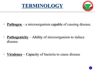
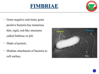
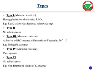
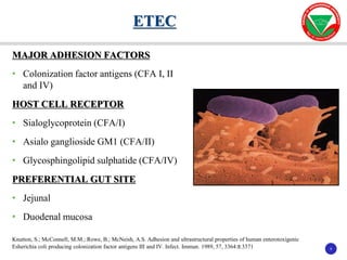
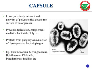
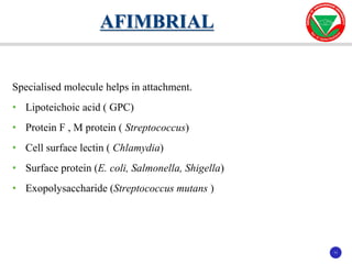
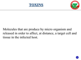
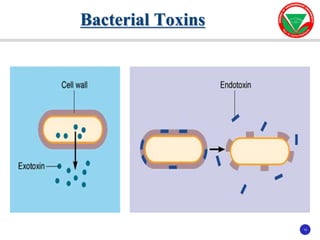
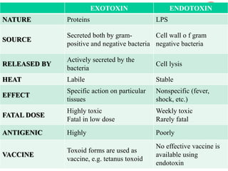
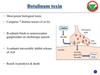
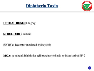
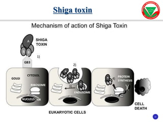
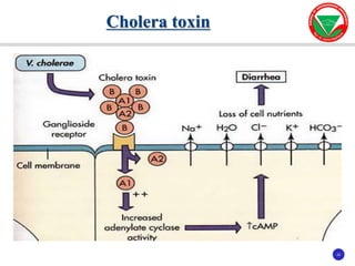
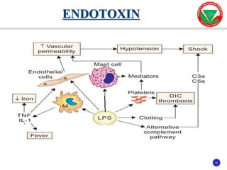
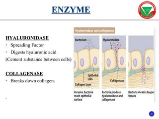
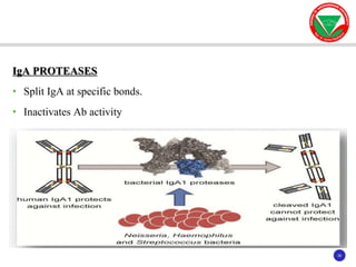
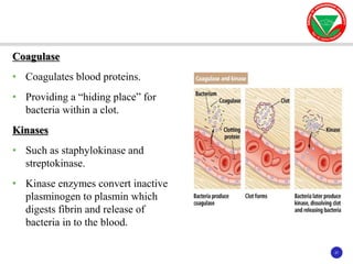
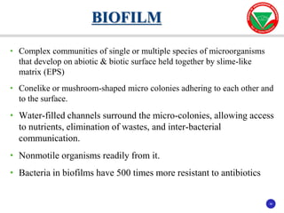
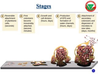
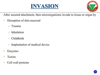
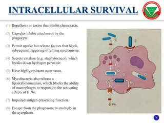
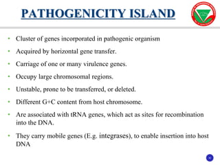
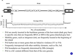
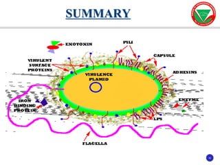
BACTERIAL VIRULENCE
PRESENTED BY:
Dr. Sumesh Kumar Dash
MODERATOR:
Dr. Sunita Kabi
2
2
INTRODUCTION
3
3
• Pathogen – a microorganism capable of causing disease.
• Pathogenicity – Ability of microorganism to induce
disease.
• Virulence – Capacity of bacteria to cause disease
TERMINOLOGY
4
4
• Different strains of same species - varying degree of virulence
• Highly, low, avirulent
• Virulent strains may undergo spontaneous or induced variation
 Exaltation
 Attenuation
VIRULENCE
5
5
Extra cellular factor
1. Fimbriae or Pilli
2. Capsule
3. Afimbrial
Intra cellular factor
1. Toxins
2. Enzyme
3. Secretion system
4. Iron
VIRULENCE FACTOR
6
6
FIMBRIAE or PILLI
CAPSULE
AFIMBRIAL
EXTRA CELLULAR FACTOR
7
7
• Gram negative and many gram
positive bacteria has numerous
thin, rigid, rod-like structures
called fimbriae or pili.
• Made of protein.
• Mediate attachment of bacteria to
cell surface.
FIMBRIAE
8
8
• Type I (Mannose sensitive)
Hemagglutination of untreated RBCs.
E.g. E.coli ,klebsiella ,Serratia, salmonella spp.
• Type II
No adhesiveness
• Type III (Mannose resistant)
Adhesive to RBCs treated with tannic acid/heated to 70 ° C
E.g. klebsiella, serratia
• Type IV (Mannose resistant)
P.aeruginosa
• Type VI
No adhesiveness
E.g. Non fimbriated strains of K.ozaenae
Types
9
9
MAJOR ADHESION FACTORS
• Colonization factor antigens (CFA I, II
and IV)
HOST CELL RECEPTOR
• Sialoglycoprotein (CFA/I)
• Asialo ganglioside GM1 (CFA/II)
• Glycosphingolipid sulphatide (CFA/IV)
PREFERENTIAL GUT SITE
• Jejunal
• Duodenal mucosa
ETEC
Knutton, S.; McConnell, M.M.; Rowe, B.; McNeish, A.S. Adhesion and ultrastructural properties of human enterotoxigenic
Esherichia coli producing colonization factor antigens III and IV. Infect. Immun. 1989, 57, 3364±3371
10
10
• Loose, relatively unstructured
network of polymers that covers the
surface of an organism.
• Prevents desiccation, complement
mediated bacterial cell lysis
• Protects from phagocytosis & action
of lysozyme and bacteriophages
• Eg- Pneumococcus, Meningococcus,
H.influenzae, Klebsiella,
Pseudomonas, Bacillus etc
CAPSULE
11
11
Specialised molecule helps in attachment.
• Lipoteichoic acid ( GPC)
• Protein F , M protein ( Streptococcus)
• Cell surface lectin ( Chlamydia)
• Surface protein (E. coli, Salmonella, Shigella)
• Exopolysaccharide (Streptococcus mutans )
AFIMBRIAL
12
12
 TOXINS
 ENZYME
 SECRETION SYSTEM
 IRON
INTRA CELLULAR FACTOR
13
13
Molecules that are produce by micro organism and
released in order to affect, at distance, a target cell and
tissue in the infected host.
TOXINS
14
14
Bacterial Toxins
15
15
EXOTOXIN ENDOTOXIN
NATURE Proteins LPS
SOURCE
Secreted both by gram-
positive and negative bacteria
Cell wall o f gram
negative bacteria
RELEASED BY
Actively secreted by the
bacteria
Cell lysis
HEAT Labile Stable
EFFECT
Specific action on particular
tissues
Nonspecific (fever,
shock, etc.)
FATAL DOSE
Highly toxic
Fatal in low dose
Weekly toxic
Rarely fatal
ANTIGENIC Highly Poorly
VACCINE
Toxoid forms are used as
vaccine, e.g. tetanus toxoid
No effective vaccine is
available using
endotoxin
16
16
• Typically proteins secreted or release by bacteria during growth.
• Usually produce by single bacterial species of a genus preferably by
virulent strains.
• Strongly antigenic.
• Toxoid are prepared.
EXOTOXIN
17
17
Consist of 2 component
Subunit A
• Responsible for the enzymatic activity.
• Enzymatically active but lack binding & cell entry capability.
Subunit B
• Facilitates binding to a specific receptor and transferring the enzyme.
• Bind to target cells & even block the binding of the native toxin, but they
are nontoxic
Structure
18
18
TOXIN SUBUNIT
Botulinum toxin A/B
Diphtheria toxin A-B
Cholera toxin A-5B
Shiga toxin A-5B
Shiga like toxin A-5B
Pertussis toxin A-5B
Anthrax toxin LF 2A+B
19
19
2 Mechanism. In both case, a large protein molecule insert and cross the
membrane lipid bilayer.
DIRECT ENTRY
• Subunit B binds to a specific receptor on the target cell
• Formation of a pore in the membrane.
• Through which subunit A is transferred into the cell cytoplasm
ALTERNATIVE MECHANISM
• Native toxin binds to the target cell
• Structure is taken into the cell by the process of receptor-mediated
endocytosis (RME )
Attachment & Entry
20
20
• Most potent biological toxin
• Comprise 7 distinct toxins (A to G)
• B-subunit binds to neuroreceptor
gangliosides on cholinergic neuron.
• A-subunit irreversibly inhibit release
of Ach
• Result in paralysis & death
Botulinum toxin
21
21
LETHAL DOSE: 0.1ug/kg
STRUCTUR: 2 subunit
ENTRY: Receptor-mediated endocytosis
MOA: A-subunit inhibit the cell protein synthesis by inactivating EF-2
Diphtheria Toxin
22
22
Shiga toxin
23
23
Cholera toxin
24
24
ENDOTOXIN
25
25
HYALURONIDASE
• Spreading Factor
• Digests hyaluronic acid
(Cement substance between cells)
COLLAGENASE
• Breaks down collagen.
.
ENZYME
26
26
IgA PROTEASES
• Split IgA at specific bonds.
• Inactivates Ab activity
27
27
Coagulase
• Coagulates blood proteins.
• Providing a “hiding place” for
bacteria within a clot.
Kinases
• Such as staphylokinase and
streptokinase.
• Kinase enzymes convert inactive
plasminogen to plasmin which
digests fibrin and release of
bacteria in to the blood.
28
28
ENZYMES ORGANISMS INVOLVED MECHANISM OFACTION
Hemolysins
Staphylococci
Streptococci
E. coli
Lyse erythrocyte, make iron
available for microbial growth
Phospholipase
Clostridium perfringenes
Destroys lecithin
(phosphatidylcholine) in
cell membranes.
Staphylococcus aureus Lyse red blood cells
Protein A
Staphylococcus aureus
Binds to IgA and prevent
the activation of
compliment system.
29
29
• Protein secretion is a critical element of toxin mediated virulence
• Secretion of proteins or toxins occur in several pathway.
BACTERIAL SECRETION SYSTEM
TYPE ORGANISM TOXIN/PROTEINS
Type 1 E coli Hemolysin
Type 2 Vibrio cholerae Cholera toxin
Pseudomonas aeruginosa Exotoxin A
Type 3 Yersinia Yops
Type 4 B pertussis Pertussis toxin
H pylori CagA
Type 5 H pylori Vacuolating cytotoxin
Type 6 Pseudomonas aeruginosa, Vibrio cholerae
Type 7 Mycobacterium tuberculosis
30
30
• Essential nutrient for growth and metabolism.
• Also affects the virulence.
• Extracellular iron chelators called Siderophores for scavenging iron.
REQUIREDMENT FOR IRON
31
31
• C.jejuni & N.meningitidis obtain iron by siderophores produced by other
species.
• H.influenzae type B, H.parainfluenzae, S.aureus & S.epidermidis have
specific receptors for transferrin and lactoferrin on their surface which
bind these protein and remove bound iron.
• Bacteroid species remove iron by proteolytic cleavage of the chelator.
32
32
• Route of transmission
• Infective dose
• Adherence
• Invasion
• Intracellular survival
• Pathogenicity island
DETERMINANT OF PATHOGENICITY
33
33
• Important role in pathogenicity
• Any mode of entry.
• Introduced by the optimal routes.
ROUTE OF TRANSMISSION
34
34
• Minimum inoculum size capable of initiating an infection
o Low Infective dose
o Large Infective dose
• Higher the virulence, lower is the infective dose
INFECTIVE DOSE
35
35
NONSPECIFIC
Attractive forces, which allow approach of the bacterium to the eukaryotic
cell surface
• Hydrophobic and electrostatic attractions
• Atomic and molecular vibration
• Brownian movement
• Recruitment and trapping by biofilm
SPECIFIC ADHERENCE
• Permanent attachment.
• Irreversible noncovalent bond between adhesin & receptor molecule.
ADHERENCE
36
36
• Complex communities of single or multiple species of microorganisms
that develop on abiotic & biotic surface held together by slime-like
matrix (EPS)
• Conelike or mushroom-shaped micro colonies adhering to each other and
to the surface.
• Water-filled channels surround the micro-colonies, allowing access
to nutrients, elimination of wastes, and inter-bacterial
communication.
• Nonmotile organisms readily from it.
• Bacteria in biofilms have 500 times more resistant to antibiotics
BIOFILM
37
37
Stages
38
38
After secured attachment, then microorganisms invade to tissue or organ by
• Disruption of skin mucosal
 Trauma
 Inhalation
 Childbirth
 Implantation of medical device
• Enzymes
• Toxins
• Cell wall proteins
INVASION
39
39
(1) Repellents or toxins that inhibit chemotaxis.
(2) Capsules inhibit attachment by the
phagocyte
(3) Permit uptake but release factors that block,
subsequent triggering of killing mechanisms.
(4) Secrete catalase (e.g. staphylococci), which
breaks down hydrogen peroxide.
(5) Have highly resistant outer coats.
(6) Mycobacteria also release a
lipoarabinomannan, which blocks the ability
of macrophages to respond to the activating
effects of IFNy.
(7) Impaired antigen-presenting function.
(8) Escape from the phagosome to multiply in
the cytoplasm.
INTRACELLULAR SURVIVAL
40
40
• Cluster of genes incorporated in pathogenic organism
• Acquired by horizontal gene transfer.
• Carriage of one or many virulence genes.
• Occupy large chromosomal regions.
• Unstable, prone to be transferred, or deleted.
• Different G+C content from host chromosome.
• Are associated with tRNA genes, which act as sites for recombination
into the DNA.
• They carry mobile genes (E.g. integrases), to enable insertion into host
DNA
PATHOGENICITY ISLAND
41
41
• PAI are mostly inserted in the backbone genome of the host strain (dark grey bars)
in specific sites that are frequently tRNA or tRNA-like genes (hatched grey bar)
• Mobility genes, such as integrases (int), are frequently located at the beginning of
the island
• PAI harbor one or more genes that are linked to virulence (V1 to V4)
• Frequently interspersed with other mobility elements, such as ISc, Isd.
• PAI boundaries are frequently determined by DRs (triangle)
• PAI is a GC content different from that of the core genome.
42
42
SUMMARY
Bacterial virulence (dr.sumesh)

Bacterial virulence (dr.sumesh)

  • 1.
    BACTERIAL VIRULENCE PRESENTED BY: Dr.Sumesh Kumar Dash MODERATOR: Dr. Sunita Kabi
  • 2.
  • 3.
    3 3 • Pathogen –a microorganism capable of causing disease. • Pathogenicity – Ability of microorganism to induce disease. • Virulence – Capacity of bacteria to cause disease TERMINOLOGY
  • 4.
    4 4 • Different strainsof same species - varying degree of virulence • Highly, low, avirulent • Virulent strains may undergo spontaneous or induced variation  Exaltation  Attenuation VIRULENCE
  • 5.
    5 5 Extra cellular factor 1.Fimbriae or Pilli 2. Capsule 3. Afimbrial Intra cellular factor 1. Toxins 2. Enzyme 3. Secretion system 4. Iron VIRULENCE FACTOR
  • 6.
  • 7.
    7 7 • Gram negativeand many gram positive bacteria has numerous thin, rigid, rod-like structures called fimbriae or pili. • Made of protein. • Mediate attachment of bacteria to cell surface. FIMBRIAE
  • 8.
    8 8 • Type I(Mannose sensitive) Hemagglutination of untreated RBCs. E.g. E.coli ,klebsiella ,Serratia, salmonella spp. • Type II No adhesiveness • Type III (Mannose resistant) Adhesive to RBCs treated with tannic acid/heated to 70 ° C E.g. klebsiella, serratia • Type IV (Mannose resistant) P.aeruginosa • Type VI No adhesiveness E.g. Non fimbriated strains of K.ozaenae Types
  • 9.
    9 9 MAJOR ADHESION FACTORS •Colonization factor antigens (CFA I, II and IV) HOST CELL RECEPTOR • Sialoglycoprotein (CFA/I) • Asialo ganglioside GM1 (CFA/II) • Glycosphingolipid sulphatide (CFA/IV) PREFERENTIAL GUT SITE • Jejunal • Duodenal mucosa ETEC Knutton, S.; McConnell, M.M.; Rowe, B.; McNeish, A.S. Adhesion and ultrastructural properties of human enterotoxigenic Esherichia coli producing colonization factor antigens III and IV. Infect. Immun. 1989, 57, 3364±3371
  • 10.
    10 10 • Loose, relativelyunstructured network of polymers that covers the surface of an organism. • Prevents desiccation, complement mediated bacterial cell lysis • Protects from phagocytosis & action of lysozyme and bacteriophages • Eg- Pneumococcus, Meningococcus, H.influenzae, Klebsiella, Pseudomonas, Bacillus etc CAPSULE
  • 11.
    11 11 Specialised molecule helpsin attachment. • Lipoteichoic acid ( GPC) • Protein F , M protein ( Streptococcus) • Cell surface lectin ( Chlamydia) • Surface protein (E. coli, Salmonella, Shigella) • Exopolysaccharide (Streptococcus mutans ) AFIMBRIAL
  • 12.
    12 12  TOXINS  ENZYME SECRETION SYSTEM  IRON INTRA CELLULAR FACTOR
  • 13.
    13 13 Molecules that areproduce by micro organism and released in order to affect, at distance, a target cell and tissue in the infected host. TOXINS
  • 14.
  • 15.
    15 15 EXOTOXIN ENDOTOXIN NATURE ProteinsLPS SOURCE Secreted both by gram- positive and negative bacteria Cell wall o f gram negative bacteria RELEASED BY Actively secreted by the bacteria Cell lysis HEAT Labile Stable EFFECT Specific action on particular tissues Nonspecific (fever, shock, etc.) FATAL DOSE Highly toxic Fatal in low dose Weekly toxic Rarely fatal ANTIGENIC Highly Poorly VACCINE Toxoid forms are used as vaccine, e.g. tetanus toxoid No effective vaccine is available using endotoxin
  • 16.
    16 16 • Typically proteinssecreted or release by bacteria during growth. • Usually produce by single bacterial species of a genus preferably by virulent strains. • Strongly antigenic. • Toxoid are prepared. EXOTOXIN
  • 17.
    17 17 Consist of 2component Subunit A • Responsible for the enzymatic activity. • Enzymatically active but lack binding & cell entry capability. Subunit B • Facilitates binding to a specific receptor and transferring the enzyme. • Bind to target cells & even block the binding of the native toxin, but they are nontoxic Structure
  • 18.
    18 18 TOXIN SUBUNIT Botulinum toxinA/B Diphtheria toxin A-B Cholera toxin A-5B Shiga toxin A-5B Shiga like toxin A-5B Pertussis toxin A-5B Anthrax toxin LF 2A+B
  • 19.
    19 19 2 Mechanism. Inboth case, a large protein molecule insert and cross the membrane lipid bilayer. DIRECT ENTRY • Subunit B binds to a specific receptor on the target cell • Formation of a pore in the membrane. • Through which subunit A is transferred into the cell cytoplasm ALTERNATIVE MECHANISM • Native toxin binds to the target cell • Structure is taken into the cell by the process of receptor-mediated endocytosis (RME ) Attachment & Entry
  • 20.
    20 20 • Most potentbiological toxin • Comprise 7 distinct toxins (A to G) • B-subunit binds to neuroreceptor gangliosides on cholinergic neuron. • A-subunit irreversibly inhibit release of Ach • Result in paralysis & death Botulinum toxin
  • 21.
    21 21 LETHAL DOSE: 0.1ug/kg STRUCTUR:2 subunit ENTRY: Receptor-mediated endocytosis MOA: A-subunit inhibit the cell protein synthesis by inactivating EF-2 Diphtheria Toxin
  • 22.
  • 23.
  • 24.
  • 25.
    25 25 HYALURONIDASE • Spreading Factor •Digests hyaluronic acid (Cement substance between cells) COLLAGENASE • Breaks down collagen. . ENZYME
  • 26.
    26 26 IgA PROTEASES • SplitIgA at specific bonds. • Inactivates Ab activity
  • 27.
    27 27 Coagulase • Coagulates bloodproteins. • Providing a “hiding place” for bacteria within a clot. Kinases • Such as staphylokinase and streptokinase. • Kinase enzymes convert inactive plasminogen to plasmin which digests fibrin and release of bacteria in to the blood.
  • 28.
    28 28 ENZYMES ORGANISMS INVOLVEDMECHANISM OFACTION Hemolysins Staphylococci Streptococci E. coli Lyse erythrocyte, make iron available for microbial growth Phospholipase Clostridium perfringenes Destroys lecithin (phosphatidylcholine) in cell membranes. Staphylococcus aureus Lyse red blood cells Protein A Staphylococcus aureus Binds to IgA and prevent the activation of compliment system.
  • 29.
    29 29 • Protein secretionis a critical element of toxin mediated virulence • Secretion of proteins or toxins occur in several pathway. BACTERIAL SECRETION SYSTEM TYPE ORGANISM TOXIN/PROTEINS Type 1 E coli Hemolysin Type 2 Vibrio cholerae Cholera toxin Pseudomonas aeruginosa Exotoxin A Type 3 Yersinia Yops Type 4 B pertussis Pertussis toxin H pylori CagA Type 5 H pylori Vacuolating cytotoxin Type 6 Pseudomonas aeruginosa, Vibrio cholerae Type 7 Mycobacterium tuberculosis
  • 30.
    30 30 • Essential nutrientfor growth and metabolism. • Also affects the virulence. • Extracellular iron chelators called Siderophores for scavenging iron. REQUIREDMENT FOR IRON
  • 31.
    31 31 • C.jejuni &N.meningitidis obtain iron by siderophores produced by other species. • H.influenzae type B, H.parainfluenzae, S.aureus & S.epidermidis have specific receptors for transferrin and lactoferrin on their surface which bind these protein and remove bound iron. • Bacteroid species remove iron by proteolytic cleavage of the chelator.
  • 32.
    32 32 • Route oftransmission • Infective dose • Adherence • Invasion • Intracellular survival • Pathogenicity island DETERMINANT OF PATHOGENICITY
  • 33.
    33 33 • Important rolein pathogenicity • Any mode of entry. • Introduced by the optimal routes. ROUTE OF TRANSMISSION
  • 34.
    34 34 • Minimum inoculumsize capable of initiating an infection o Low Infective dose o Large Infective dose • Higher the virulence, lower is the infective dose INFECTIVE DOSE
  • 35.
    35 35 NONSPECIFIC Attractive forces, whichallow approach of the bacterium to the eukaryotic cell surface • Hydrophobic and electrostatic attractions • Atomic and molecular vibration • Brownian movement • Recruitment and trapping by biofilm SPECIFIC ADHERENCE • Permanent attachment. • Irreversible noncovalent bond between adhesin & receptor molecule. ADHERENCE
  • 36.
    36 36 • Complex communitiesof single or multiple species of microorganisms that develop on abiotic & biotic surface held together by slime-like matrix (EPS) • Conelike or mushroom-shaped micro colonies adhering to each other and to the surface. • Water-filled channels surround the micro-colonies, allowing access to nutrients, elimination of wastes, and inter-bacterial communication. • Nonmotile organisms readily from it. • Bacteria in biofilms have 500 times more resistant to antibiotics BIOFILM
  • 37.
  • 38.
    38 38 After secured attachment,then microorganisms invade to tissue or organ by • Disruption of skin mucosal  Trauma  Inhalation  Childbirth  Implantation of medical device • Enzymes • Toxins • Cell wall proteins INVASION
  • 39.
    39 39 (1) Repellents ortoxins that inhibit chemotaxis. (2) Capsules inhibit attachment by the phagocyte (3) Permit uptake but release factors that block, subsequent triggering of killing mechanisms. (4) Secrete catalase (e.g. staphylococci), which breaks down hydrogen peroxide. (5) Have highly resistant outer coats. (6) Mycobacteria also release a lipoarabinomannan, which blocks the ability of macrophages to respond to the activating effects of IFNy. (7) Impaired antigen-presenting function. (8) Escape from the phagosome to multiply in the cytoplasm. INTRACELLULAR SURVIVAL
  • 40.
    40 40 • Cluster ofgenes incorporated in pathogenic organism • Acquired by horizontal gene transfer. • Carriage of one or many virulence genes. • Occupy large chromosomal regions. • Unstable, prone to be transferred, or deleted. • Different G+C content from host chromosome. • Are associated with tRNA genes, which act as sites for recombination into the DNA. • They carry mobile genes (E.g. integrases), to enable insertion into host DNA PATHOGENICITY ISLAND
  • 41.
    41 41 • PAI aremostly inserted in the backbone genome of the host strain (dark grey bars) in specific sites that are frequently tRNA or tRNA-like genes (hatched grey bar) • Mobility genes, such as integrases (int), are frequently located at the beginning of the island • PAI harbor one or more genes that are linked to virulence (V1 to V4) • Frequently interspersed with other mobility elements, such as ISc, Isd. • PAI boundaries are frequently determined by DRs (triangle) • PAI is a GC content different from that of the core genome.
  • 42.

Editor's Notes

  • #19 A/B – Found from single protein A–B – Synthesis separately & bound by noncovalent bound A+B - Separate subunit interact at target cell surface and from the toxin
  • #23 STRUCTUR: Enzymatically active (A) subunit 5 binding (B) subunits ENTRY Gb3 (Globotriasylceramide) glycopeptide receptor MOA N-glycosidase activity of the A-subunit then cleaves an adenosine residue from 28S ribosomal RNA, which halts protein synthesis
  • #24 Five B subunits and one A subunit A subunit is synthesized as single chain Then, after secretion, cleaved into two fragments (A1 and A2; held together by disulfide bonds)  B-subunit binds to GM1 ganglioside receptors in small intestine A ADP-ribosylates GTPase (part of complex that makes cAMP) Synthesis of cAMP becomes unregulated; made in large amounts Provokes loss of fluids and copious diarrhea