Morphology of Bacteria and Anatomy of Bacterial Cell.pptx
•Download as PPTX, PDF•
1 like•502 views

Detailed discussion on bacterial anatomy and cell structure for theory class for undergraduates and post graduates

Recommended
More Related Content
What's hot
What's hot (20)
Similar to Morphology of Bacteria and Anatomy of Bacterial Cell.pptx
Similar to Morphology of Bacteria and Anatomy of Bacterial Cell.pptx (20)
More from Prasad Gunjal
More from Prasad Gunjal (20)
Recently uploaded
Recently uploaded (20)
Morphology of Bacteria and Anatomy of Bacterial Cell.pptx
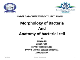
- 1. Morphology of Bacteria And Anatomy of bacterial cell UNDER GARADUATE STUDENT’S LECTUER ON BY GUNJAL PN ASSIST. PROF. DEPT OF MICROBIOLOGY DVVPF’S MEDICAL COLLEGE & HOSPITAL AHMENDAGAR 6/7/2022 Dept. of Microbiology 1
- 2. 6/7/2022 Dept. of Microbiology 2 GENERAL BACTERIOLOGY: MORPHOLOGY OF BACTERIA
- 3. At the end of the Lecture, the students will be able to understand: • Classification of bacteria depending on their morphology and Gram staining property. • Anatomy of Bacterial Cell. • Bacterial cell wall. • Various Bacterial cell appendages and their functions. 6/7/2022 Dept. of Microbiology 3 LEARNING OBJECTIVES
- 4. 6/7/2022 Dept. of Microbiology 4 MICROORGANISMS EUKARYOTIC • Parasites, fungi • Membrane enclosed organelles • Cytoskeleton PROKARYOTIC • Bacterial cell • Do not contain organelles • Cell wall, peptidoglycan
- 5. • SIZE - 0.25-1 µm width • 1-3 µm length Depending - on shape, bacteria - classified into: • Cocci (meaning berry) - oval or spherical cells – Staphylococcus. • Bacilli or rods - rod shaped –E.coli • Comma shaped- Vibrio. • Spiral- coiled – Spirillum/Spirochetes. • Branching filaments – Actinomycetes. 6/7/2022 Dept. of Microbiology 5 SHAPE OF BACTERIA
- 6. 6/7/2022 Dept. of Microbiology 6 • Cocci - arranged in groups (clusters), pair or chains. • Bacilli - arranged in chain, pair, and some bacilli are curved, comma shaped, or cuneiform shaped. SHAPE OF BACTERIA
- 7. 6/7/2022 Dept. of Microbiology 7 SHAPE OF BACTERIA
- 8. 6/7/2022 Dept. of Microbiology 8 SHAPE OF BACTERIA Cocci and bacilli - further classified - based on Gram staining into: • Gram-positive cocci • Gram-negative cocci • Gram-positive bacilli • Gram-negative bacilli.
- 9. 6/7/2022 Dept. of Microbiology 9 SHAPE OF BACTERIA Based on Gram’s staining differentiation
- 10. Bacterial cell anatomy: • The outer layer/envelope - consists - (1)rigid cell wall (2) plasma membrane • Cytoplasm contains - cytoplasmic inclusions (mesosomes, ribosomes, etc) and a diffuse nucleoid containing single circular chromosome. • Some bacteria - possess additional cell wall appendages - capsule, flagella and fimbriae. 6/7/2022 Dept. of Microbiology 10 ANATOMY OF BACTERIAL CELL
- 11. 6/7/2022 Dept. of Microbiology 11 CLASSIFICATION OF BACTERIA DEPENDING ON THEIR MORPHOLOGY AND GRAM STAINING PROPERTY Bacteria Example Gram-positive cocci arranged in Cluster Staphylococcus Chain Streptococcus Pairs, lanceolate shaped Pneumococcus Pair or in short chain, spectacle shaped Enterococcus Tetrads Micrococcus Octate Sarcina Gram-negative cocci arranged in Pairs, lens shaped Meningococcus Pairs, kidney shaped Gonococcus
- 12. 6/7/2022 Dept. of Microbiology 12 Bacteria Example Gram-positive bacilli arranged in Chain (bamboo stick appearance) Bacillus anthracis Chinese letter or cuneiform pattern Corynebacterium diphtheriae Palisade pattern Diphtheroids Branched and filamentous form Actinomyces and Nocardia CLASSIFICATION OF BACTERIA DEPENDING ON THEIR MORPHOLOGY AND GRAM STAINING PROPERTY
- 13. 6/7/2022 Dept. of Microbiology 13 CLASSIFICATION OF BACTERIA DEPENDING ON THEIR MORPHOLOGY AND GRAM STAINING PROPERTY Bacteria Example Gram-negative bacilli arranged in Pleomorphic (various shapes) Haemophilus, Proteus Thumb print appearance Bordetella pertussis Comma shaped (fish in stream appearance) Vibrio cholerae Curved Campylobacter (Gull-wing shaped) and Helicobacter Chain Streptobacillus Spirally coiled, flexible Spirochetes Rigid spiral forms Spirillum Bacteria that lack cell wall Mycoplasma
- 14. 6/7/2022 Dept. of Microbiology 14 BACTERIAL CELL WALL
- 15. 6/7/2022 Dept. of Microbiology 15 STRUCTURE OF BACTERIAL CELL
- 16. • Tough and rigid structure - surrounding the bacterium. • 10–25 nm thickness • Weighs about 20–25% of the dry weight of the cell. 6/7/2022 Dept. of Microbiology 16 BACTERIAL CELL WALL
- 17. FUNCTIONS OF CELL WALL • Protection to cell against osmotic lysis • Confers rigidity - presence of peptidoglycan layer in the cell wall • Protect cell from toxic substances • Site of action of several antibiotics • Contains - virulence factors (e.g. endotoxin) - contribute to pathogenicity • Antibody against specific cell wall antigens (e.g. antibody to LPS) - provide immunity against bacterial infection. 6/7/2022 Dept. of Microbiology 17
- 18. 6/7/2022 Dept. of Microbiology 18 DIFFERENCES BETWEEN GRAM-POSITIVE AND GRAM NEGATIVE CELL WALL
- 19. GRAM- POSITIVE CELL WALL 6/7/2022 Dept. of Microbiology 19 PEPTIDOGLYCAN: Thicker (50–100 layers thick, 16–80 nm) than gram- negative cell wall • Each layer - Mucopeptide (murein) chain - alternate units of N-acetyl muramic acid (NAMA) and N-acetyl glucosamine (NAGA) molecules - cross linked via tetrapeptide side chains and pentaglycine bridges. • Tetrapeptide side chain - from NAMA molecule - composed of L-alanine-D-glutamine - L-lysine - D-alanine.
- 20. 6/7/2022 Dept. of Microbiology 20 GRAM-POSITIVE CELL WALL Structure of Gram-Positive cell wall
- 21. 6/7/2022 Dept. of Microbiology 21 PEPTIDOGLYCAN LAYER OF GRAM-POSITIVE CELL WALL
- 22. 6/7/2022 Dept. of Microbiology 22 Teichoic Acid: • Polymers of glycerol or ribitol - joined by phosphate groups. • Maintain - structure of cell wall. • Two types: • (i) Cell wall teichoic acid and • (ii) Lipoteichoic acid. • Absent in gram-negative bacteria. GRAM-POSITIVE CELL WALL
- 23. 6/7/2022 Dept. of Microbiology 23 GRAM-NEGATIVE CELL WALL Peptidoglycan layer: • Very thin (1–2 layer, 2 nm thick) - composed of a mucopeptide chain - similar to gram-positive cell wall. • Consists - alternate NAMA and NAGA molecules. • Meso-diaminopimelic acid - present at third position of the tetrapeptide side chain . • Pentaglycine bridge is absent.
- 24. 6/7/2022 Dept. of Microbiology 24
- 25. 6/7/2022 Dept. of Microbiology 25 Outer Membrane: • Phospholipid layer - lies outside the thin peptidoglycan layer • Serves as - protective barrier to the cell • Outer membrane proteins (OMP) or porin proteins - specialized proteins GRAM-NEGATIVE CELL WALL
- 26. 6/7/2022 Dept. of Microbiology 26 Lipopolysaccharide (LPS): Consists of three parts: • Lipid A or the endotoxin- pyrogen- fever-causing agent. • Core polysaccharide • O side chain (or O antigen or somatic antigen) Periplasmic Space • Space between the inner cell membrane and outer membrane. It encompasses the peptidoglycan layer. GRAM-NEGATIVE CELL WALL
- 27. 6/7/2022 Dept. of Microbiology 27 CELL MEMBRANE
- 28. 6/7/2022 Dept. of Microbiology 28 CELL MEMBRANE • Essential for - survival of the bacteria. • Fluid mosaic model - most widely accepted model • 5–10 nm thick -bilayered phospholipid - several proteins are embedded - integral proteins and peripheral proteins • Lacks sterols -such as cholesterol (except in Mycoplasma). • Contain Pentacyclic sterol-like molecules – hopanoids- used as biomarkers.
- 29. 6/7/2022 Dept. of Microbiology 29 CELL MEMBRANE
- 30. • Semi permeable membrane • Transport system -nutrient uptake, and waste excretion • Site for metabolic processes 6/7/2022 Dept. of Microbiology 30 FUNCTIONS OF CELL MEMBRANE
- 31. 6/7/2022 Dept. of Microbiology 31 CYTOPLASMIC MATRIX • Bacterial cytoplasm, lacks membrane-bound organelles. • Composed of water (70%), salt and proteins. • Packed with ribosomes, storage granules - inclusions and cell membrane invaginations - mesosomes. • Plasma membrane and everything within it - called as protoplast.
- 32. 6/7/2022 Dept. of Microbiology 32 RIBOSOMES • Sites for protein synthesis. • Composed of rRNA and ribosomal proteins. • Integrated with the mRNA to form polysomes. • At this site - genetic codons of the mRNA - translated into peptide sequences. • Each 70 S unit - consists of a 30 S and a 50 S subunits.
- 33. • Storage sites of nutrients/energy. • Formed by bacteria under nutritional deficiency conditions and disappear when the deficient nutrients are supplied. • Two types of inclusions: Organic inclusion bodies. Inorganic inclusion bodies. 6/7/2022 Dept. of Microbiology 33 INTRACYTOPLASMIC INCLUSIONS
- 34. • Invaginations of plasma membrane - in the shape of vesicles, tubules. • Prominent in gram-positive bacteria. • Involved in: Bacterial respiration Cell wall formation Chromosome replication 6/7/2022 Dept. of Microbiology 34 MESOSOMES
- 35. • Bacteria do not have a true nucleus - genetic material - located in an irregularly shaped region called - nucleoid. • Bacteria possess a single haploid chromosome. • Comprises of super coiled circular double stranded DNA • Seen by electron microscopy or on staining with Feulgen stain • Bacteria also possess extrachromosomal DNA - plasmids 6/7/2022 Dept. of Microbiology 35 NUCLEOID
- 36. 6/7/2022 Dept. of Microbiology 36 CELL WALL APPENDAGES
- 37. 6/7/2022 Dept. of Microbiology 37 CELL WALL APPENDAGES Composed of: • Capsule and Slime Layer • Flagella • Fimbriae or Pili
- 38. • Layer of amorphous viscid material lying outside the cell wall called glycocalyx. • Capsule -well organized and not easily washed off • Slime layer - diffuse, unorganized loose material that can be removed easily 6/7/2022 Dept. of Microbiology 38 CAPSULE AND SLIME LAYER
- 39. 6/7/2022 Dept. of Microbiology 39 CAPSULE AND SLIME LAYER Capsulated bacteria Composition Pneumococcus Polysaccharide Meningococcus Polysaccharide Haemophilus influenzae Polysaccharide Klebsiella pneumoniae Polysaccharide Pseudomonas aeruginosa Polysaccharide Bacteroides fragilis Polysaccharide Bacillus anthracis Polypeptide (glutamate) Streptococcus pyogenes (some strains) Hyaluronic acid
- 40. 6/7/2022 Dept. of Microbiology 40 FUNCTIONS OF CAPSULES Bacterial virulence : • Prevent cell from drying out (desiccation) • Protects the bacterium from the action of lysozyme and bacteriophages. • Toxic to the host cells and induces abscess formation (e.g. Bacteroides fragilis) Capsules as vaccine: • Capsular vaccines are available for bacteria such as Pneumococcus, Meningococcus and Haemophilus influenzae serotype-b.
- 41. Biofilm Formation • Biofilm - Living ecosystem made up of millions of adherent bacterial cells, which are embedded within a self-produced matrix of extracellular polymeric substance. • Capable of adherence to damaged tissues and plastic surfaces. • Adhesion is a first step in colonization and sometimes leads to disease. 6/7/2022 Dept. of Microbiology 41 FUNCTIONS OF CAPSULES
- 42. Capsule can be detected by various methods: • Negative staining by India ink and Nigrosin stain. • M’Faydean capsule stain. • Serological test Quellung reaction Capsular antigen 6/7/2022 Dept. of Microbiology 42 DEMONSTRATION OF CAPSULE S.pneumoniae capsule seen by India ink staining
- 43. • Thread-like appendages - protruding from the cell wall. • Confer motility to the bacteria. • 5–20 µm in length and 0.01 - 0.02 µm in thickness. • ARRANGEMENT OF FLAGELLA • Monotrichous - e.g. Vibrio cholerae, Pseudomonas and Campylobacter. • Lophotrichous - e.g. Spirillum. • Peritrichous - e.g. Salmonella typhi, Escherichia coli . • Amphitrichous - e.g. Alcaligenes faecalis. 6/7/2022 Dept. of Microbiology 43 FLAGELLA
- 44. ARRANGEMENT OF FLAGELLA 6/7/2022 Dept. of Microbiology 44
- 45. On electron microscope - bacterial flagellum is - composed of three parts. 1. Filament - longest portion of the flagellum that extends from the cell surface to the tip. 2. The basal body 3. Hook 6/7/2022 Dept. of Microbiology 45 ULTRASTRUCTURE OF FLAGELLA Hook
- 46. Direct demonstration of flagella: Tannic acid staining (Leifson’s method) Electron microscopy. Indirect means by demonstrating the motility: • Craigie tube method • Hanging drop method • Semisolid medium, e.g. mannitol motility medium • Dark ground or phase contrast microscopy. 6/7/2022 Dept. of Microbiology 46 DETECTION OF FLAGELLA
- 47. 6/7/2022 Dept. of Microbiology 47 TYPES OF MOTILITY SHOWN BY DIFFERENT BACTERIA Types of motility Bacteria Tumbling motility Listeria Gliding motility Mycoplasma Stately motility Clostridium Darting motility Vibrio cholerae, Campylobacter Swarming on agar plate Proteus, Clostridium tetani Corkscrew, lashing, flexion extension motility Spirochete
- 48. • Short, fine, hair-like appendages - help in bacterial adhesion. • Special type of pili (called sex pilus) - helps in conjugation. • Pili - made up of protein called pilin. • Antigenic; but, the antibodies against pilin antigens are not protective. • Not related to motility 6/7/2022 Dept. of Microbiology 48 FIMBRIAE OR PILI
- 49. 6/7/2022 Dept. of Microbiology 49 TYPE OF PILI According to functions, pili are of two types. 1. Common pili or fimbriae 2. Sex pili
- 50. 6/7/2022 Dept. of Microbiology 50 DETECTION OF FIMBRIAE • Electron microscope • Surface pellicle - thin layer formed at the surface of a liquid culture of strongly aerobic bacteria such as Pseudomonas.
- 51. • Involution forms: Swollen and aberrant forms of bacteria (e.g. gonococci and Yersinia pestis) formed in ageing cultures in high salt concentration. • Pleomorphic bacteria: Exhibit great variation in the shape and size of individual cells, e.g. Proteus and Haemophilus. L Form (Cell Wall Deficient Forms) - Cell wall deficient bacteria. • Discovered by E. Klieneberger, while studying Streptobacillus moniliformis. • Named it as L form - after its place of discovery, i.e. Lister Institute, London (1935) • L forms play a role in the persistence of pyelonephritis and other chronic infections. 6/7/2022 Dept. of Microbiology 51 ATYPICAL FORMS OF BACTERIA
- 52. 6/7/2022 Dept. of Microbiology 52 Unstable L forms: Bacteria lose their cell wall in presence of penicillin, a mechanism of resistance shown by the bacteria against penicillin. • Maintained only in presence of penicillin - can revert to the original morphology once penicillin is removed. • Protoplasts - Gram-positive bacteria whose cell wall is entirely removed. • Spheroplasts - Derived from gram-negative bacteria whose cell wall is partially removed. Stable L forms: Mycoplasmas lack cell wall permanently - may represent stable L-forms of bacteria. ATYPICAL FORMS OF BACTERIA
- 53. 6/7/2022 Dept. of Microbiology 53 BACTERIAL SPORES
- 54. • Spores are highly resistant resting (or dormant) stage of the bacteria. • Formed in unfavorable environmental conditions - as a result of the depletion of exogenous nutrients. • Bacterial spore comprises of several layers. • From innermost towards the outermost, the layers are: core → cortex → coat → exosporium 6/7/2022 Dept. of Microbiology 54 BACTERIAL SPORES
- 55. • Refers to - process of formation of spores from vegetative stage of bacteria. • Not a method of reproduction - because bacteria do not divide during sporulation. • Complex process and takes about 10 hours. • Mature spore formed is extremely resistant to heat and disinfectant • Transformation of dormant spores into active vegetative cells when grown in a nutrient-rich medium. 6/7/2022 Dept. of Microbiology 55 SPORULATION GERMINATION
- 56. • For a given species, the precise position, shape and relative size of the spore are constant. • Position: Central, subterminal or terminal • Shape: Oval or spherical in shape • Width: Diameter of spore may be same or less than the width of bacteria 6/7/2022 Dept. of Microbiology 56 SHAPE AND POSITION OF SPORES A. Non-bulging, oval and terminal; B. Non-bulging, round, and subterminal; C. Non-bulging, oval and central; D. Bulging, round and terminal; E. Bulging, oval and terminal; F. Bulging, oval, and central
- 57. • Spores are resistant to most of the routinely used disinfectants. • Only limited agents called as sterilants are capable of killing the spores, e.g. autoclave, or ethylene oxide sterilizer, etc. DEMONSTRATION OF SPORES • Gram staining: Spores appear as unstained retractile bodies within the cells • Modified Ziehl–Neelsen staining: Spores are weakly acid-fast and appear red color. • Special techniques for endospore staining include the Schaeffer–Fulton stain and the Moeller stain. 6/7/2022 Dept. of Microbiology 57 SPORICIDAL AGENTS
- 58. • Indicators of proper sterilization. Spores of Geobacillus stearothermophilus - sterilization control for autoclave and plasma sterilizer. Spores of Bacillus atrophaeus - sterilization control for hot air oven and ethylene oxide sterilizer. • Used as agents of bioterrorism 6/7/2022 Dept. of Microbiology 58 APPLICATIONS OF SPORES
- 59. Q1. Bamboo stick appearance arrangement is characteristic of: a. Staphylococcus b. Streptococcus c. Corynebacterium diphtheriae d. Bacillus anthracis Q2. Bacterial capsule can be best demonstrated by: a. Gram staining b. Acid-fast staining c. Negative staining d. Albert staining 6/7/2022 Dept. of Microbiology 59 QUESTIONS:
- 60. Q3. Lipopolysaccharide is a component of cell wall of: a. Gram-positive bacteria b. Gram-negative bacteria c. Virus d. Fungi Q4. Bacterial structure involved in respiration is: a. Ribosome b. Pili c. Mesosome d. Flagella 6/7/2022 Dept. of Microbiology 60
- 61. Thank you all ! 6/7/2022 Dept. of Microbiology 61