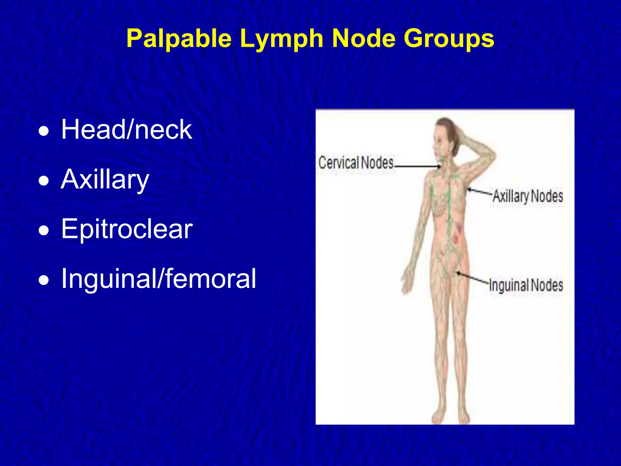
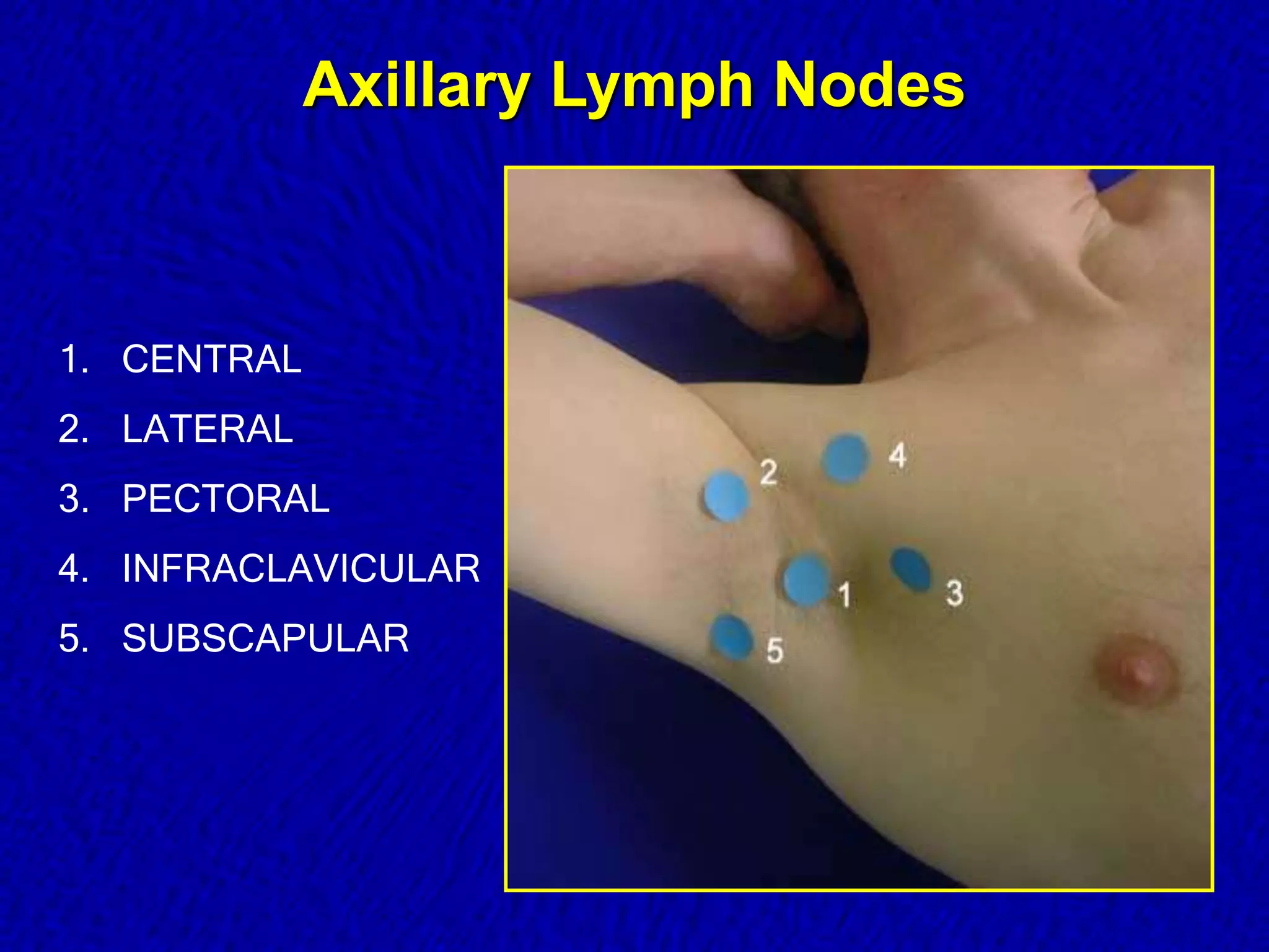
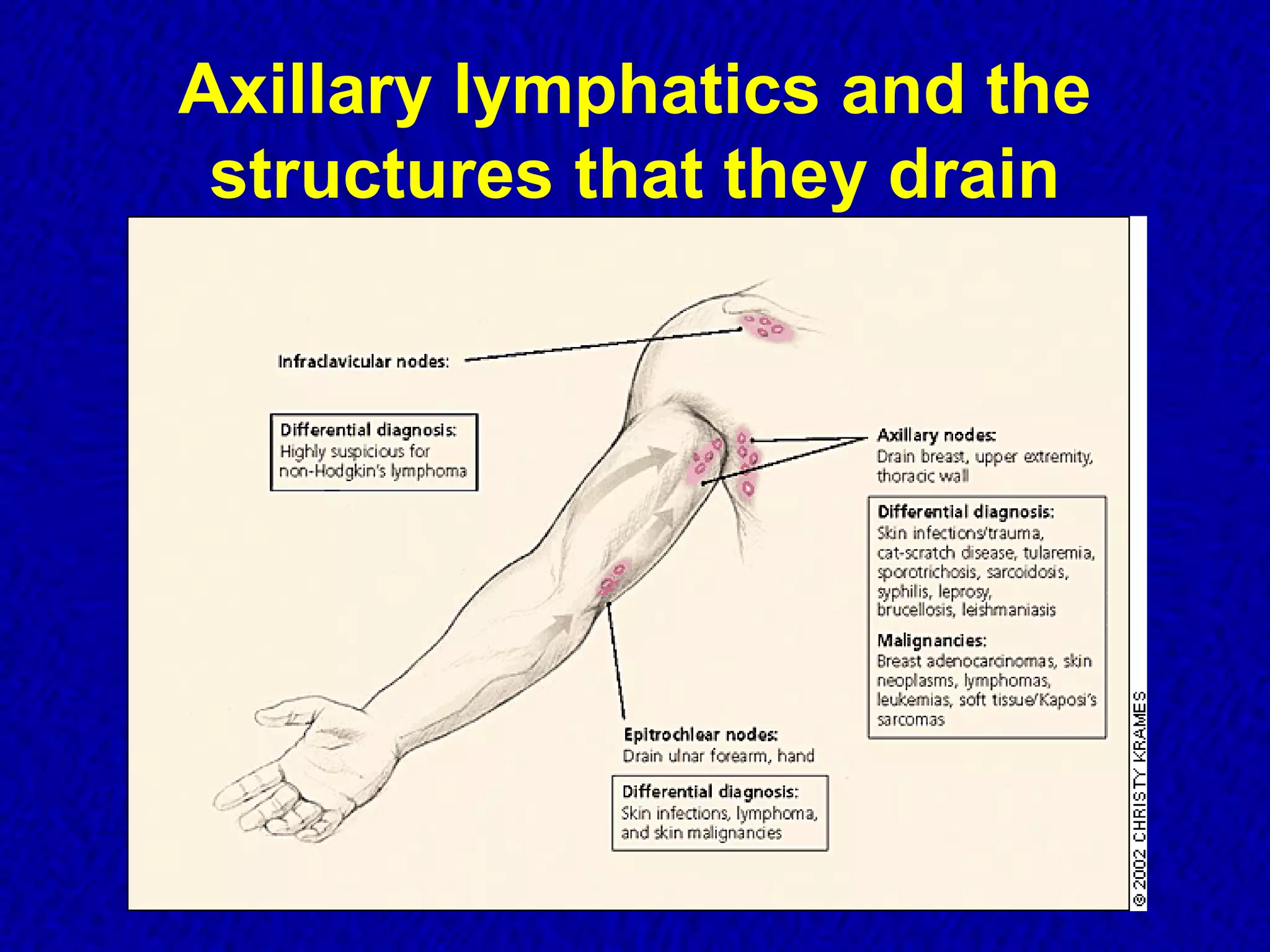
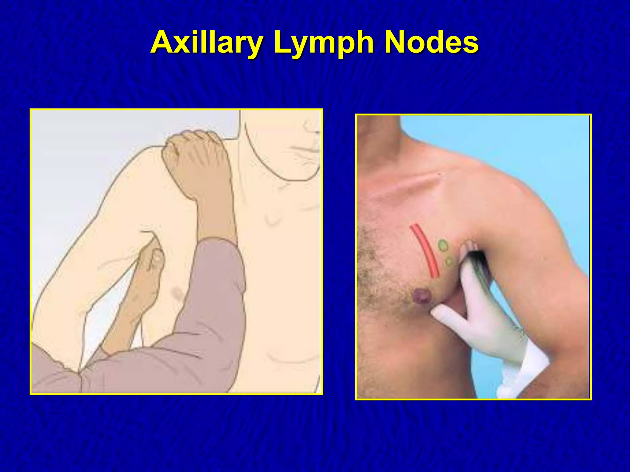
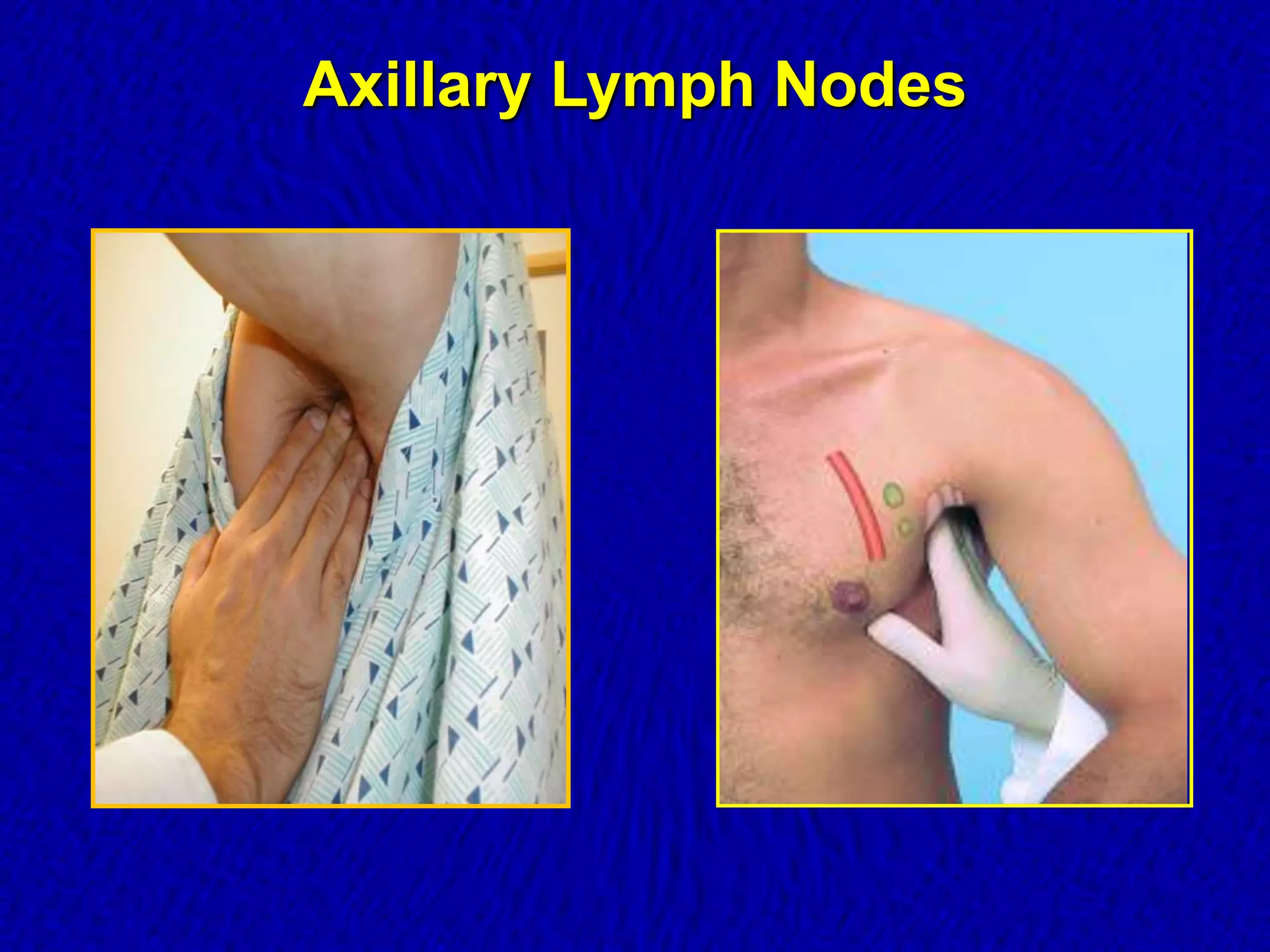
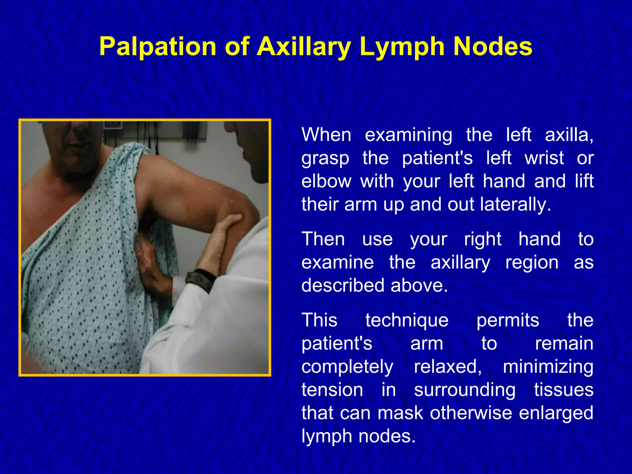
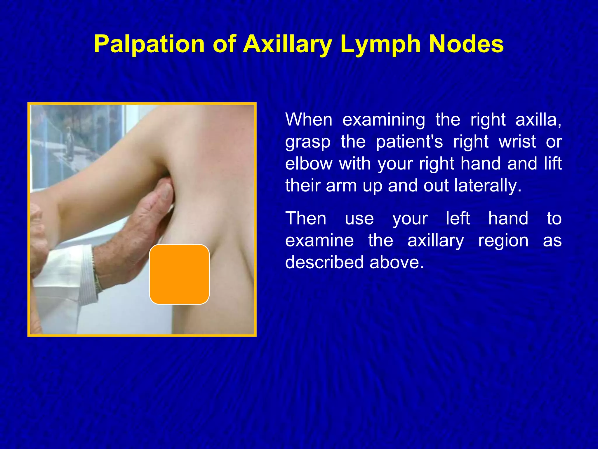
Axillary Lymph
Nodes Examination
Section 1
Examination of the
Lymphatic System
 Utilizes inspection and palpation.
 Generally examined region by region during
the examination of the other body systems.
 Always ask patients if they are aware of any
“lumps”.
Examination of the Lymphatic System
 Inspect:
 any visible nodes for:
• edema
• erythema
 Palpate:
 the superficial nodes
 compare side to side for:
• size
• consistency
• mobility
• discrete borders or
matted
• tenderness
• warmth
Nodal Character and Size
 Hard and painless nodes have higher suspicion of
malignancy or granulomatous disease.
 Viral infection typically produces hyperplastic nodes
that are bilateral, mobile, nontender, and clearly
demarcated.
 Increasing size and persistence over time are of
greater concern for malignancy than a specific level
of nodal enlargement.
Differential Diagnosis of Nodes
CANCER
 Firm, hard
 Non-movable
 No fever
 Not painful
INFECTION
 Soft
 Movable
 Fever
 Painful
Examination of the
Lymphatic System
 If an enlarged lymph node is found,
examine:
P Primary site
A All associated nodes
L Liver
S Spleen
Palpable Lymph Node Groups
 Head/neck
 Axillary
 Epitroclear
 Inguinal/femoral
Examination of Lymph Nodes
 Small, mobile, discrete, nontender nodes are common
and termed shotty
 Nodes are abnormal if greater than 1 cm and/or present
greater than one month
 Hard nodes suggest malignancy
 Tender nodes suggest infection
 Rubbery nodes suggest lymphoma
Age Related Variations
Infants and Children
 Commonly find small, discreet, firm, movable nodes in
occipital, postauricular, cervical and inguinal chains . . .
 should not be warm or tender
 shape usually ovoid or globular
 often referred to as “shotty nodes”
 May find enlarged postauricular and occipital nodes in
children < 2 years old
Axillary Lymph Nodes
1. CENTRAL
2. LATERAL
3. PECTORAL
4. INFRACLAVICULAR
5. SUBSCAPULAR
Axillary Lymph Nodes
Axillary lymphatics and the
structures that they drain
Axillary Lymph Nodes
Palpation of Axillary Lymph Nodes
When examining the left axilla,
grasp the patient's left wrist or
elbow with your left hand and lift
their arm up and out laterally.
Then use your right hand to
examine the axillary region as
described above.
This technique permits the
patient's arm to remain
completely relaxed, minimizing
tension in surrounding tissues
that can mask otherwise enlarged
lymph nodes.
Palpation of Axillary Lymph Nodes
When examining the right axilla,
grasp the patient's right wrist or
elbow with your right hand and lift
their arm up and out laterally.
Then use your left hand to
examine the axillary region as
described above.
Left Axillary Adenopathy
Axillary Lymphadenopathy
 Most of cases are nonspecific or reactive to local
injury/infection in etiology.
 Persistent lymphadenopathy is less commonly found
in the axillary nodes than in the inguinal chain.
 Breast adenocarcinoma often metastasis initially
to the anterior and central axillary nodes, which may
be palpable before discovery of the primary tumor.
Generalized Lymphadenopathy
 Generalized lymphadenopathy : lymphadenopathy
found in two or more distinct anatomic regions
 More likely to result from serious infections,
autoimmune diseases, and disseminated
malignancies.
 Specific testing is usually required.
 Generalized adenopathy infrequently occurs in pt’s
with neoplasms, but it is occasionally seen in
patients with leukemias and lymphomas, or
advanced disseminated metastatic solid tumors.
Causes of Generalized Lymphadenopathy
 Malignancy: lymphoma, leukemia or metastases.
 Autoimmune: SLE, RA or Sjogren’s syndrome.
 Infectious: Brucellosis, Cat-scratch disease, CMV,
HIV, EBV, Rubella, Tuberculosis, Typhoid Fever,
Syphilis or viral hepatitis.
 Other: Kawasaki’s disease, sarcoidosis,
amyloidosis, lipid storage diseases or
hyperthyroidism
Drugs which induces Lymphadenopathy
 Allopurinol
 Atenolol
 Captopril
 Carbamazepine
 Gold
 Hydralazine
 Penicillins
 Phenytoin
 Primidone
 Pyrimethamine
 Quinidine
 Trimethoprim/Sulfame
thozole
 Cervical nodes most commonly involved
 Usual course of lymph node disease is as follows:
Firm, discrete nodes

fluctuant nodes matted together

skin breakdown, abscesses, chronic sinuses

healing and scarring
Tuberculosis lymphadenopathy
Sarcoidosis
Bilateral symmetric hilar and is the most common
pattern of lymphadenopathy in sarcoidosis.
Thank you!

Axillary Lymph Nodes Examination.pptx

  • 1.
  • 2.
    Examination of the LymphaticSystem  Utilizes inspection and palpation.  Generally examined region by region during the examination of the other body systems.  Always ask patients if they are aware of any “lumps”.
  • 3.
    Examination of theLymphatic System  Inspect:  any visible nodes for: • edema • erythema  Palpate:  the superficial nodes  compare side to side for: • size • consistency • mobility • discrete borders or matted • tenderness • warmth
  • 4.
    Nodal Character andSize  Hard and painless nodes have higher suspicion of malignancy or granulomatous disease.  Viral infection typically produces hyperplastic nodes that are bilateral, mobile, nontender, and clearly demarcated.  Increasing size and persistence over time are of greater concern for malignancy than a specific level of nodal enlargement.
  • 5.
    Differential Diagnosis ofNodes CANCER  Firm, hard  Non-movable  No fever  Not painful INFECTION  Soft  Movable  Fever  Painful
  • 6.
    Examination of the LymphaticSystem  If an enlarged lymph node is found, examine: P Primary site A All associated nodes L Liver S Spleen
  • 7.
    Palpable Lymph NodeGroups  Head/neck  Axillary  Epitroclear  Inguinal/femoral
  • 8.
    Examination of LymphNodes  Small, mobile, discrete, nontender nodes are common and termed shotty  Nodes are abnormal if greater than 1 cm and/or present greater than one month  Hard nodes suggest malignancy  Tender nodes suggest infection  Rubbery nodes suggest lymphoma
  • 9.
    Age Related Variations Infantsand Children  Commonly find small, discreet, firm, movable nodes in occipital, postauricular, cervical and inguinal chains . . .  should not be warm or tender  shape usually ovoid or globular  often referred to as “shotty nodes”  May find enlarged postauricular and occipital nodes in children < 2 years old
  • 10.
  • 11.
    1. CENTRAL 2. LATERAL 3.PECTORAL 4. INFRACLAVICULAR 5. SUBSCAPULAR Axillary Lymph Nodes
  • 12.
    Axillary lymphatics andthe structures that they drain
  • 13.
  • 14.
  • 15.
    Palpation of AxillaryLymph Nodes When examining the left axilla, grasp the patient's left wrist or elbow with your left hand and lift their arm up and out laterally. Then use your right hand to examine the axillary region as described above. This technique permits the patient's arm to remain completely relaxed, minimizing tension in surrounding tissues that can mask otherwise enlarged lymph nodes.
  • 16.
    Palpation of AxillaryLymph Nodes When examining the right axilla, grasp the patient's right wrist or elbow with your right hand and lift their arm up and out laterally. Then use your left hand to examine the axillary region as described above.
  • 17.
  • 18.
    Axillary Lymphadenopathy  Mostof cases are nonspecific or reactive to local injury/infection in etiology.  Persistent lymphadenopathy is less commonly found in the axillary nodes than in the inguinal chain.  Breast adenocarcinoma often metastasis initially to the anterior and central axillary nodes, which may be palpable before discovery of the primary tumor.
  • 19.
    Generalized Lymphadenopathy  Generalizedlymphadenopathy : lymphadenopathy found in two or more distinct anatomic regions  More likely to result from serious infections, autoimmune diseases, and disseminated malignancies.  Specific testing is usually required.  Generalized adenopathy infrequently occurs in pt’s with neoplasms, but it is occasionally seen in patients with leukemias and lymphomas, or advanced disseminated metastatic solid tumors.
  • 20.
    Causes of GeneralizedLymphadenopathy  Malignancy: lymphoma, leukemia or metastases.  Autoimmune: SLE, RA or Sjogren’s syndrome.  Infectious: Brucellosis, Cat-scratch disease, CMV, HIV, EBV, Rubella, Tuberculosis, Typhoid Fever, Syphilis or viral hepatitis.  Other: Kawasaki’s disease, sarcoidosis, amyloidosis, lipid storage diseases or hyperthyroidism
  • 21.
    Drugs which inducesLymphadenopathy  Allopurinol  Atenolol  Captopril  Carbamazepine  Gold  Hydralazine  Penicillins  Phenytoin  Primidone  Pyrimethamine  Quinidine  Trimethoprim/Sulfame thozole
  • 22.
     Cervical nodesmost commonly involved  Usual course of lymph node disease is as follows: Firm, discrete nodes  fluctuant nodes matted together  skin breakdown, abscesses, chronic sinuses  healing and scarring Tuberculosis lymphadenopathy
  • 23.
    Sarcoidosis Bilateral symmetric hilarand is the most common pattern of lymphadenopathy in sarcoidosis.
  • 24.