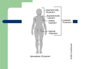
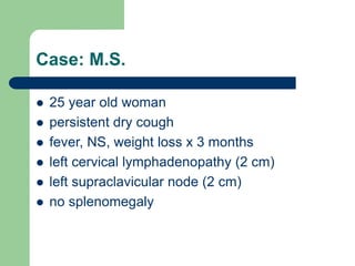
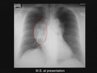
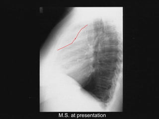
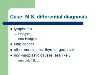
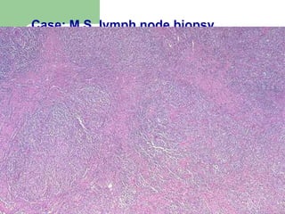
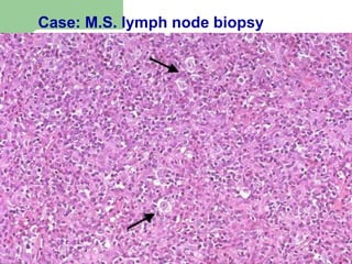
Lymphadenopathy
Soheir Adam , MD, MSc, MRCPath
The Lymphatic System
 The body has approximately 600 lymph nodes, but
only those in the submandibular, axillary or inguinal
regions may normally be palpable in healthy
people.1 Lymphadenopathy refers to nodes that are
abnormal in either size, consistency or number.
There are various classifications of
lymphadenopathy, but a simple and clinically useful
system is to classify lymphadenopathy as
"generalized" if lymph nodes are enlarged in two or
more noncontiguous areas or "localized" if only one
area is involved.
 Distinguishing between localized and
generalized lymphadenopathy is important in
formulating a differential diagnosis.
 In primary care patients with unexplained
lymphadenopathy, approximately 3/4 of
patients will present with localized
lymphadenopathy and 1/4 with generalized
lymphadenopathy.
Lympahdenopathy
 Findings from a Dutch study revealed a 0.6%
annual incidence of unexplained lymphadenopathy
in the general population.
 Of 2,556 patients in the study who presented with
unexplained lymphadenopathy to their family
physicians, 256 (10 %) were referred to a
subspecialist and 82 (3.2 %) required a biopsy, but
only 29 (1.1 %) had a malignancy.
Lymphadenopathy
History
 First, are there localizing symptoms or signs to
suggest infection or neoplasm in a specific site?
 Second, are there constitutional symptoms such as
fever, weight loss, fatigue or night sweats to suggest
disorders such as tuberculosis, lymphoma, collagen
vascular diseases, unrecognized infection or
malignancy?
History
 Third, are there epidemiologic clues such as
occupational exposures, recent travel or high-risk
behaviors that suggest specific disorders?
 Fourth, is the patient taking a medication that may
cause lymphadenopathy? Some medications are
known to specifically cause lymphadenopathy (e.g.,
phenytoin ), while others, such as cephalosporins,
penicillins or sulfonamides, are more likely to cause
a serum sickness-like syndrome with fever,
arthralgias and rash in addition to lymphadenopathy.
Medications That May Cause Lymphadenopathy
Allopurinol (Zyloprim)
Atenolol (Tenormin)
Captopril (Capozide)
Carbamazepine (Tegretol)
Cephalosporins
Gold
Hydralazine (Apresoline)
Penicillin
Phenytoin (Dilantin)
Primidone (Mysoline)
Pyrimethamine (Daraprim)
Quinidine
Sulfonamides
Sulindac (Clinoril)
Adapted with permission from Pangalis GA, Vassilakopoulos TP, Boussiotis VA, Fessas P. Clinical approach to lymphadenopathy. Semin Oncol 1993; 20:570-82.
Physical Examination
 Size.
 Pain/Tenderness :The presence or absence of tenderness
does not reliably differentiate benign from malignant nodes.
 Consistency: Stony-hard nodes are typically a sign of cancer,
usually metastatic. Very firm, rubbery nodes suggest
lymphoma. Softer nodes are the result of infections or
inflammatory conditions. Suppurant nodes may be fluctuant.
The term "shotty" refers to small nodes that feel like buckshot
under the skin, as found in the cervical nodes of children with
viral illnesses.
Physical Examination
 Matting : can be either benign (e.g.,
tuberculosis, sarcoidosis) or malignant (e.g.,
metastatic carcinoma or lymphomas
 Location : infectious mononucleosis causes
cervical adenopathy and a number of
sexually transmitted diseases are associated
with inguinal adenopathy
Physical Examination
 Supraclavicular lymphadenopathy has the highest risk of
malignancy, estimated as 90 percent in patients older than 40
years and 25 percent in those younger than age.
 Lymphadenopathy of the right supraclavicular node is
associated with cancer in the mediastinum, lungs or
esophagus.
 The left supraclavicular (Virchow's) node receives lymphatic
flow from the thorax and abdomen, and may signal pathology in
the testes, ovaries, kidneys, pancreas, prostate, stomach or
gallbladder. Although rarely present
Evaluation of Suggestive S & S Associated with Lymphadenopathy
Mononucleosis-type
syndromes
Fatigue, malaise, fever, atypical
lymphocytosis
Epstein-Barr virus* Splenomegaly in 50% of patients Monospot, IgM EA or VCA
Toxoplasmosis* 80 to 90% of patients are
asymptomatic
IgM toxoplasma antibody
Cytomegalovirus* Often mild symptoms; patients may
have hepatitis
IgM CMV antibody, viral
culture of urine or blood
Initial stages of HIV
infection*
"Flu-like" illness, rash HIV antibody
Cat-scratch disease Fever in one third of patients; cervical
or axillary nodes
Usually clinical criteria; biopsy
if necessary
Pharyngitis due to group A
streptococcus,
gonococcus
Fever, pharyngeal exudates, cervical
nodes
Throat culture on appropriate
medium
Tuberculosis lymphadenitis* Painless, matted cervical nodes PPD, biopsy
Secondary syphilis* Rash RPR
Hepatitis B* Fever, nausea, vomiting, icterus Liver function tests, HBsAg
Lymphogranuloma venereum Tender, matted inguinal nodes Serology
Chancroid Painful ulcer, painful inguinal nodes Clinical criteria, culture
Lupus erythematosus* Arthritis, rash, serositis, renal, neurologic, hematologic
disorders
Clinical criteria, antinuclear antibodies,
complement levels
Rheumatoid arthritis* Arthritis Clinical criteria, rheumatoid factor
Lymphoma* Fever, night sweats, weight loss in 20 to 30% of patients Biopsy
Leukemia* Blood dyscrasias, bruising Blood smear, bone marrow
Serum sickness* Fever, malaise, arthralgia, urticaria; exposure to antisera
or medications
Clinical criteria, complement assays
Sarcoidosis Hilar nodes, skin lesions, dyspnea Biopsy
Kawasaki disease* Fever, conjunctivitis, rash, mucous membrane lesions Clinical criteria
Less common causes of lymphadenopathy
Lyme disease* Rash, arthritis IgM serology
Measles* Fever, conjunctivitis, rash, cough Clinical criteria, serology
Rubella* Rash Clinical criteria, serology
Tularemiala* Fever, ulcer at inoculation site Blood culture, serology
Brucellosis* Fever, sweats, malaise Blood culture, serology
Plague Febrile, acutely ill with cluster of tender nodes Blood culture, serology
Typhoid fever* Fever, chills, headache, abdominal complaints Blood culture, serology
Still's disease* Fever, rash, arthritis Clinical criteria, antinuclear antibody,
rheumatoid factor
Dermatomyositis* Proximal weakness, skin changes Muscle enzymes, EMG, muscle biopsy
Amyloidosis* Fatigue, weight loss Biopsy
*--Causes of generalized lymphadenopathy.
EA=early antibody; VCA=viral capsid antigen; CMV=cytomegalovirus; HIV=human immunodeficiency virus; PPD=purified protein derivative; RPR=rapid plasma reagin; HBsAg=hepatitis B
surface antigen; EMG=electromyelography.
Unexplained Lymphadenopathy
Generalized Lymphadenopathy
 almost always indicates a systemic disease is
present, proceed with specific testing as indicated.
 If a diagnosis cannot be made, the clinician should
obtain a biopsy of the node.
 The diagnostic yield of the biopsy can be maximized
by obtaining an excisional biopsy of the largest and
most abnormal node
 The physician should not select inguinal and axillary
nodes for biopsy, since they frequently show only
reactive hyperplasia
Unexplained Lymphadenopathy
Localized Lymphadenopathy
 The decision about when to biopsy is more difficult.
 Patients with a benign clinical history, an unremarkable
physical examination and no constitutional symptoms
should be reexamined in three to four weeks to see if the
lymph nodes have regressed or disappeared.
 Patients with unexplained localized lymphadenopathy who
have constitutional symptoms or signs, risk factors for
malignancy or lymphadenopathy that persists for three to
four weeks should undergo a biopsy.
Unexplained Lymphadenopathy
Localized Lymphadenopathy
 Biopsy should be avoided in patients with
probable viral illness because lymph node
pathology in these patients may
sometimes simulate lymphoma and lead
to a false-positive diagnosis of
malignancy.
Lymphoma
Risk factors for NHL
 immunosuppression or immunodeficiency
 connective tissue disease
 family history of lymphoma
 infectious agents
 ionizing radiation
A practical way to think of lymphoma
Category Survival of
untreated
patients
Curability To treat or
not to treat
Non-
Hodgkin
lymphoma
Indolent Years Generally
not curable
Generally
defer Rx if
asymptomatic
Aggressive Months Curable in
some
Treat
Very
aggressive
Weeks Curable in
some
Treat
Hodgkin
lymphoma
All types Variable –
months to
years
Curable in
most
Treat
Diagnosis requires an adequate
biopsy
 Diagnosis should be biopsy-proven
before treatment is initiated
 Need enough tissue to assess cells and
architecture
– open bx vs core needle bx vs FNA
Stage I Stage II Stage III Stage IV
Staging of lymphoma
A: absence of B symptoms
B: fever, night sweats, weight loss
Case: M.S.
 25 year old woman
 persistent dry cough
 fever, NS, weight loss x 3 months
 left cervical lymphadenopathy (2 cm)
 left supraclavicular node (2 cm)
 no splenomegaly
M.S. at presentation
Case: M.S. differential diagnosis
 lymphoma
– Hodgkin
– non-Hodgkin
 lung cancer
 other neoplasms: thyroid, germ cell
 non-neoplastic causes less likely
– sarcoid, TB, ...
What next?
 Needle aspirate of LN: a few necrotic cells
 Needle biopsy of LN: admixture of B- and T-
lymphocytes. A few atypical cells.
Case: M.S. lymph node biopsy
Case: M.S. staging investigations
 CT chest / abdo / pelvis
 bone marrow
 gallium scan
 Blood work: normal
Staging Investigations
 bone marrow normal
 CT scan: L supraclavicular adenopathy; large
mediastinal mass; R hilum; no disease below
diaphragm
 gallium avid
What is her diagnosis and stage?
 nodular sclerosis HD
 stage IIB
 with bulky mediastinal mass

32577_Lymphadenopathy.ppt

  • 1.
  • 2.
    The Lymphatic System The body has approximately 600 lymph nodes, but only those in the submandibular, axillary or inguinal regions may normally be palpable in healthy people.1 Lymphadenopathy refers to nodes that are abnormal in either size, consistency or number. There are various classifications of lymphadenopathy, but a simple and clinically useful system is to classify lymphadenopathy as "generalized" if lymph nodes are enlarged in two or more noncontiguous areas or "localized" if only one area is involved.
  • 3.
     Distinguishing betweenlocalized and generalized lymphadenopathy is important in formulating a differential diagnosis.  In primary care patients with unexplained lymphadenopathy, approximately 3/4 of patients will present with localized lymphadenopathy and 1/4 with generalized lymphadenopathy.
  • 5.
    Lympahdenopathy  Findings froma Dutch study revealed a 0.6% annual incidence of unexplained lymphadenopathy in the general population.  Of 2,556 patients in the study who presented with unexplained lymphadenopathy to their family physicians, 256 (10 %) were referred to a subspecialist and 82 (3.2 %) required a biopsy, but only 29 (1.1 %) had a malignancy.
  • 7.
  • 8.
    History  First, arethere localizing symptoms or signs to suggest infection or neoplasm in a specific site?  Second, are there constitutional symptoms such as fever, weight loss, fatigue or night sweats to suggest disorders such as tuberculosis, lymphoma, collagen vascular diseases, unrecognized infection or malignancy?
  • 9.
    History  Third, arethere epidemiologic clues such as occupational exposures, recent travel or high-risk behaviors that suggest specific disorders?  Fourth, is the patient taking a medication that may cause lymphadenopathy? Some medications are known to specifically cause lymphadenopathy (e.g., phenytoin ), while others, such as cephalosporins, penicillins or sulfonamides, are more likely to cause a serum sickness-like syndrome with fever, arthralgias and rash in addition to lymphadenopathy.
  • 10.
    Medications That MayCause Lymphadenopathy Allopurinol (Zyloprim) Atenolol (Tenormin) Captopril (Capozide) Carbamazepine (Tegretol) Cephalosporins Gold Hydralazine (Apresoline) Penicillin Phenytoin (Dilantin) Primidone (Mysoline) Pyrimethamine (Daraprim) Quinidine Sulfonamides Sulindac (Clinoril) Adapted with permission from Pangalis GA, Vassilakopoulos TP, Boussiotis VA, Fessas P. Clinical approach to lymphadenopathy. Semin Oncol 1993; 20:570-82.
  • 11.
    Physical Examination  Size. Pain/Tenderness :The presence or absence of tenderness does not reliably differentiate benign from malignant nodes.  Consistency: Stony-hard nodes are typically a sign of cancer, usually metastatic. Very firm, rubbery nodes suggest lymphoma. Softer nodes are the result of infections or inflammatory conditions. Suppurant nodes may be fluctuant. The term "shotty" refers to small nodes that feel like buckshot under the skin, as found in the cervical nodes of children with viral illnesses.
  • 12.
    Physical Examination  Matting: can be either benign (e.g., tuberculosis, sarcoidosis) or malignant (e.g., metastatic carcinoma or lymphomas  Location : infectious mononucleosis causes cervical adenopathy and a number of sexually transmitted diseases are associated with inguinal adenopathy
  • 13.
    Physical Examination  Supraclavicularlymphadenopathy has the highest risk of malignancy, estimated as 90 percent in patients older than 40 years and 25 percent in those younger than age.  Lymphadenopathy of the right supraclavicular node is associated with cancer in the mediastinum, lungs or esophagus.  The left supraclavicular (Virchow's) node receives lymphatic flow from the thorax and abdomen, and may signal pathology in the testes, ovaries, kidneys, pancreas, prostate, stomach or gallbladder. Although rarely present
  • 14.
    Evaluation of SuggestiveS & S Associated with Lymphadenopathy Mononucleosis-type syndromes Fatigue, malaise, fever, atypical lymphocytosis Epstein-Barr virus* Splenomegaly in 50% of patients Monospot, IgM EA or VCA Toxoplasmosis* 80 to 90% of patients are asymptomatic IgM toxoplasma antibody Cytomegalovirus* Often mild symptoms; patients may have hepatitis IgM CMV antibody, viral culture of urine or blood Initial stages of HIV infection* "Flu-like" illness, rash HIV antibody Cat-scratch disease Fever in one third of patients; cervical or axillary nodes Usually clinical criteria; biopsy if necessary Pharyngitis due to group A streptococcus, gonococcus Fever, pharyngeal exudates, cervical nodes Throat culture on appropriate medium Tuberculosis lymphadenitis* Painless, matted cervical nodes PPD, biopsy Secondary syphilis* Rash RPR Hepatitis B* Fever, nausea, vomiting, icterus Liver function tests, HBsAg
  • 15.
    Lymphogranuloma venereum Tender,matted inguinal nodes Serology Chancroid Painful ulcer, painful inguinal nodes Clinical criteria, culture Lupus erythematosus* Arthritis, rash, serositis, renal, neurologic, hematologic disorders Clinical criteria, antinuclear antibodies, complement levels Rheumatoid arthritis* Arthritis Clinical criteria, rheumatoid factor Lymphoma* Fever, night sweats, weight loss in 20 to 30% of patients Biopsy Leukemia* Blood dyscrasias, bruising Blood smear, bone marrow Serum sickness* Fever, malaise, arthralgia, urticaria; exposure to antisera or medications Clinical criteria, complement assays Sarcoidosis Hilar nodes, skin lesions, dyspnea Biopsy Kawasaki disease* Fever, conjunctivitis, rash, mucous membrane lesions Clinical criteria
  • 16.
    Less common causesof lymphadenopathy Lyme disease* Rash, arthritis IgM serology Measles* Fever, conjunctivitis, rash, cough Clinical criteria, serology Rubella* Rash Clinical criteria, serology Tularemiala* Fever, ulcer at inoculation site Blood culture, serology Brucellosis* Fever, sweats, malaise Blood culture, serology Plague Febrile, acutely ill with cluster of tender nodes Blood culture, serology Typhoid fever* Fever, chills, headache, abdominal complaints Blood culture, serology Still's disease* Fever, rash, arthritis Clinical criteria, antinuclear antibody, rheumatoid factor Dermatomyositis* Proximal weakness, skin changes Muscle enzymes, EMG, muscle biopsy Amyloidosis* Fatigue, weight loss Biopsy *--Causes of generalized lymphadenopathy. EA=early antibody; VCA=viral capsid antigen; CMV=cytomegalovirus; HIV=human immunodeficiency virus; PPD=purified protein derivative; RPR=rapid plasma reagin; HBsAg=hepatitis B surface antigen; EMG=electromyelography.
  • 17.
    Unexplained Lymphadenopathy Generalized Lymphadenopathy almost always indicates a systemic disease is present, proceed with specific testing as indicated.  If a diagnosis cannot be made, the clinician should obtain a biopsy of the node.  The diagnostic yield of the biopsy can be maximized by obtaining an excisional biopsy of the largest and most abnormal node  The physician should not select inguinal and axillary nodes for biopsy, since they frequently show only reactive hyperplasia
  • 18.
    Unexplained Lymphadenopathy Localized Lymphadenopathy The decision about when to biopsy is more difficult.  Patients with a benign clinical history, an unremarkable physical examination and no constitutional symptoms should be reexamined in three to four weeks to see if the lymph nodes have regressed or disappeared.  Patients with unexplained localized lymphadenopathy who have constitutional symptoms or signs, risk factors for malignancy or lymphadenopathy that persists for three to four weeks should undergo a biopsy.
  • 19.
    Unexplained Lymphadenopathy Localized Lymphadenopathy Biopsy should be avoided in patients with probable viral illness because lymph node pathology in these patients may sometimes simulate lymphoma and lead to a false-positive diagnosis of malignancy.
  • 20.
  • 21.
    Risk factors forNHL  immunosuppression or immunodeficiency  connective tissue disease  family history of lymphoma  infectious agents  ionizing radiation
  • 22.
    A practical wayto think of lymphoma Category Survival of untreated patients Curability To treat or not to treat Non- Hodgkin lymphoma Indolent Years Generally not curable Generally defer Rx if asymptomatic Aggressive Months Curable in some Treat Very aggressive Weeks Curable in some Treat Hodgkin lymphoma All types Variable – months to years Curable in most Treat
  • 23.
    Diagnosis requires anadequate biopsy  Diagnosis should be biopsy-proven before treatment is initiated  Need enough tissue to assess cells and architecture – open bx vs core needle bx vs FNA
  • 24.
    Stage I StageII Stage III Stage IV Staging of lymphoma A: absence of B symptoms B: fever, night sweats, weight loss
  • 25.
    Case: M.S.  25year old woman  persistent dry cough  fever, NS, weight loss x 3 months  left cervical lymphadenopathy (2 cm)  left supraclavicular node (2 cm)  no splenomegaly
  • 26.
  • 27.
  • 28.
    Case: M.S. differentialdiagnosis  lymphoma – Hodgkin – non-Hodgkin  lung cancer  other neoplasms: thyroid, germ cell  non-neoplastic causes less likely – sarcoid, TB, ...
  • 29.
    What next?  Needleaspirate of LN: a few necrotic cells  Needle biopsy of LN: admixture of B- and T- lymphocytes. A few atypical cells.
  • 30.
    Case: M.S. lymphnode biopsy
  • 31.
    Case: M.S. lymphnode biopsy
  • 32.
    Case: M.S. staginginvestigations  CT chest / abdo / pelvis  bone marrow  gallium scan  Blood work: normal
  • 33.
    Staging Investigations  bonemarrow normal  CT scan: L supraclavicular adenopathy; large mediastinal mass; R hilum; no disease below diaphragm  gallium avid
  • 34.
    What is herdiagnosis and stage?  nodular sclerosis HD  stage IIB  with bulky mediastinal mass