Download to read offline


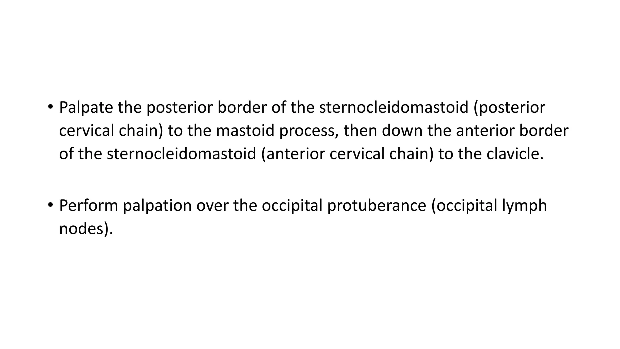

![If the finding is cervical nodes only, the head and neck need to be examined
thoroughly.
o The ears, nose, and throat must be examined with the auroscope, including
carefully inspecting the teeth and gums.
o If any teeth appear carious, then wear gloves to palpate them for tenderness.
o Look at the external aspects of the eyes, for conjunctivitis (which can occur
with Kawasaki disease, Parinaud's oculo-glandular syndrome [with
preauricular lymphadenopathy] or leptospirosis), and for Horner's syndrome,
which can occur with neuroblastoma.
o Inspect and palpate the scalp for infected areas (hiding under the hair, such
as tinea capitis or a kerion).](https://image.slidesharecdn.com/lymphadenopathy-210811152441/75/Lymphadenopathy-11-2048.jpg)


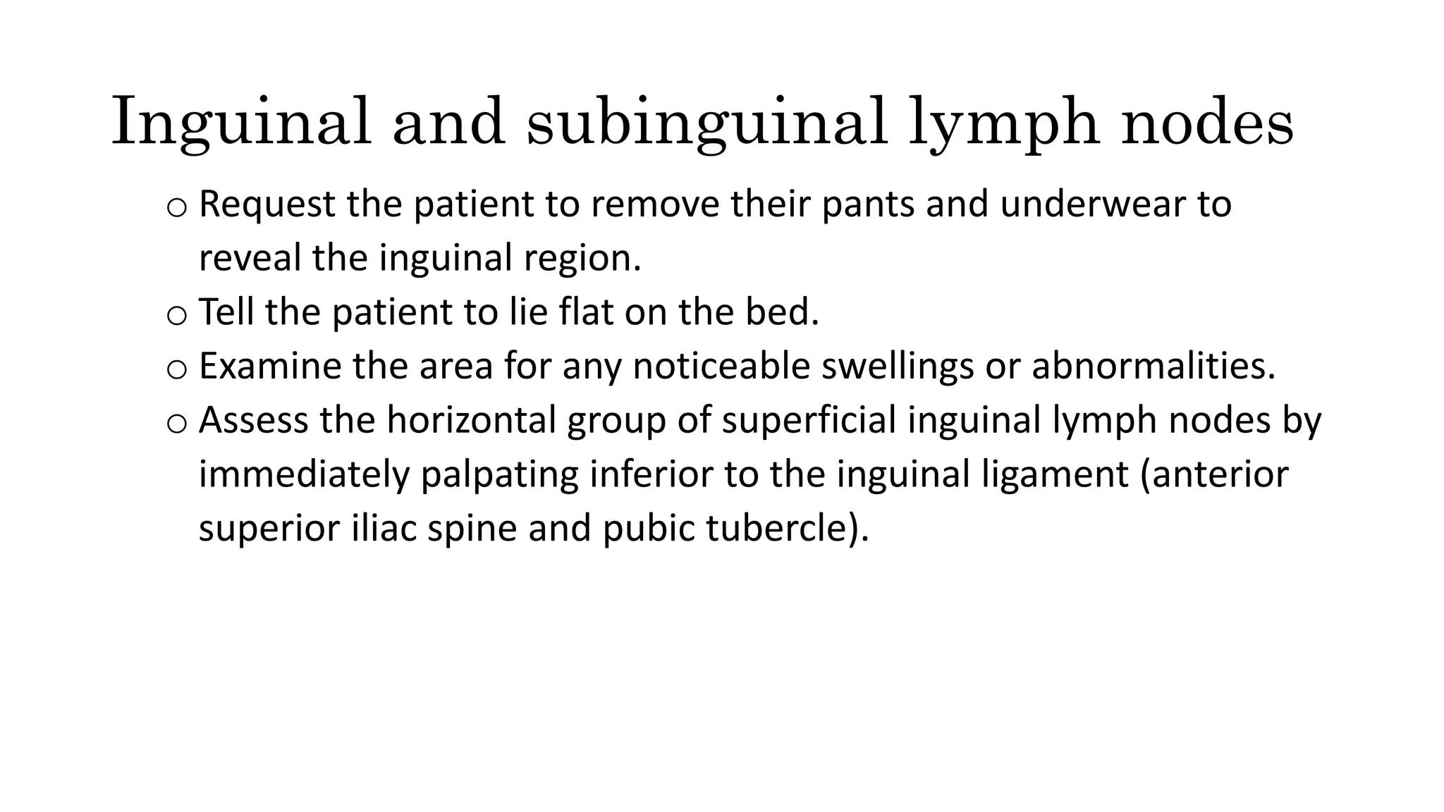

![Examine the skin for any local lesions (such as herpetic infections [HSV I
or II, HVZ], cat-scratch disease's papular lesions on hands/ fingers
[Bartonella henselae], any inflamed areas, reddened, cellulitic, or
purulent from staphylococcal or streptococcal infection; any
generalized rash [rubella classically causing suboccipital
lymphadenopathy; Kawasaki disease can have many types of rash; SLE
causes a malar rash]) or discoloration over the nodes themselves
(purplish discoloration classic for MAIS).](https://image.slidesharecdn.com/lymphadenopathy-210811152441/75/Lymphadenopathy-16-2048.jpg)
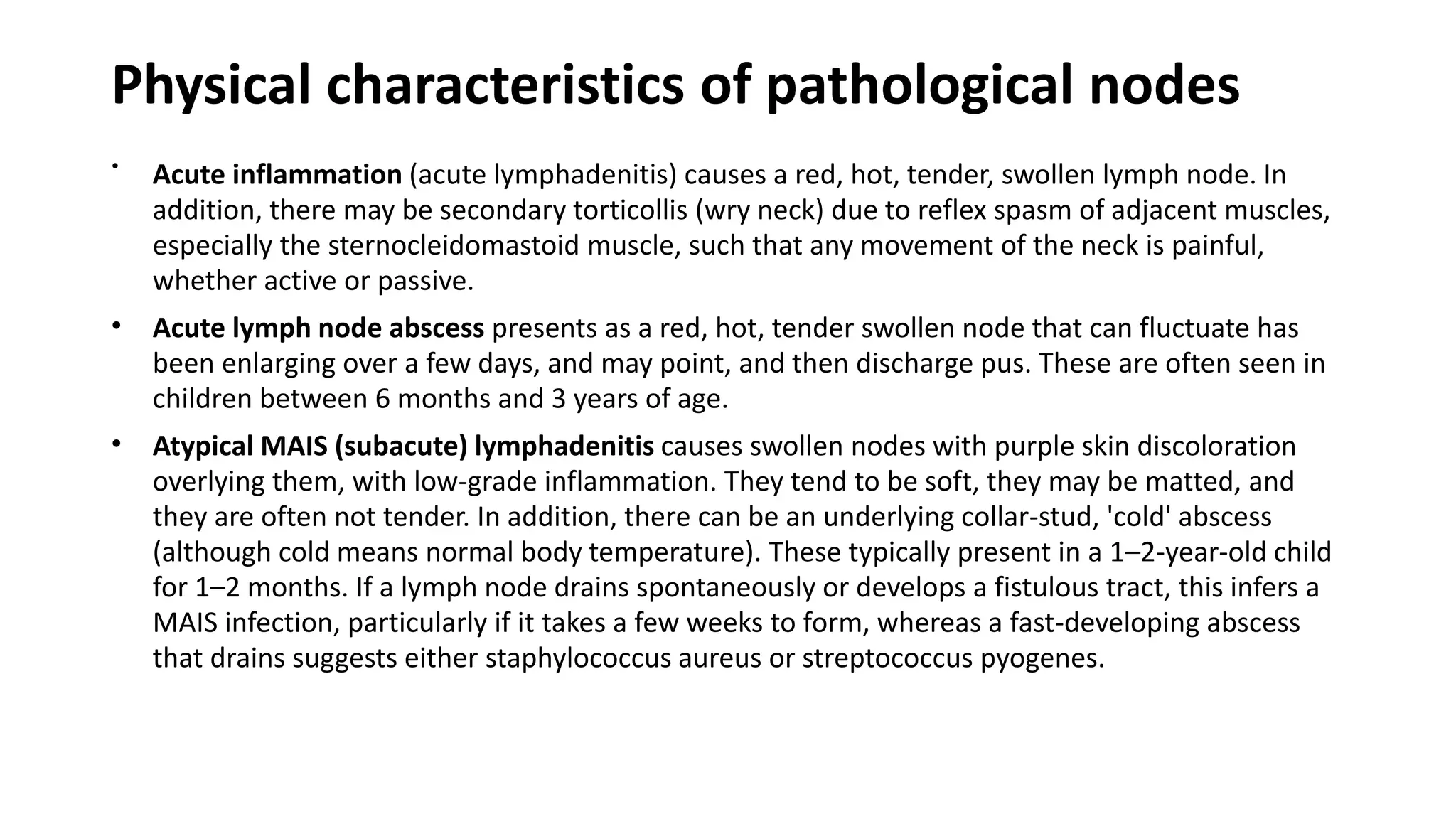
![• Examine the chest for any evidence of asthma (for underlying Churg–
Strauss syndrome, or diffuse pulmonary Langerhans cell
histiocytosis) or histoplasmosis (from inhaling fungal spores,
Histoplasma capsulatum).
• Examine the abdomen for hepatosplenomegaly (Malignancy
[neuroblastoma, lymphoma], ALL/AML, Toxoplasmosis, CMV,
Connective tissue disorders, HIV, EBV, Syphilis).](https://image.slidesharecdn.com/lymphadenopathy-210811152441/75/Lymphadenopathy-17-2048.jpg)

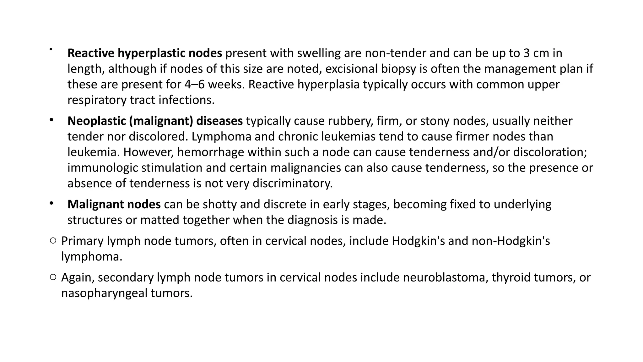
![A brief list of typical causes for typical locations of nodes
• Cervical:
1. oropharyngeal/scalp infection (viral [usual URTI pathogens, EBV, CMV, HSV, HHV-6],
streptococcus, staphylococcus, mycobacteria [TB, MAIS]).
2. cat-derived: cat-scratch disease; toxoplasmosis.
3. Kawasaki disease.
4. Dental caries (an infectious dental hard tissues, decalcification of inorganic parts of the
tooth, then a breakdown of the organic matrix, dietary carbohydrate-modified, saliva-
regulated).
• Supraclavicular:
1. (a) left side: intraabdominal malignancy; (b) right side: intra-mediastinal malignancy or
infection.
2. Lymphoma.
3. TB.
• Epitrochlear:
1. hand or arm infection.
2. Cat-scratch disease.
3. Lymphoma](https://image.slidesharecdn.com/lymphadenopathy-210811152441/75/Lymphadenopathy-19-2048.jpg)


The document outlines the examination and characteristics of lymphadenopathy in children, including normal lymph node sizes by region and indications for further investigation, such as size and specific lymph node locations. It describes the examination process step-by-step, emphasizing the need to check vital signs, assess growth parameters, and conduct thorough palpation of all relevant lymph node groups. Various causes of lymphadenopathy are also detailed, along with physical characteristics of pathological nodes, to help determine underlying health conditions.


