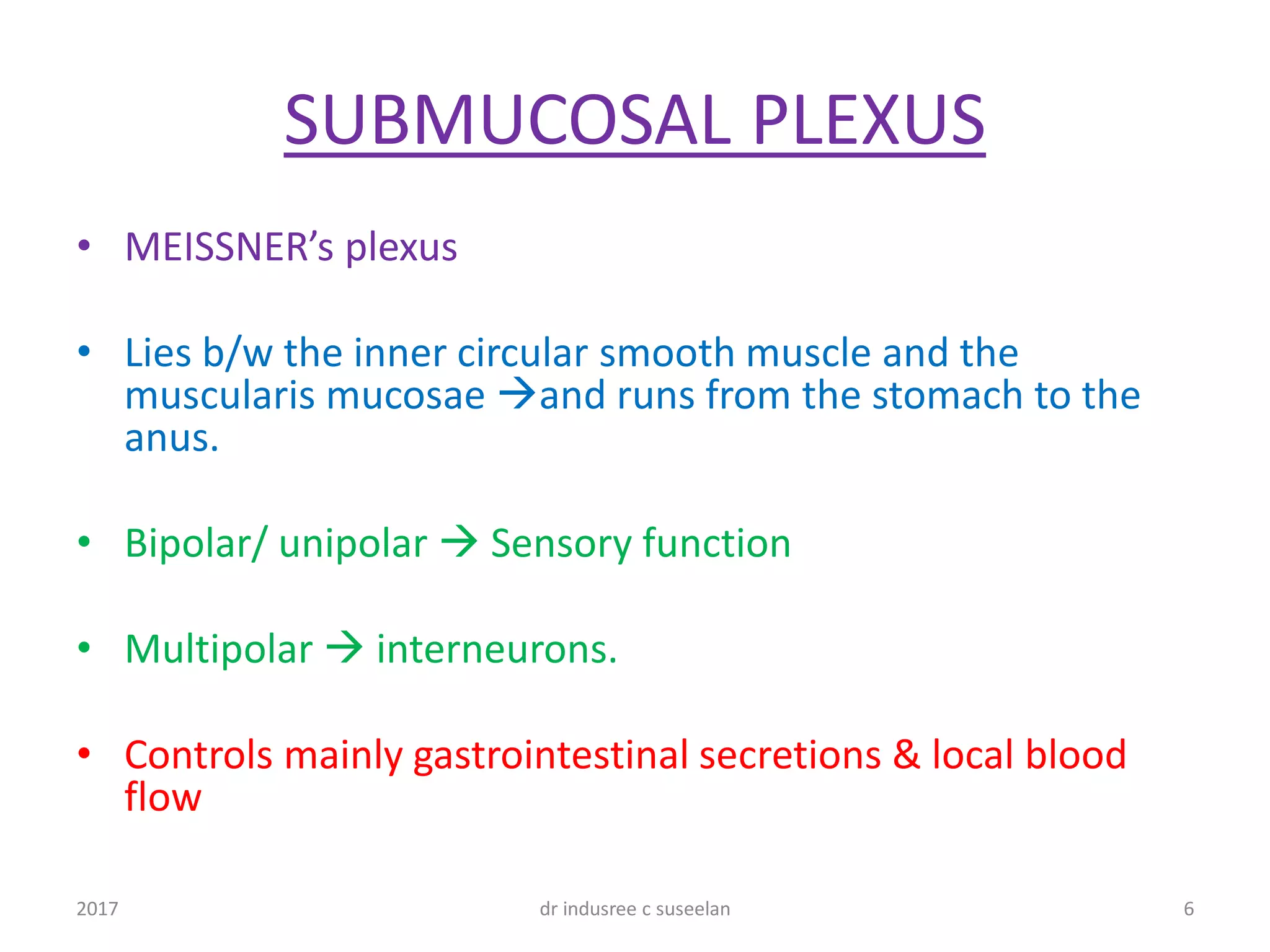
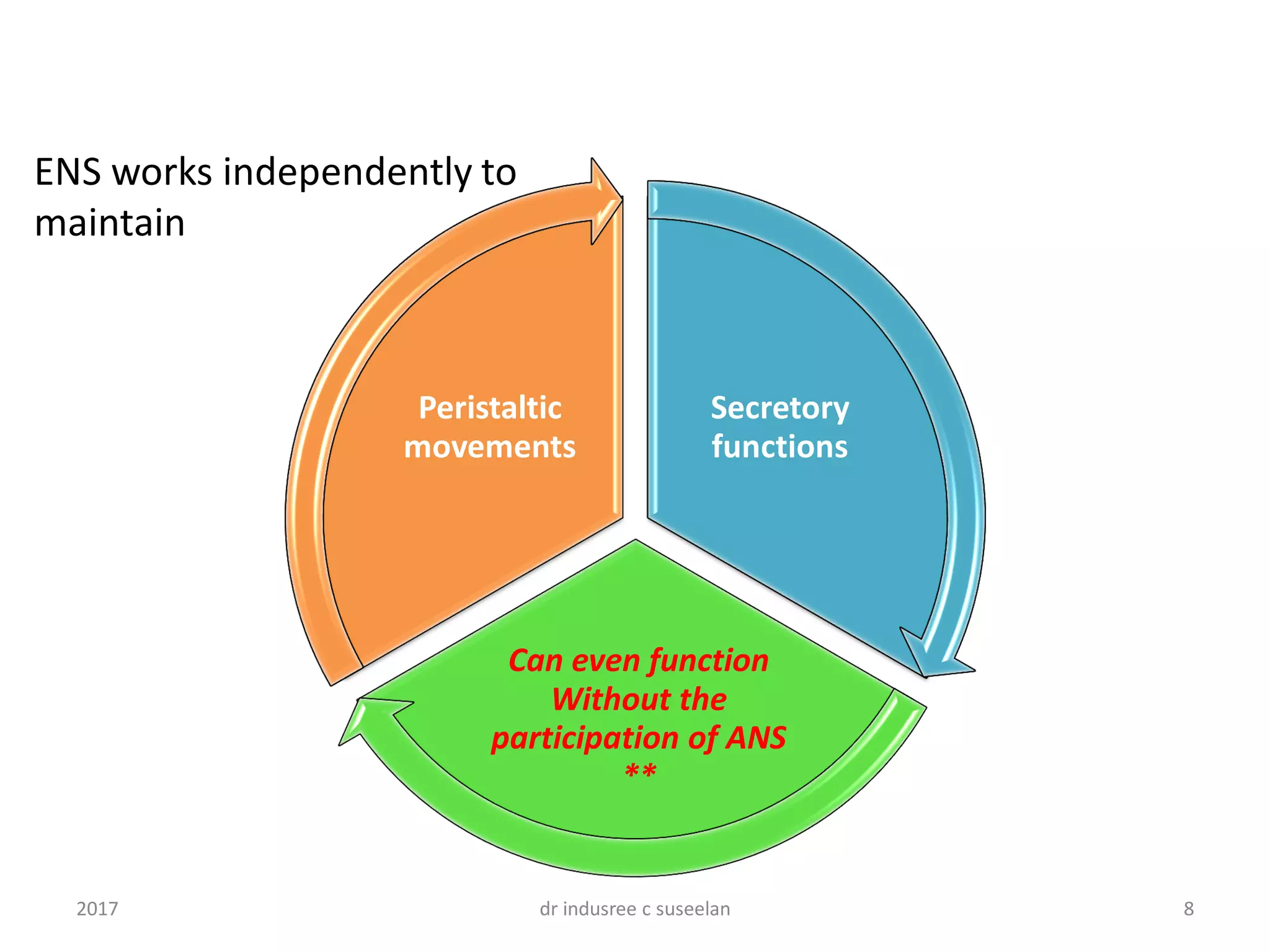
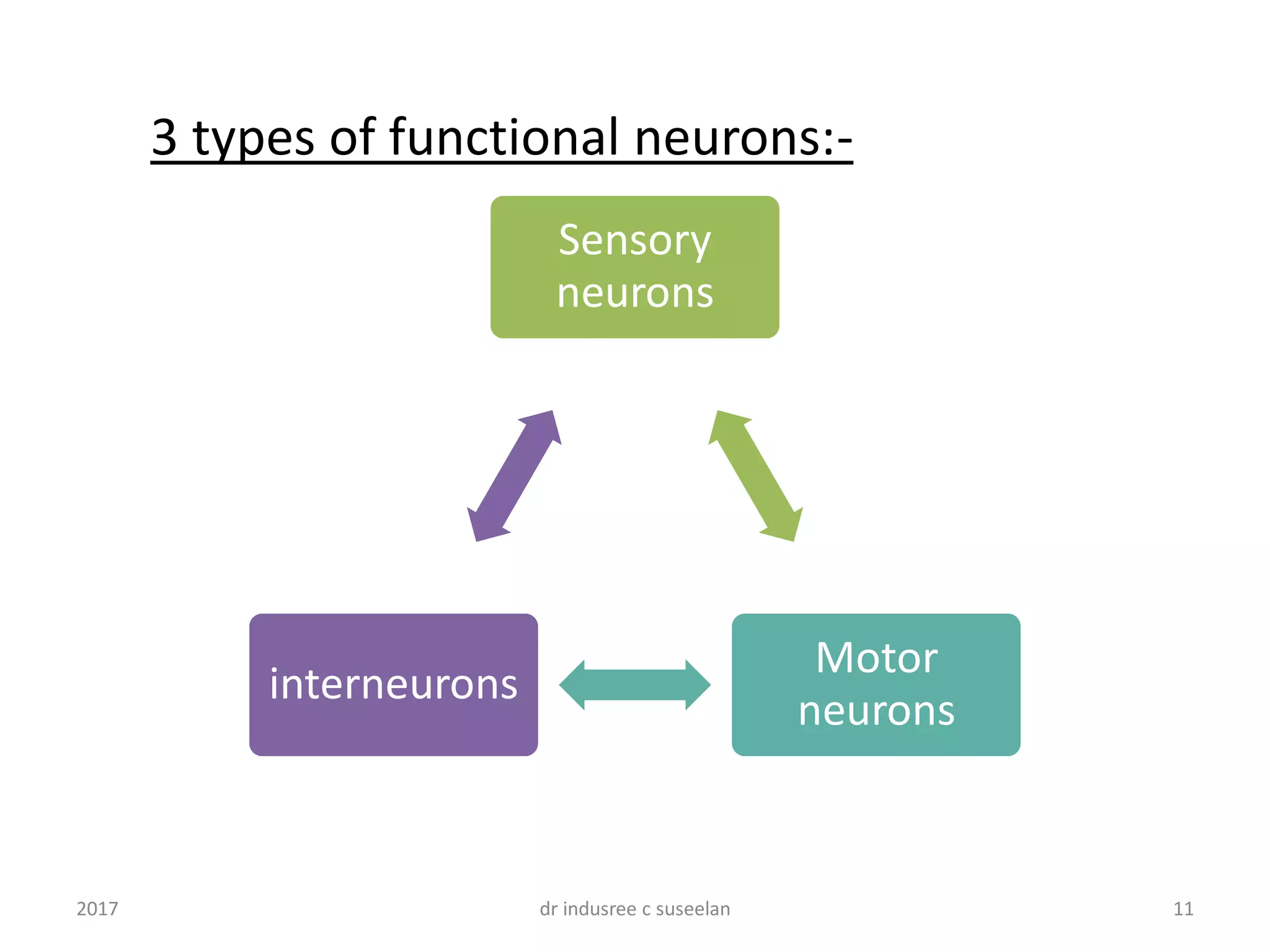
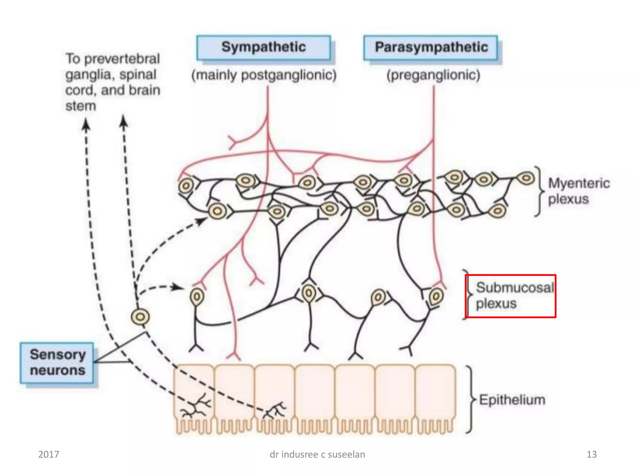
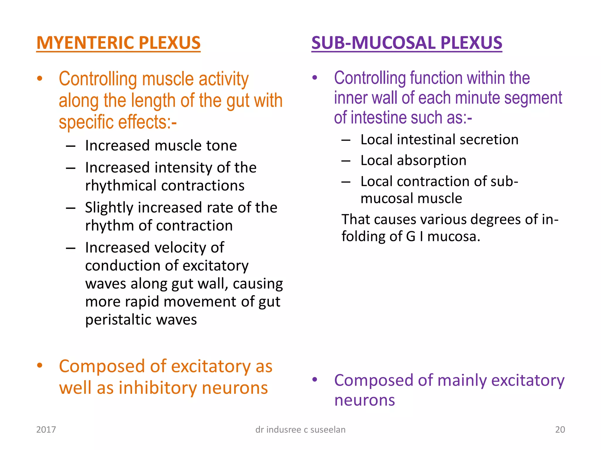
By
Dr. Indusree. C. Suseelan
3rd yr PG
Dept. of Shareera Rachana,
Sri Dharmastala Manjunatheswara college of Ayurveda,
udupi 12017 dr indusree c suseelan
2
Peripheral nervous system by J.N Langley
2017 dr indusree c suseelan
32017 dr indusree c suseelan
Enteric nervous system
• Confined within the wall of the GUT
• Extends from oral part of the Esophagus to
the Internal anal sphincter.
• 2 sets of intercommunicating nerve plexus
– MYENTERIC / AUERBACH plexus
– SUBMUCOUS / MESSINER plexus
Both plexus are composed of intricate
network of fine un-myelinated nerve fibers
intermingled with clusters of neuronal cell
bodies arranged in the form of ganglia
42017 dr indusree c suseelan
MYENTERIC PLEXUS
• AUERBACH’s plexus
• Located b/w the outer longitudinal and the inner
circular layers of smooth muscles extend from the
upper esophagus to the anus.
• Multipolar
• Motor function
• Controls mainly the gastrointestinal movements
52017 dr indusree c suseelan
SUBMUCOSAL PLEXUS
• MEISSNER’s plexus
• Lies b/w the inner circular smooth muscle and the
muscularis mucosae and runs from the stomach to the
anus.
• Bipolar/ unipolar  Sensory function
• Multipolar  interneurons.
• Controls mainly gastrointestinal secretions & local blood
flow
62017 dr indusree c suseelan
72017 dr indusree c suseelan
Secretory
functions
Can even function
Without the
participation of ANS
**
Peristaltic
movements
ENS works independently to
maintain
82017 dr indusree c suseelan
Equivalent to
total no: of
neurons in
spinal cord
Derived
from
specific
area of
neural crest
cells
10-100
million
neurons
92017 dr indusree c suseelan
Supported
by astrocyte
like glial
cells
BLOOD –
GANGLION
BARRIER
No
endoneurium
102017 dr indusree c suseelan
Sensory
neurons
Motor
neurons
interneurons
3 types of functional neurons:-
112017 dr indusree c suseelan
Sensory neurons
• Bipolar / unipolar neurons
• Confined in the Sub-mucous plexus
• Activated by- distension of gut wall- stretch receptors
• Receive chemical stimulation from mucosa- chemical
receptors
• Axons synapses with interneurons located close to the
circular muscle layer.
122017 dr indusree c suseelan
132017 dr indusree c suseelan
interneurons
• Their axons are projected longitudinally
up & down to a series of motor neurons
which are located mostly in the
myenteric plexus & some in submucosal
plexus
142017 dr indusree c suseelan
Motor neurons
EXCITATORY
*Acetylcholine
(Ach)
*Substance-P (SP)
INHIBITORY
*adenosine triposphate
(ATP)
*vasoactive intestinal
polypeptide(VIP)
*Nitric oxide (NO)
152017 dr indusree c suseelan
162017 dr indusree c suseelan
Motor neurons project fibers
o Smooth muscle fibers of outer longitudinal and inner circular
layers,
o muscularis mucosae,
o glands &
o blood vessels of the gut.
Some fibers projected outside the gut
o to the ANS for modulation of the neural output &
o to the mesenteric blood vessels for regulation of the blood
flow.
172017 dr indusree c suseelan
18
Gastro-intestinal reflex pathway
2017 dr indusree c suseelan
192017 dr indusree c suseelan
MYENTERIC PLEXUS
• Controlling muscle activity
along the length of the gut with
specific effects:-
– Increased muscle tone
– Increased intensity of the
rhythmical contractions
– Slightly increased rate of the
rhythm of contraction
– Increased velocity of
conduction of excitatory
waves along gut wall, causing
more rapid movement of gut
peristaltic waves
• Composed of excitatory as
well as inhibitory neurons
SUB-MUCOSAL PLEXUS
• Controlling function within the
inner wall of each minute segment
of intestine such as:-
– Local intestinal secretion
– Local absorption
– Local contraction of sub-
mucosal muscle
That causes various degrees of in-
folding of G I mucosa.
• Composed of mainly excitatory
neurons
202017 dr indusree c suseelan
Applied anatomy:-
• Injury to the ENS neurons or its pathway
may leaded to chances of slow recovery
due to absence of endoneurium
212017 dr indusree c suseelan
222017 dr indusree c suseelan
232017 dr indusree c suseelan
242017 dr indusree c suseelan
252017 dr indusree c suseelan
262017 dr indusree c suseelan
272017 dr indusree c suseelan

Enteric nervous system

  • 1.
    By Dr. Indusree. C.Suseelan 3rd yr PG Dept. of Shareera Rachana, Sri Dharmastala Manjunatheswara college of Ayurveda, udupi 12017 dr indusree c suseelan
  • 2.
    2 Peripheral nervous systemby J.N Langley 2017 dr indusree c suseelan
  • 3.
  • 4.
    Enteric nervous system •Confined within the wall of the GUT • Extends from oral part of the Esophagus to the Internal anal sphincter. • 2 sets of intercommunicating nerve plexus – MYENTERIC / AUERBACH plexus – SUBMUCOUS / MESSINER plexus Both plexus are composed of intricate network of fine un-myelinated nerve fibers intermingled with clusters of neuronal cell bodies arranged in the form of ganglia 42017 dr indusree c suseelan
  • 5.
    MYENTERIC PLEXUS • AUERBACH’splexus • Located b/w the outer longitudinal and the inner circular layers of smooth muscles extend from the upper esophagus to the anus. • Multipolar • Motor function • Controls mainly the gastrointestinal movements 52017 dr indusree c suseelan
  • 6.
    SUBMUCOSAL PLEXUS • MEISSNER’splexus • Lies b/w the inner circular smooth muscle and the muscularis mucosae and runs from the stomach to the anus. • Bipolar/ unipolar  Sensory function • Multipolar  interneurons. • Controls mainly gastrointestinal secretions & local blood flow 62017 dr indusree c suseelan
  • 7.
  • 8.
    Secretory functions Can even function Withoutthe participation of ANS ** Peristaltic movements ENS works independently to maintain 82017 dr indusree c suseelan
  • 9.
    Equivalent to total no:of neurons in spinal cord Derived from specific area of neural crest cells 10-100 million neurons 92017 dr indusree c suseelan
  • 10.
    Supported by astrocyte like glial cells BLOOD– GANGLION BARRIER No endoneurium 102017 dr indusree c suseelan
  • 11.
    Sensory neurons Motor neurons interneurons 3 types offunctional neurons:- 112017 dr indusree c suseelan
  • 12.
    Sensory neurons • Bipolar/ unipolar neurons • Confined in the Sub-mucous plexus • Activated by- distension of gut wall- stretch receptors • Receive chemical stimulation from mucosa- chemical receptors • Axons synapses with interneurons located close to the circular muscle layer. 122017 dr indusree c suseelan
  • 13.
  • 14.
    interneurons • Their axonsare projected longitudinally up & down to a series of motor neurons which are located mostly in the myenteric plexus & some in submucosal plexus 142017 dr indusree c suseelan
  • 15.
    Motor neurons EXCITATORY *Acetylcholine (Ach) *Substance-P (SP) INHIBITORY *adenosinetriposphate (ATP) *vasoactive intestinal polypeptide(VIP) *Nitric oxide (NO) 152017 dr indusree c suseelan
  • 16.
  • 17.
    Motor neurons projectfibers o Smooth muscle fibers of outer longitudinal and inner circular layers, o muscularis mucosae, o glands & o blood vessels of the gut. Some fibers projected outside the gut o to the ANS for modulation of the neural output & o to the mesenteric blood vessels for regulation of the blood flow. 172017 dr indusree c suseelan
  • 18.
  • 19.
  • 20.
    MYENTERIC PLEXUS • Controllingmuscle activity along the length of the gut with specific effects:- – Increased muscle tone – Increased intensity of the rhythmical contractions – Slightly increased rate of the rhythm of contraction – Increased velocity of conduction of excitatory waves along gut wall, causing more rapid movement of gut peristaltic waves • Composed of excitatory as well as inhibitory neurons SUB-MUCOSAL PLEXUS • Controlling function within the inner wall of each minute segment of intestine such as:- – Local intestinal secretion – Local absorption – Local contraction of sub- mucosal muscle That causes various degrees of in- folding of G I mucosa. • Composed of mainly excitatory neurons 202017 dr indusree c suseelan
  • 21.
    Applied anatomy:- • Injuryto the ENS neurons or its pathway may leaded to chances of slow recovery due to absence of endoneurium 212017 dr indusree c suseelan
  • 22.
  • 23.
  • 24.
  • 25.
  • 26.
  • 27.