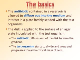
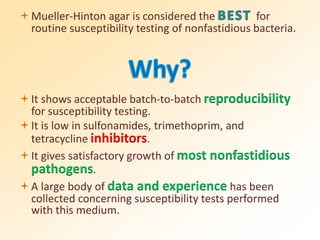
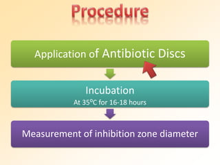
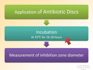
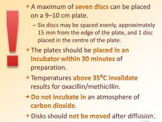
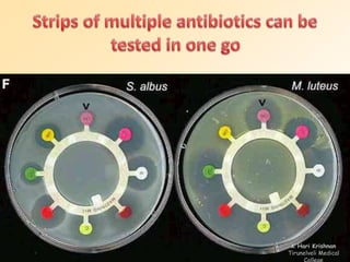
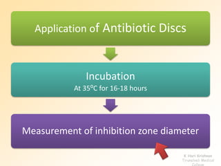
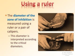
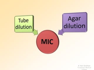
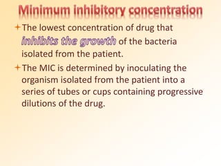
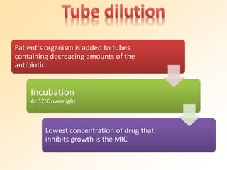
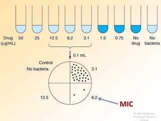
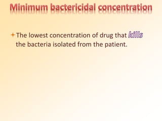
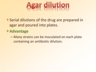
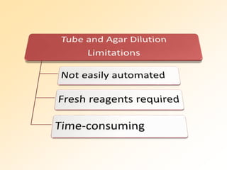
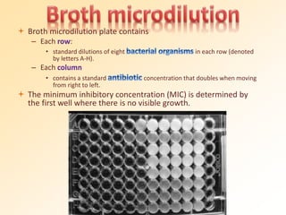
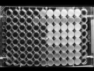
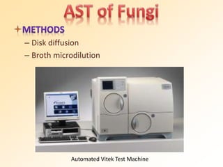
y
A test done to check the
effectiveness of a drug against a
bacterium and to select the best
drug that acts against the
bacterium.
The in vitro testing of bacterial cultures
with antibiotics to determine
.
Antibiotic
Sensitivity
Testing
l
To guide the clinician in selecting the best
antibiotic agent for an individual patient.
To control the use of
in clinical practice.
To accumulate epidemiological information on
the resistance of microorganisms of public
health importance within the community.
To reveal the changing trends in the
local isolates.
Bacteria have the ability to develop
resistance following repeated or subclinical
(insufficient) doses, so more advanced
antibiotics and synthetic antibiotics are
continually required to overcome them.
AST is essential for the selection of the
Tt
– For the testing of isolates from “healthy” patients with
intact immune defenses.
– For such as uncomplicated
urinary tract infections.
– In the treatment of serious infections such as
endocarditis or osteomyelitis.
– For infections in high-risk patient groups such as
immunocompromised patients (e.g.. transplant
patients).
– Those who are critically ill.
College
Antibiotic Sensitivity Tests
Diffusion
Kirby-Bauer
Method
Stokes
Method
Dilution
Tube
Dilution
Agar
Dilution
Diffusion &
Dilution
E-Test
Qualitative Methods Quantitative Methods
Staphylococcus
Enterobacteriaceae
Pseudomonas
aeruginosa
Intestinal Urinary
Blood &
Tissues
Drugs
Benzylpenicillin
Oxacillin
Erythromycin
Tetracycline
Chloramphenicol
Ampicillin
Chloramphenicol
Cotrimoxazole
Tetracycline
Sulfonamides
Trimethoprim
Cotrimoxazole
Ampicillin
Nitrofurantoin
Tetracycline
Ampicillin
Chloramphenicol
Cotrimoxazole
Tetracycline
Cefalotin
Gentamycin
Piperacillin
Gentamycin
Tobramycin
– A paper disk with a defined amount of antibiotic is
used to generate a dynamically changing gradient
of antibiotic concentrations in the agar in the
vicinity of the disk.
The contained in a reservoir is
allowed to and
interact in a plate freshly seeded with the test
organisms.
The disk is applied to the surface of an agar
plate inoculated with the test organism.
– The diffuses out of the disk to form the
gradient.
– The starts to divide and grow and
progresses toward a critical mass of cells.
is formed at the critical
time where a particular concentration of the
antibiotic is just able to inhibit the organism
before it reaches an overwhelming cell mass
or critical mass.
Medium containing beef
infusion, peptone, and
starch.
Used primarily for the disk-
diffusion method.
Robust red algae (Solieria robusta)
Source of Agar
Mueller-Hinton agar is considered the for
routine susceptibility testing of nonfastidious bacteria.
It shows acceptable batch-to-batch reproducibility
for susceptibility testing.
It is low in sulfonamides, trimethoprim, and
tetracycline inhibitors.
It gives satisfactory growth of most nonfastidious
pathogens.
A large body of data and experience has been
collected concerning susceptibility tests performed
with this medium.
to 45–50 ⁰C and pour into
the plates. Allow to , to a
depth of approximately 4 mm.
– A 9-cm plate requires approximately 25 ml of
medium.
When the agar has solidified,
for 10–30 minutes at 35 ⁰C by placing them in
the upright position in the incubator with the
lids tilted.
– If it is not to be used immediately, the agar
medium can be stored in a refrigerator (2 to 8C)
for 2 weeks.
Any commercially available discs with the
proper diameter and potency can be used.
Stocks of antibiotic discs can be stored at
-20 ⁰C for 1 month.
– On removal from the refrigerator, the containers
should be left at room temperature for about 1
hour to allow the temperature to equilibrate.
Prepared by pouring 0.6 ml of
a 1% (10 g/l) solution of
into a 100-ml graduated
cylinder, and filling to 100ml
with 1% (10 ml/l)
.
A supply of cotton wool swabs on wooden
applicator sticks should be prepared.
They can be sterilized in tins, culture tubes, or
on paper, either in the autoclave or by dry
heat.
Application of Antibiotic Discs
Incubation
At 35⁰C for 16-18 hours
Measurement of inhibition zone diameter
To prepare the inoculum from the primary
culture plate,
, of similar appearance,
of the organism to be tested.
Transfer this growth to a tube of saline.
Compare the
tube with the
turbidity standard
and
of the test
suspension to that
of the standard by
adding more
bacteria or more
sterile saline.
Inoculate the plates by
into the inoculum.
Remove excess inoculum by pressing and rotating
the swab firmly against the side of the tube above
the level of the liquid.
the swab all over the surface of the
medium three times, rotating the plate
through an angle of 60⁰ after each application.
Finally, pass the swab round the edge of the
agar surface.
Leave the inoculum to for a few
minutes at room temperature with the lid
closed.
The may be placed on the
inoculated plates using
– sterile forceps.
– a template.
– a sterile needle tip.
– antibiotic disc dispenser.
K Hari Krishnan
Tirunelveli Medical
College
Application of Antibiotic Discs
Incubation
At 35⁰C for 16-18 hours
Measurement of inhibition zone diameter
K Hari Krishnan
Tirunelveli Medical
College
A maximum of can be placed
on a 9–10 cm plate.
– Six discs may be spaced evenly, approximately
15 mm from the edge of the plate, and 1 disc
placed in the centre of the plate.
The plates should be
of
preparation.
Temperatures
results for oxacillin/methicillin.
 an atmosphere of
.
Disks should after diffusion.
K Hari Krishnan
Tirunelveli Medical
College
K Hari Krishnan
Tirunelveli Medical
College
Application of Antibiotic Discs
Incubation
At 35⁰C for 16-18 hours
Measurement of inhibition zone diameter
K Hari Krishnan
Tirunelveli Medical
College
Using a ruler
– on the under-surface
of the plate containing
transparent medium.
Using a pair of
calipers
– on the plate
containing opaque
medium.
K Hari Krishnan
Tirunelveli Medical
College
Using automated zone readers
– BIOMIC
– Aura
– Protozone
K Hari Krishnan
Tirunelveli Medical
College
Standard templates are available for each
antibiotic.
K Hari Krishnan
Tirunelveli Medical
College
Result interpretation
• When the edge of the zone of inhibition is the
black circle.
• When there is no zone, or when it lies the
white circle.
• When the edge of the zone of inhibition lies the
black circle.
The diameter of the
zone of inhibition is
measured using a
ruler or a pair of
calipers.
– This diameter is
interpreted according
to the critical
diameters.
K Hari Krishnan
Tirunelveli Medical
College
Interpretative chart of zone sizes
Antibiotic
Diameter of zone inhibition (mm)
Resistant Intermediate Susceptible
Tetracycline <14 15-18 >19
Chloramphenicol <12 13-17 >18
Cotrimoxazole <10 11-15 ≥16
Nitrofurantoin <14 15-16 >17
Erythromycin <13 14-22 >23
Gentamycin <12 13-14 >15
– An organism is called susceptible to an antibiotic when the
infection caused by it is likely to respond to treatment
with this antibiotic, at the recommended dosage.
– An organism is called resistant if it is expected not to
respond to a given antibiotic, irrespective of the
dosage and of the location of the infection.
– Strains that are “moderately susceptible” to an antibiotic
that can be used for treatment at a higher dosage
(e.g. b-lactams) because of its low toxicity.
– Strains that show “intermediate susceptibility” to a more
toxic antibiotic (e.g. aminoglycoside) that cannot be used
at a higher dosage. K Hari Krishnan
Tirunelveli Medical
College
– Too inoculum
• Inhibition zones will be larger even though the
sensitivity of the organism is unchanged
Relatively resistant strains may be falsely reported as
susceptible.
– Too inoculum
• Inhibition zones will be smaller
Relatively susceptible strains may then be falsely
reported as resistant.
K Hari Krishnan
Tirunelveli Medical
College
• of disk application
of incubation
– If the temperature is , the time
required for effective growth is extended and
result.
Potency of
– If the owing to
deterioration during storage, the
.
Standardised inoculum is replaced by the
pathological specimen itself, e.g. urine, a
positive blood culture, or a swab of pus.
Advantage
– Results are obtained 24 hours earlier.
Disadvantage
– Density of the inoculum cannot be properly
controlled.
• The results of the primary test should be verified by
testing the isolates subsequently.
Used to determine the
of antibiotic to inhibit or kill the
microorganism.
Achieved by dilution of antibiotic in either
agar or broth media.
K Hari Krishnan
Tirunelveli Medical
College
The lowest concentration of drug that
of the bacteria
isolated from the patient.
The MIC is determined by inoculating the
organism isolated from the patient into a
series of tubes or cups containing progressive
dilutions of the drug.
Patient's organism is added to tubes
containing decreasing amounts of the
antibiotic
Incubation
At 37°C overnight
Lowest concentration of drug that
inhibits growth is the MIC
MIC
K Hari Krishnan
Tirunelveli Medical
College
The lowest concentration of drug that
the bacteria isolated from the patient.
Serial dilutions of the drug are prepared in
agar and poured into plates.
– Many strains can be inoculated on each plate
containing an antibiotic dilution.
 Broth microdilution plate contains
– Each row:
• standard dilutions of eight in each row (denoted
by letters A-H).
– Each column
• contains a standard concentration that doubles when moving
from right to left.
 The minimum inhibitory concentration (MIC) is determined by
the first well where there is no visible growth.
Epsilometer Test
Quantitative method of antibiotic sensitivity
testing.
Applies both dilution of antibiotic and
diffusion of antibiotic into the medium.
Combines the principles of disk diffusion and
agar dilution methods
Diffusion
Dilution
E-Test
A predefined stable antibiotic gradient is
present on a thin inert carrier strip.
Using innovative dry chemistry technology,
E-Test is used to determine the on-scale
Minimum Inhibitory Concentration (MIC).
The intersection of the inhibitory zone edge and the
calibrated carrier strip indicates the MIC with inherent
precision and accuracy.
MIC
Over 100 are now available in the
product range for testing of aerobic bacteria
and fastidious organisms such as
– Pneumococci
– Haemophilus
– Helicobacter pylori
– Meningococci
– Gonococci
– Fungi
– Mycobacteria
Determining the MIC of
, or for
a specific type of patient or infection.
Detecting
– Glycopeptide-resistant Enterococci (GRE)
– Glycopeptide-intermediate S. aureus (GISA)
– Resistant Mycobacterium tuberculosis
– Extended spectrum beta lactamases (ESBL)
Detecting .
Testing an antibiotic not performed in routine use
or a new, antibiotic agent.
 an equivocal AST result.
K Hari Krishnan
Tirunelveli Medical
College
Most fastidious organisms do not grow well
enough in routine antibiotic testing systems
and require some type of supplementation.
Pathogen Medium
Streptococcus pneumoniae Mueller-Hinton sheep blood agar
Haemophilus sp. Haemophilus Test Medium (Mueller-
Hinton Agar, β NAD, bovine hematin,
yeast extract)
Neisseria gonorrheae Thayer-Martin agar
Antibiotic resistance among many clinically important
species of anaerobes has increased, which has made
empiric therapy choices unpredictable.
– E.g.. Metronidazole resistance in Propionibacterium and
Bacteroides
– Agar dilution
– Broth microdilution
– Brucella agar
(or)
– Broth supplemented with vitamin K and hemin
– Disk diffusion
– Broth microdilution
Automated Vitek Test Machine
Antibiotic
Sensitivity Testing
antibioticsensitivitytesting.pdf

antibioticsensitivitytesting.pdf

  • 1.
  • 2.
    A test doneto check the effectiveness of a drug against a bacterium and to select the best drug that acts against the bacterium.
  • 3.
    The in vitrotesting of bacterial cultures with antibiotics to determine .
  • 4.
  • 5.
    To guide theclinician in selecting the best antibiotic agent for an individual patient. To control the use of in clinical practice. To accumulate epidemiological information on the resistance of microorganisms of public health importance within the community. To reveal the changing trends in the local isolates.
  • 6.
    Bacteria have theability to develop resistance following repeated or subclinical (insufficient) doses, so more advanced antibiotics and synthetic antibiotics are continually required to overcome them.
  • 7.
    AST is essentialfor the selection of the Tt
  • 8.
    – For thetesting of isolates from “healthy” patients with intact immune defenses. – For such as uncomplicated urinary tract infections. – In the treatment of serious infections such as endocarditis or osteomyelitis. – For infections in high-risk patient groups such as immunocompromised patients (e.g.. transplant patients). – Those who are critically ill. College
  • 9.
  • 10.
  • 12.
    – A paperdisk with a defined amount of antibiotic is used to generate a dynamically changing gradient of antibiotic concentrations in the agar in the vicinity of the disk.
  • 13.
    The contained ina reservoir is allowed to and interact in a plate freshly seeded with the test organisms. The disk is applied to the surface of an agar plate inoculated with the test organism. – The diffuses out of the disk to form the gradient. – The starts to divide and grow and progresses toward a critical mass of cells.
  • 14.
    is formed atthe critical time where a particular concentration of the antibiotic is just able to inhibit the organism before it reaches an overwhelming cell mass or critical mass.
  • 17.
    Medium containing beef infusion,peptone, and starch. Used primarily for the disk- diffusion method. Robust red algae (Solieria robusta) Source of Agar
  • 18.
    Mueller-Hinton agar isconsidered the for routine susceptibility testing of nonfastidious bacteria. It shows acceptable batch-to-batch reproducibility for susceptibility testing. It is low in sulfonamides, trimethoprim, and tetracycline inhibitors. It gives satisfactory growth of most nonfastidious pathogens. A large body of data and experience has been collected concerning susceptibility tests performed with this medium.
  • 19.
    to 45–50 ⁰Cand pour into the plates. Allow to , to a depth of approximately 4 mm. – A 9-cm plate requires approximately 25 ml of medium. When the agar has solidified, for 10–30 minutes at 35 ⁰C by placing them in the upright position in the incubator with the lids tilted. – If it is not to be used immediately, the agar medium can be stored in a refrigerator (2 to 8C) for 2 weeks.
  • 20.
    Any commercially availablediscs with the proper diameter and potency can be used. Stocks of antibiotic discs can be stored at -20 ⁰C for 1 month. – On removal from the refrigerator, the containers should be left at room temperature for about 1 hour to allow the temperature to equilibrate.
  • 22.
    Prepared by pouring0.6 ml of a 1% (10 g/l) solution of into a 100-ml graduated cylinder, and filling to 100ml with 1% (10 ml/l) .
  • 23.
    A supply ofcotton wool swabs on wooden applicator sticks should be prepared. They can be sterilized in tins, culture tubes, or on paper, either in the autoclave or by dry heat.
  • 25.
    Application of AntibioticDiscs Incubation At 35⁰C for 16-18 hours Measurement of inhibition zone diameter
  • 26.
    To prepare theinoculum from the primary culture plate, , of similar appearance, of the organism to be tested.
  • 27.
    Transfer this growthto a tube of saline.
  • 28.
    Compare the tube withthe turbidity standard and of the test suspension to that of the standard by adding more bacteria or more sterile saline.
  • 29.
    Inoculate the platesby into the inoculum. Remove excess inoculum by pressing and rotating the swab firmly against the side of the tube above the level of the liquid.
  • 30.
    the swab allover the surface of the medium three times, rotating the plate through an angle of 60⁰ after each application. Finally, pass the swab round the edge of the agar surface.
  • 31.
    Leave the inoculumto for a few minutes at room temperature with the lid closed.
  • 32.
    The may beplaced on the inoculated plates using – sterile forceps. – a template. – a sterile needle tip. – antibiotic disc dispenser. K Hari Krishnan Tirunelveli Medical College
  • 33.
    Application of AntibioticDiscs Incubation At 35⁰C for 16-18 hours Measurement of inhibition zone diameter K Hari Krishnan Tirunelveli Medical College
  • 34.
    A maximum ofcan be placed on a 9–10 cm plate. – Six discs may be spaced evenly, approximately 15 mm from the edge of the plate, and 1 disc placed in the centre of the plate. The plates should be of preparation. Temperatures results for oxacillin/methicillin.  an atmosphere of . Disks should after diffusion. K Hari Krishnan Tirunelveli Medical College
  • 35.
  • 36.
    Application of AntibioticDiscs Incubation At 35⁰C for 16-18 hours Measurement of inhibition zone diameter K Hari Krishnan Tirunelveli Medical College
  • 37.
    Using a ruler –on the under-surface of the plate containing transparent medium. Using a pair of calipers – on the plate containing opaque medium. K Hari Krishnan Tirunelveli Medical College
  • 38.
    Using automated zonereaders – BIOMIC – Aura – Protozone K Hari Krishnan Tirunelveli Medical College
  • 40.
    Standard templates areavailable for each antibiotic. K Hari Krishnan Tirunelveli Medical College
  • 41.
    Result interpretation • Whenthe edge of the zone of inhibition is the black circle. • When there is no zone, or when it lies the white circle. • When the edge of the zone of inhibition lies the black circle.
  • 42.
    The diameter ofthe zone of inhibition is measured using a ruler or a pair of calipers. – This diameter is interpreted according to the critical diameters. K Hari Krishnan Tirunelveli Medical College
  • 43.
    Interpretative chart ofzone sizes Antibiotic Diameter of zone inhibition (mm) Resistant Intermediate Susceptible Tetracycline <14 15-18 >19 Chloramphenicol <12 13-17 >18 Cotrimoxazole <10 11-15 ≥16 Nitrofurantoin <14 15-16 >17 Erythromycin <13 14-22 >23 Gentamycin <12 13-14 >15
  • 44.
    – An organismis called susceptible to an antibiotic when the infection caused by it is likely to respond to treatment with this antibiotic, at the recommended dosage. – An organism is called resistant if it is expected not to respond to a given antibiotic, irrespective of the dosage and of the location of the infection. – Strains that are “moderately susceptible” to an antibiotic that can be used for treatment at a higher dosage (e.g. b-lactams) because of its low toxicity. – Strains that show “intermediate susceptibility” to a more toxic antibiotic (e.g. aminoglycoside) that cannot be used at a higher dosage. K Hari Krishnan Tirunelveli Medical College
  • 46.
    – Too inoculum •Inhibition zones will be larger even though the sensitivity of the organism is unchanged Relatively resistant strains may be falsely reported as susceptible. – Too inoculum • Inhibition zones will be smaller Relatively susceptible strains may then be falsely reported as resistant. K Hari Krishnan Tirunelveli Medical College
  • 47.
    • of diskapplication
  • 48.
    of incubation – Ifthe temperature is , the time required for effective growth is extended and result. Potency of – If the owing to deterioration during storage, the .
  • 49.
    Standardised inoculum isreplaced by the pathological specimen itself, e.g. urine, a positive blood culture, or a swab of pus. Advantage – Results are obtained 24 hours earlier. Disadvantage – Density of the inoculum cannot be properly controlled. • The results of the primary test should be verified by testing the isolates subsequently.
  • 51.
    Used to determinethe of antibiotic to inhibit or kill the microorganism. Achieved by dilution of antibiotic in either agar or broth media.
  • 52.
  • 53.
    The lowest concentrationof drug that of the bacteria isolated from the patient. The MIC is determined by inoculating the organism isolated from the patient into a series of tubes or cups containing progressive dilutions of the drug.
  • 54.
    Patient's organism isadded to tubes containing decreasing amounts of the antibiotic Incubation At 37°C overnight Lowest concentration of drug that inhibits growth is the MIC
  • 55.
  • 56.
    The lowest concentrationof drug that the bacteria isolated from the patient.
  • 57.
    Serial dilutions ofthe drug are prepared in agar and poured into plates. – Many strains can be inoculated on each plate containing an antibiotic dilution.
  • 59.
     Broth microdilutionplate contains – Each row: • standard dilutions of eight in each row (denoted by letters A-H). – Each column • contains a standard concentration that doubles when moving from right to left.  The minimum inhibitory concentration (MIC) is determined by the first well where there is no visible growth.
  • 62.
    Epsilometer Test Quantitative methodof antibiotic sensitivity testing. Applies both dilution of antibiotic and diffusion of antibiotic into the medium.
  • 63.
    Combines the principlesof disk diffusion and agar dilution methods Diffusion Dilution E-Test
  • 64.
    A predefined stableantibiotic gradient is present on a thin inert carrier strip. Using innovative dry chemistry technology, E-Test is used to determine the on-scale Minimum Inhibitory Concentration (MIC).
  • 65.
    The intersection ofthe inhibitory zone edge and the calibrated carrier strip indicates the MIC with inherent precision and accuracy.
  • 66.
  • 68.
    Over 100 arenow available in the product range for testing of aerobic bacteria and fastidious organisms such as – Pneumococci – Haemophilus – Helicobacter pylori – Meningococci – Gonococci – Fungi – Mycobacteria
  • 69.
    Determining the MICof , or for a specific type of patient or infection. Detecting – Glycopeptide-resistant Enterococci (GRE) – Glycopeptide-intermediate S. aureus (GISA) – Resistant Mycobacterium tuberculosis – Extended spectrum beta lactamases (ESBL) Detecting . Testing an antibiotic not performed in routine use or a new, antibiotic agent.  an equivocal AST result. K Hari Krishnan Tirunelveli Medical College
  • 71.
    Most fastidious organismsdo not grow well enough in routine antibiotic testing systems and require some type of supplementation. Pathogen Medium Streptococcus pneumoniae Mueller-Hinton sheep blood agar Haemophilus sp. Haemophilus Test Medium (Mueller- Hinton Agar, β NAD, bovine hematin, yeast extract) Neisseria gonorrheae Thayer-Martin agar
  • 72.
    Antibiotic resistance amongmany clinically important species of anaerobes has increased, which has made empiric therapy choices unpredictable. – E.g.. Metronidazole resistance in Propionibacterium and Bacteroides – Agar dilution – Broth microdilution – Brucella agar (or) – Broth supplemented with vitamin K and hemin
  • 73.
    – Disk diffusion –Broth microdilution Automated Vitek Test Machine
  • 76.