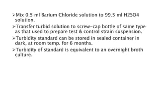
DR. REENA KULSHRESTHA
AST is
This misleads physician and patient to
receive necessary therapy.
 Such therapy leads to side-effects &
resistance of pathogens.
AST measures only in vitro not in vivo drug activity.
Selection of best drug depends on:
a) Patients clinical condition.
b) Type and site of infection.
c) History of drug hypersensitivity.
d) Drug activity : absorption, diffusion in tissues,
metabolism, excretion, toxicity, effect on patient
normal flora are not known by sensitivity testing.
SENSITIVITY TESTING AGAR:
 Best is Mueller - Hinton Agar / Broth.
 Depth of medium is 4mm (25 ml).
 Pour plates on a level surface using Pour plate method.
 Too thin & too thick media gives false inhibition zones.
 Plates can be stored in plastic bags at 2 - 8°C for up to 2
weeks.
• Used in routine sensitivity testing.
• A disc of filter paper is impregnated with a known volume & concentration of a drug
& placed on an agar medium inoculated with a test organism.
 -
• Control organisms are inoculated on a separate plate.
• Disc is placed so that Drug diffuses into medium.
• After an overnight incubation, culture is examined for areas of no growth (inhibition
zones) around discs.
• Sensitive bacteria are inhibited at a distance from disc.
• Resistant bacteria grow up to the edge of disc.
 :
• A disc of filter paper is impregnated with a known volume
& concentration of a drug & placed on an agar medium
inoculated with a test organism.
• Both test and control organisms are inoculated on same
plate
• Disc is placed so that Drug diffuses into medium.
• Inhibition zone is compared directly with that of control.
• Inoculum gives semi-confluent growth.
Filter paper discs impregnated with
antibiotics
• Filter paper discs
6mm in diameter are
charged with
required
concentration of
drugs and stored dry
in the cold.
Antifungal
Disc
preparation
• Using a sterile a 4mm loop, apply the organism suspension
to center of sensitivity plate.
• Using a sterile swab, spread inoculum across the center
third of the plate.
• Control / Test
• Do not use same swab for both application and spreading.
• For Inoculum - Judge your turbidity with turbidity standard,
not by naked eye.
Similarly, inoculate the broth culture of control
strain across the upper and lower thirds of plate,
leaving 5 mm. on each side of test organism.
 Control suspension must be standardized against
the standard.
 Allow the inocula to dry for few minutes, with the
Petri dish closed.
 Place the antibiotic discs (after warming to room temp.)
between test and control inocula.
 Press disc down a little and do not move once it is
once it is placed.
 After ½ hr. incubate at 37°C .
(For methicillin, incubate at 35°C)
 Read test when : Growth of both test and
control strains is not too heavy or too light.
 Control inhibition zones measure 8-15 mm
radius.
 Measurement is from edge of disc to edge
of zone.
 If growth of test & control strains is not
semi confluent, sensitivity test is repeated.
 :
It is a standard of Barium Chloride for matching
turbidity of test & control strains inoculum.
 Add 1 ml of H2SO4 to 99 ml water to make 1%
H2SO4.
 Dissolve 2.35g Barium Chloride in 200 ml water .
Mix 0.5 ml Barium Chloride solution to 99.5 ml H2SO4
solution.
Transfer turbid solution to screw-cap bottle of same type
as that used to prepare test & control strain suspension.
Turbidity standard can be stored in sealed container in
dark, at room temp. for 6 months.
Turbidity of standard is equivalent to an overnight broth
culture.
To select drugs for sensitivity, consult clinicians.
 Drug list must be limited & reviewed at regular
intervals.*
 If resistance developed, one member of each
drug group is selected.
:
 Control strains.
 Select according to Site of infection in
patient.
 Drug concentration at this site.
 Strain must respond to treatment with
normal doses .
 Strain must grow at same rate as test
organism.
S. aureus – Oxford strain - NCTC 6571
• Used for all except polymyxins & for pathogens of
all specimens except urine.
• E.coli – NCTC 10418 - is used for all drugs against
pathogens from urine.
• P. aeruginosa – NCTC 13921- is used for
controlling all drugs against Pseudomonas.
:
 Strain is cultivated on NA slopes.
 Strains are stored in dark at room temp.(20-
28°C).
 Subculture is made every 3 – 6 months.
 Each week, a nutrient broth or agar culture is
made & stored at 2 – 8°C & from this culture,
suspensions are prepared for daily use.
• In Kirby-Bauer technique: zone is measured & compared
against a previously prepared scale that correlates zone
size with MIC.
• In Stokes technique: Inhibition zone is compared directly
with that of control.
• Test is reported: Sensitive, Intermediate & Resistant.
 : Test zone is wider than control zone or equal to
control zone or not less than 3 mm smaller of control zone.
: Test zone is more than 3 mm smaller of
control zone but not less than 3 mm in diameter.
 : Test zone is 2 mm or less or No zone of
inhibition.
 Intermediate zone drugs must be prescribed
in high doses to cure infection or when drug is
concentrated at site of infection, e.g. UTI.
 With sulphonamides & trimethoprim, slight
growth may occur within inhibition zone.
(This is due to presence of inhibitors
(thymidine) and it must be ignored)
• Strains are considered resistant if:
 Growth is heaped-up at zone edge without gradual fading
(penicillin-resistant Staph.)
Large colonies are seen growing within inhibition zone
(Staph. aureus).
Colistin & polymyxin give smaller zones of inhibition because
of their large molecular size. (control zone must be at least
3-4mm)
 Check your discs daily for any decrease in zone size
resulting from drug deterioration.
• Inhibition zones vary in size due to:
Difference in molecular structures of drugs.
When bacterial growth is heavy.
Factors affecting the medium - volume, moisture, pH &
constituents
Factors affecting the disc- drug concentration, storage &
application.
• Performed under conditions:
Patient not responding to therapy.
Patient is immunosuppressed.
•METHODS : 2 methods
Measures the Minimum Bactericidal concentration (MBC)
Measures the Minimum Inhibitory Concentration (MIC)
It is useful in verifying adequate Drug concentration in
blood and body fluids.
It also guards us against excessive blood levels of toxic
drugs.
:
• Dilutions of drug are added to a medium.
• A standard inoculum of organism is added.
• After overnight incubation, MIC is reported .
• Clinical response is assessed by comparing MIC obtained
with already known concentrations of the drug.
• MBC may be determined by subculturing last tube in the
dilution series to show visible growth.
• Other tubes should detect no growth on subculture.
:
Good standardization
Good control of: inoculum, medium, drugs ,
incubation time, diluting techniques, reading
of results.
MIC may be determined by automated
machines.
• Before use, dry the plates with lids slightly open
for ½ hr at 37°C.
• 5% blood is added to M-H agar to test for
fastidious organisms (Neisseria, Haemophilus,
Streptococcus).
 Media containing substances inhibiting action of
aminoglycosides, tetracyclines, trimethoprim, e.g:
the substance thymidine.
pH of media: False large zones are formed if
medium is acidic (tetracycline), or false small zones
if medium is alkaline (aminoglycosides).
Fermentable sugars are not added to medium to
avoid production of acid and change of pH.
Manufacturer instructions regarding discs: store
temp., expiry date, etc. are followed accordingly.
 Bring discs to room temp. (at least one hour
before use)
 Do not expose discs to sunlight.
 Quality control of discs is essential.
 Avoid dryness & heat that decrease control zone
size.
• :
Direct (Primary) test: Inoculum is a specimen.
Indirect (Secondary) test: Inoculum is a pure culture.
:
Apply Stokes technique as follows: Emulsify colonies of
organism in Muller-Hinton broth.
Match turbidity developed against standard turbidity.
No incubation required.
:
 Performed when Gram smear stain show large
organism.
 To obtain a presumptive result for serious cases.
 For urine, pus, blood cultures.
 If it is difficult to isolate & identify a pathogen or
to detect a resistant strain.
• Sensitivity plate must not replace routine
culture plate
 Blood is added to Muller Hinton agar to be
used for direct sensitivity.
 Procedure for direct sensitivity is same as for
indirect sensitivity.
 Result of direct sensitivity must be confirmed
by indirect sensitivity .
•Do not report direct sensitivity result if:
 Growth of bacteria is too heavy or too light.
 Zone size is smaller than that of the control.
1) Active against –
• Penicillin (G & V)
• Methicillin
• Cloxacillin
• Erythromycin
• Novobiocin
• Vancomycin
• Bacitracin
• Fucidin
2) Active against –
•Polymyxin
• Aminoglycoside
3) Active against –
•Tetracycline
• Chloramphenicol
•Ampicillin
• Cephalosporins
4) Active against –
• Greseofulvin
• Iodides
• Nystatin
• Amphotericin B
• MicroScan Walk Away - report ready in 3.5 – 7 hours
• Vitek 2 – Turbidimetric detection - report ready in 4 – 15 hours
• BD Phoenix automated System
• Sensiture system
• Optical Imaging
• Micro-Channel Resonators
• Biosensors
• Quantitating molecular or Biochemical markers
Antibiotic-Presentation.pptx

Antibiotic-Presentation.pptx

  • 1.
  • 7.
    AST is This misleadsphysician and patient to receive necessary therapy.  Such therapy leads to side-effects & resistance of pathogens.
  • 8.
    AST measures onlyin vitro not in vivo drug activity. Selection of best drug depends on: a) Patients clinical condition. b) Type and site of infection. c) History of drug hypersensitivity. d) Drug activity : absorption, diffusion in tissues, metabolism, excretion, toxicity, effect on patient normal flora are not known by sensitivity testing.
  • 10.
    SENSITIVITY TESTING AGAR: Best is Mueller - Hinton Agar / Broth.  Depth of medium is 4mm (25 ml).  Pour plates on a level surface using Pour plate method.  Too thin & too thick media gives false inhibition zones.  Plates can be stored in plastic bags at 2 - 8°C for up to 2 weeks.
  • 11.
    • Used inroutine sensitivity testing. • A disc of filter paper is impregnated with a known volume & concentration of a drug & placed on an agar medium inoculated with a test organism.  - • Control organisms are inoculated on a separate plate. • Disc is placed so that Drug diffuses into medium. • After an overnight incubation, culture is examined for areas of no growth (inhibition zones) around discs. • Sensitive bacteria are inhibited at a distance from disc. • Resistant bacteria grow up to the edge of disc.
  • 12.
     : • Adisc of filter paper is impregnated with a known volume & concentration of a drug & placed on an agar medium inoculated with a test organism. • Both test and control organisms are inoculated on same plate • Disc is placed so that Drug diffuses into medium. • Inhibition zone is compared directly with that of control. • Inoculum gives semi-confluent growth.
  • 13.
    Filter paper discsimpregnated with antibiotics • Filter paper discs 6mm in diameter are charged with required concentration of drugs and stored dry in the cold.
  • 14.
  • 15.
    • Using asterile a 4mm loop, apply the organism suspension to center of sensitivity plate. • Using a sterile swab, spread inoculum across the center third of the plate. • Control / Test • Do not use same swab for both application and spreading. • For Inoculum - Judge your turbidity with turbidity standard, not by naked eye.
  • 16.
    Similarly, inoculate thebroth culture of control strain across the upper and lower thirds of plate, leaving 5 mm. on each side of test organism.  Control suspension must be standardized against the standard.  Allow the inocula to dry for few minutes, with the Petri dish closed.
  • 17.
     Place theantibiotic discs (after warming to room temp.) between test and control inocula.  Press disc down a little and do not move once it is once it is placed.  After ½ hr. incubate at 37°C . (For methicillin, incubate at 35°C)
  • 18.
     Read testwhen : Growth of both test and control strains is not too heavy or too light.  Control inhibition zones measure 8-15 mm radius.  Measurement is from edge of disc to edge of zone.  If growth of test & control strains is not semi confluent, sensitivity test is repeated.
  • 19.
     : It isa standard of Barium Chloride for matching turbidity of test & control strains inoculum.  Add 1 ml of H2SO4 to 99 ml water to make 1% H2SO4.  Dissolve 2.35g Barium Chloride in 200 ml water .
  • 20.
    Mix 0.5 mlBarium Chloride solution to 99.5 ml H2SO4 solution. Transfer turbid solution to screw-cap bottle of same type as that used to prepare test & control strain suspension. Turbidity standard can be stored in sealed container in dark, at room temp. for 6 months. Turbidity of standard is equivalent to an overnight broth culture.
  • 21.
    To select drugsfor sensitivity, consult clinicians.  Drug list must be limited & reviewed at regular intervals.*  If resistance developed, one member of each drug group is selected.
  • 22.
    :  Control strains. Select according to Site of infection in patient.  Drug concentration at this site.  Strain must respond to treatment with normal doses .  Strain must grow at same rate as test organism.
  • 23.
    S. aureus –Oxford strain - NCTC 6571 • Used for all except polymyxins & for pathogens of all specimens except urine. • E.coli – NCTC 10418 - is used for all drugs against pathogens from urine. • P. aeruginosa – NCTC 13921- is used for controlling all drugs against Pseudomonas.
  • 24.
    :  Strain iscultivated on NA slopes.  Strains are stored in dark at room temp.(20- 28°C).  Subculture is made every 3 – 6 months.  Each week, a nutrient broth or agar culture is made & stored at 2 – 8°C & from this culture, suspensions are prepared for daily use.
  • 25.
    • In Kirby-Bauertechnique: zone is measured & compared against a previously prepared scale that correlates zone size with MIC. • In Stokes technique: Inhibition zone is compared directly with that of control.
  • 27.
    • Test isreported: Sensitive, Intermediate & Resistant.  : Test zone is wider than control zone or equal to control zone or not less than 3 mm smaller of control zone. : Test zone is more than 3 mm smaller of control zone but not less than 3 mm in diameter.  : Test zone is 2 mm or less or No zone of inhibition.
  • 28.
     Intermediate zonedrugs must be prescribed in high doses to cure infection or when drug is concentrated at site of infection, e.g. UTI.  With sulphonamides & trimethoprim, slight growth may occur within inhibition zone. (This is due to presence of inhibitors (thymidine) and it must be ignored)
  • 40.
    • Strains areconsidered resistant if:  Growth is heaped-up at zone edge without gradual fading (penicillin-resistant Staph.) Large colonies are seen growing within inhibition zone (Staph. aureus). Colistin & polymyxin give smaller zones of inhibition because of their large molecular size. (control zone must be at least 3-4mm)  Check your discs daily for any decrease in zone size resulting from drug deterioration.
  • 41.
    • Inhibition zonesvary in size due to: Difference in molecular structures of drugs. When bacterial growth is heavy. Factors affecting the medium - volume, moisture, pH & constituents Factors affecting the disc- drug concentration, storage & application.
  • 42.
    • Performed underconditions: Patient not responding to therapy. Patient is immunosuppressed.
  • 43.
  • 44.
    Measures the MinimumBactericidal concentration (MBC) Measures the Minimum Inhibitory Concentration (MIC) It is useful in verifying adequate Drug concentration in blood and body fluids. It also guards us against excessive blood levels of toxic drugs.
  • 45.
    : • Dilutions ofdrug are added to a medium. • A standard inoculum of organism is added. • After overnight incubation, MIC is reported . • Clinical response is assessed by comparing MIC obtained with already known concentrations of the drug. • MBC may be determined by subculturing last tube in the dilution series to show visible growth. • Other tubes should detect no growth on subculture.
  • 48.
    : Good standardization Good controlof: inoculum, medium, drugs , incubation time, diluting techniques, reading of results. MIC may be determined by automated machines.
  • 49.
    • Before use,dry the plates with lids slightly open for ½ hr at 37°C. • 5% blood is added to M-H agar to test for fastidious organisms (Neisseria, Haemophilus, Streptococcus).
  • 50.
     Media containingsubstances inhibiting action of aminoglycosides, tetracyclines, trimethoprim, e.g: the substance thymidine. pH of media: False large zones are formed if medium is acidic (tetracycline), or false small zones if medium is alkaline (aminoglycosides). Fermentable sugars are not added to medium to avoid production of acid and change of pH.
  • 51.
    Manufacturer instructions regardingdiscs: store temp., expiry date, etc. are followed accordingly.  Bring discs to room temp. (at least one hour before use)  Do not expose discs to sunlight.  Quality control of discs is essential.  Avoid dryness & heat that decrease control zone size.
  • 52.
    • : Direct (Primary)test: Inoculum is a specimen. Indirect (Secondary) test: Inoculum is a pure culture. : Apply Stokes technique as follows: Emulsify colonies of organism in Muller-Hinton broth. Match turbidity developed against standard turbidity. No incubation required.
  • 53.
    :  Performed whenGram smear stain show large organism.  To obtain a presumptive result for serious cases.  For urine, pus, blood cultures.  If it is difficult to isolate & identify a pathogen or to detect a resistant strain.
  • 54.
    • Sensitivity platemust not replace routine culture plate  Blood is added to Muller Hinton agar to be used for direct sensitivity.  Procedure for direct sensitivity is same as for indirect sensitivity.  Result of direct sensitivity must be confirmed by indirect sensitivity .
  • 55.
    •Do not reportdirect sensitivity result if:  Growth of bacteria is too heavy or too light.  Zone size is smaller than that of the control.
  • 56.
    1) Active against– • Penicillin (G & V) • Methicillin • Cloxacillin • Erythromycin • Novobiocin • Vancomycin • Bacitracin • Fucidin
  • 57.
    2) Active against– •Polymyxin • Aminoglycoside 3) Active against – •Tetracycline • Chloramphenicol •Ampicillin • Cephalosporins
  • 58.
    4) Active against– • Greseofulvin • Iodides • Nystatin • Amphotericin B
  • 59.
    • MicroScan WalkAway - report ready in 3.5 – 7 hours • Vitek 2 – Turbidimetric detection - report ready in 4 – 15 hours • BD Phoenix automated System • Sensiture system
  • 60.
    • Optical Imaging •Micro-Channel Resonators • Biosensors • Quantitating molecular or Biochemical markers