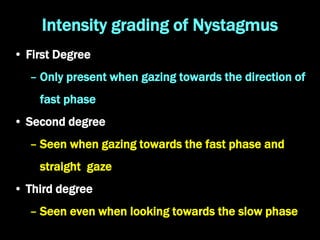
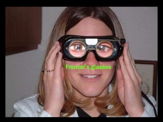
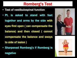
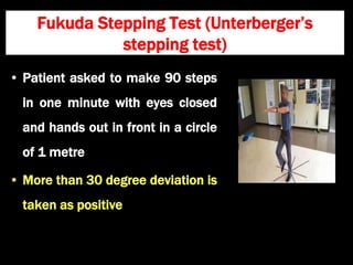
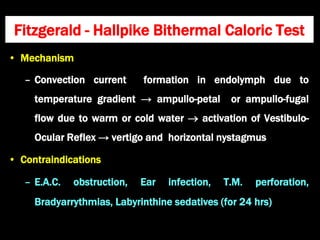
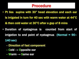
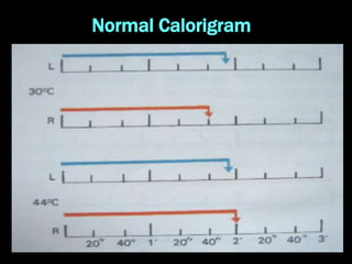
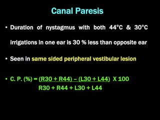
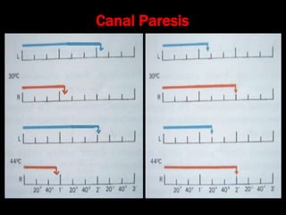
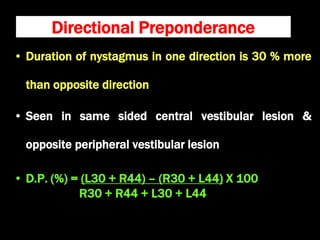
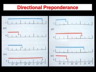
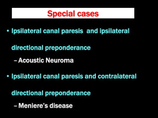
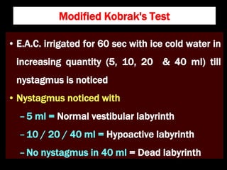
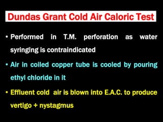
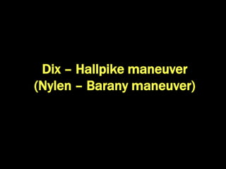
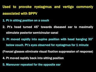
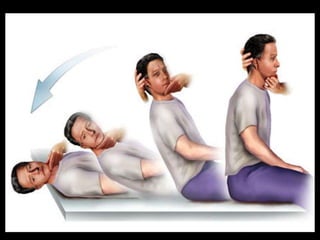
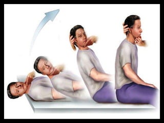
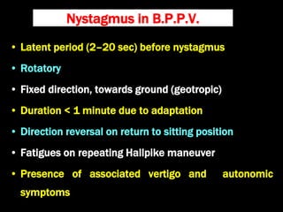
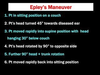
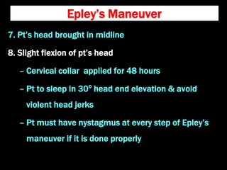
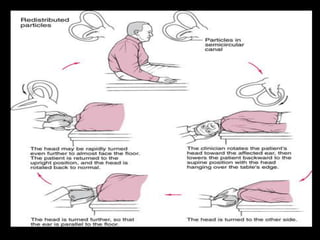
Routine Clinical Tests of
Vestibular Function
Dr. Krishna Koirala
2021-02-02
Nystagmus
• Rhythmic, involuntary, oscillatory movement of
eyeball
• Vestibular disorders cause nystagmus with slow and
fast phases
– The slow phase of the nystagmus is the direction of
flow of endolymph and is vestibular in origin
– The fast phase is most likely initiated by the
reticular formation as a compensatory mechanism
Intensity grading of Nystagmus
• First Degree
– Only present when gazing towards the direction of
fast phase
• Second degree
– Seen when gazing towards the fast phase and
straight gaze
• Third degree
– Seen even when looking towards the slow phase
• Vestibular nystagmus
– Suppressed by optic fixation
– Enhanced with use of Frenzel glasses (remove
optic fixation)
• Irritative vestibular labyrinthine lesion causes
ipsilateral nystagmus
• Paralytic vestibular labyrinthine lesion gives rise to
contralateral nystagmus
Frenzel’s glasses
Test for gaze evoked nystagmus
• Examiner’s finger / pointed objects
kept 30 cm away from pt's eyes in
centre and moved in horizontal and
vertical planes not exceeding 30°
(to avoid physiological end-point
nystagmus)
• Pt is asked to follow it with his eyes
• Examiner looks for nystagmus
Fistula test
• Transmission of increased air pressure into
inner ear through a labyrinthine fistula
causes vertigo and nystagmus towards the
affected ear
• E.A.C. pressure is increased by intermittent
tragal pressure or Siegelization
Sites of labyrinthine fistula
• Horizontal semicircular canal
– Cholesteatoma destruction
– Fenestration operation
• Oval window
– Post-stapedectomy
• Round window membrane rupture
Hennebert’s sign
• False positive fistula sign in absence of
labyrinthine fistula
• Seen in
–Meniere's disease (fibrosis between stapes
footplate and utricle)
–Hyper mobile stapes footplate (congenital
syphilis, idiopathic)
False negative fistula sign
• Negative fistula sign in presence of labyrinthine
fistula
• Seen in
– Cholesteatoma / granulation covering the
labyrinthine fistula
– Dead Labyrinth
– Total E.A.C. obstruction
Romberg’s Test
• Test of vestibulospinal function
• Pt. is asked to stand with feet
together and arms by the side with
eyes first open ( can compensate the
balance) and then closed ( cannot
compensate the balance and sways
to side of lesion )
• Sharpened Romberg’s if Romberg is
negative
Fukuda Stepping Test (Unterberger’s
stepping test)
• Patient asked to make 90 steps
in one minute with eyes closed
and hands out in front in a circle
of 1 metre
• More than 30 degree deviation is
taken as positive
• Mechanism
– Convection current formation in endolymph due to
temperature gradient → ampullo-petal or ampullo-fugal
flow due to warm or cold water  activation of Vestibulo-
Ocular Reflex → vertigo and horizontal nystagmus
• Contraindications
– E.A.C. obstruction, Ear infection, T.M. perforation,
Bradyarrythmias, Labyrinthine sedatives (for 24 hrs)
Fitzgerald - Hallpike Bithermal Caloric Test
Procedure
• Pt lies supine with 30° head elevation and each ear
is irrigated in turn for 40 sec with warm water at 44°C
& then cold water at 30°C after a gap of 8 mins
• Duration of nystagmus is counted from start of
irrigation to end point of nystagmus (Normal = 90–
140 sec)
– Direction of fast component
– Cold → Opposite ear
– Warm → Same ear
Normal Calorigram
Canal Paresis
• Duration of nystagmus with both 44°C & 30°C
irrigations in one ear is 30 % less than opposite ear
• Seen in same sided peripheral vestibular lesion
• C. P. (%) = (R30 + R44) – (L30 + L44) X 100
R30 + R44 + L30 + L44
Canal Paresis
Directional Preponderance
• Duration of nystagmus in one direction is 30 % more
than opposite direction
• Seen in same sided central vestibular lesion &
opposite peripheral vestibular lesion
• D.P. (%) = (L30 + R44) – (R30 + L44) X 100
R30 + R44 + L30 + L44
Directional Preponderance
Special cases
• Ipsilateral canal paresis and ipsilateral
directional preponderance
–Acoustic Neuroma
• Ipsilateral canal paresis and contralateral
directional preponderance
–Meniere’s disease
Modified Kobrak's Test
• E.A.C. irrigated for 60 sec with ice cold water in
increasing quantity (5, 10, 20 & 40 ml) till
nystagmus is noticed
• Nystagmus noticed with
–5 ml = Normal vestibular labyrinth
–10 / 20 / 40 ml = Hypoactive labyrinth
–No nystagmus in 40 ml = Dead labyrinth
Dundas Grant Cold Air Caloric Test
• Performed in T.M. perforation as water
syringing is contraindicated
• Air in coiled copper tube is cooled by pouring
ethyl chloride in it
• Effluent cold air is blown into E.A.C. to produce
vertigo + nystagmus
Dix – Hallpike maneuver
(Nylen – Barany maneuver)
Used to provoke nystagmus and vertigo commonly
associated with BPPV
1. Pt in sitting position on a couch
2. Pt’s head turned 45° towards diseased ear to maximally
stimulate posterior semicircular canal
3. Pt moved rapidly into supine position with head hanging 30°
below couch. Pt’s eyes observed for nystagmus for 1 minute
(Frenzel glasses eliminate visual fixation suppression of response)
4. Pt moved rapidly back into sitting position
5. Maneuver repeated for the opposite ear
Nystagmus in B.P.P.V.
• Latent period (2–20 sec) before nystagmus
• Rotatory
• Fixed direction, towards ground (geotropic)
• Duration < 1 minute due to adaptation
• Direction reversal on return to sitting position
• Fatigues on repeating Hallpike maneuver
• Presence of associated vertigo and autonomic
symptoms
Epley’s particle repositioning
maneuver
Epley’s Maneuver
1. Pt in sitting position on a couch
2. Pt’s head turned 45° towards diseased ear
3. Pt moved rapidly into supine position with head
hanging 30° below couch
4. Pt’s head rotated by 90° to opposite side
5. Further 90° head + trunk rotation
6. Pt moved rapidly back into sitting position
Epley’s Maneuver
7. Pt’s head brought in midline
8. Slight flexion of pt’s head
– Cervical collar applied for 48 hours
– Pt to sleep in 30o
head end elevation & avoid
violent head jerks
– Pt must have nystagmus at every step of Epley’s
maneuver if it is done properly
Routine clinical tests of vestibular function

Routine clinical tests of vestibular function

  • 1.
    Routine Clinical Testsof Vestibular Function Dr. Krishna Koirala 2021-02-02
  • 2.
    Nystagmus • Rhythmic, involuntary,oscillatory movement of eyeball • Vestibular disorders cause nystagmus with slow and fast phases – The slow phase of the nystagmus is the direction of flow of endolymph and is vestibular in origin – The fast phase is most likely initiated by the reticular formation as a compensatory mechanism
  • 3.
    Intensity grading ofNystagmus • First Degree – Only present when gazing towards the direction of fast phase • Second degree – Seen when gazing towards the fast phase and straight gaze • Third degree – Seen even when looking towards the slow phase
  • 4.
    • Vestibular nystagmus –Suppressed by optic fixation – Enhanced with use of Frenzel glasses (remove optic fixation) • Irritative vestibular labyrinthine lesion causes ipsilateral nystagmus • Paralytic vestibular labyrinthine lesion gives rise to contralateral nystagmus
  • 5.
  • 6.
    Test for gazeevoked nystagmus • Examiner’s finger / pointed objects kept 30 cm away from pt's eyes in centre and moved in horizontal and vertical planes not exceeding 30° (to avoid physiological end-point nystagmus) • Pt is asked to follow it with his eyes • Examiner looks for nystagmus
  • 7.
    Fistula test • Transmissionof increased air pressure into inner ear through a labyrinthine fistula causes vertigo and nystagmus towards the affected ear • E.A.C. pressure is increased by intermittent tragal pressure or Siegelization
  • 8.
    Sites of labyrinthinefistula • Horizontal semicircular canal – Cholesteatoma destruction – Fenestration operation • Oval window – Post-stapedectomy • Round window membrane rupture
  • 9.
    Hennebert’s sign • Falsepositive fistula sign in absence of labyrinthine fistula • Seen in –Meniere's disease (fibrosis between stapes footplate and utricle) –Hyper mobile stapes footplate (congenital syphilis, idiopathic)
  • 10.
    False negative fistulasign • Negative fistula sign in presence of labyrinthine fistula • Seen in – Cholesteatoma / granulation covering the labyrinthine fistula – Dead Labyrinth – Total E.A.C. obstruction
  • 11.
    Romberg’s Test • Testof vestibulospinal function • Pt. is asked to stand with feet together and arms by the side with eyes first open ( can compensate the balance) and then closed ( cannot compensate the balance and sways to side of lesion ) • Sharpened Romberg’s if Romberg is negative
  • 12.
    Fukuda Stepping Test(Unterberger’s stepping test) • Patient asked to make 90 steps in one minute with eyes closed and hands out in front in a circle of 1 metre • More than 30 degree deviation is taken as positive
  • 13.
    • Mechanism – Convectioncurrent formation in endolymph due to temperature gradient → ampullo-petal or ampullo-fugal flow due to warm or cold water  activation of Vestibulo- Ocular Reflex → vertigo and horizontal nystagmus • Contraindications – E.A.C. obstruction, Ear infection, T.M. perforation, Bradyarrythmias, Labyrinthine sedatives (for 24 hrs) Fitzgerald - Hallpike Bithermal Caloric Test
  • 14.
    Procedure • Pt liessupine with 30° head elevation and each ear is irrigated in turn for 40 sec with warm water at 44°C & then cold water at 30°C after a gap of 8 mins • Duration of nystagmus is counted from start of irrigation to end point of nystagmus (Normal = 90– 140 sec) – Direction of fast component – Cold → Opposite ear – Warm → Same ear
  • 15.
  • 16.
    Canal Paresis • Durationof nystagmus with both 44°C & 30°C irrigations in one ear is 30 % less than opposite ear • Seen in same sided peripheral vestibular lesion • C. P. (%) = (R30 + R44) – (L30 + L44) X 100 R30 + R44 + L30 + L44
  • 17.
  • 18.
    Directional Preponderance • Durationof nystagmus in one direction is 30 % more than opposite direction • Seen in same sided central vestibular lesion & opposite peripheral vestibular lesion • D.P. (%) = (L30 + R44) – (R30 + L44) X 100 R30 + R44 + L30 + L44
  • 19.
  • 20.
    Special cases • Ipsilateralcanal paresis and ipsilateral directional preponderance –Acoustic Neuroma • Ipsilateral canal paresis and contralateral directional preponderance –Meniere’s disease
  • 21.
    Modified Kobrak's Test •E.A.C. irrigated for 60 sec with ice cold water in increasing quantity (5, 10, 20 & 40 ml) till nystagmus is noticed • Nystagmus noticed with –5 ml = Normal vestibular labyrinth –10 / 20 / 40 ml = Hypoactive labyrinth –No nystagmus in 40 ml = Dead labyrinth
  • 22.
    Dundas Grant ColdAir Caloric Test • Performed in T.M. perforation as water syringing is contraindicated • Air in coiled copper tube is cooled by pouring ethyl chloride in it • Effluent cold air is blown into E.A.C. to produce vertigo + nystagmus
  • 23.
    Dix – Hallpikemaneuver (Nylen – Barany maneuver)
  • 24.
    Used to provokenystagmus and vertigo commonly associated with BPPV 1. Pt in sitting position on a couch 2. Pt’s head turned 45° towards diseased ear to maximally stimulate posterior semicircular canal 3. Pt moved rapidly into supine position with head hanging 30° below couch. Pt’s eyes observed for nystagmus for 1 minute (Frenzel glasses eliminate visual fixation suppression of response) 4. Pt moved rapidly back into sitting position 5. Maneuver repeated for the opposite ear
  • 27.
    Nystagmus in B.P.P.V. •Latent period (2–20 sec) before nystagmus • Rotatory • Fixed direction, towards ground (geotropic) • Duration < 1 minute due to adaptation • Direction reversal on return to sitting position • Fatigues on repeating Hallpike maneuver • Presence of associated vertigo and autonomic symptoms
  • 28.
  • 29.
    Epley’s Maneuver 1. Ptin sitting position on a couch 2. Pt’s head turned 45° towards diseased ear 3. Pt moved rapidly into supine position with head hanging 30° below couch 4. Pt’s head rotated by 90° to opposite side 5. Further 90° head + trunk rotation 6. Pt moved rapidly back into sitting position
  • 30.
    Epley’s Maneuver 7. Pt’shead brought in midline 8. Slight flexion of pt’s head – Cervical collar applied for 48 hours – Pt to sleep in 30o head end elevation & avoid violent head jerks – Pt must have nystagmus at every step of Epley’s maneuver if it is done properly