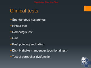
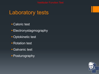
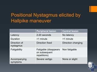
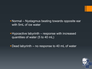
Assessment of
Vestibular Functions
Venkatesh Karthikeyan
III MBBS
Velammal Medical College
Email: 4852012@gmail.com
Tests done
Clinical Tests
Laboratory test
Vestibular Function Test
Clinical tests
Spontaneous nystagmus
Fistula test
Romberg’s test
Gait
Past pointing and falling
Dix - Hallpike manoeuver (positional test)
Test of cerebellar dysfunction
Vestibular Function Test
Laboratory tests
Caloric test
Electronystagmography
Optokinetic test
Rotation test
Galvanic test
Posturography
Vestibular Function Test
Clinical tests
Spontaneous nystagmus
Fistula test
Romberg test
Gait
Post pointing and falling
Hallpike manoeuver (positional test)
Test of cerebellar dysfunction
Vestibular Function Test
Spontaneous Nystagmus
Nystagmus:
 Rhythmical
 Involuntary
 Oscillatory movement of eyes
Types of nystagmus
Horizontal nystagmus
Vertical nystagmus
Rotatory nystagmus
Horizontal nystagmus
Direction of fast component Direction of slow component
Direction of Nystagmus
Vertical Nystagmus
Rotatory Nystagmus
Eliciting Nystagmus
Patient is seated in front of examiner or lies supine on
the bed.
Finger at 30 cm from patient’s eye in central position.
Move the finger.
Avoid Gaze nystagmus.
SPONTANEOUS NYSTAGMUS ALWAYS INDICATES
AN ORGANIC LESION.
VESTIBULAR NYSTAGMUS
 Peripheral – due to lesion in labyrinth or CN VIII
 Central – due to lesion in vestibular nuclei or brainstem or
cerebellum
Peripheral Nystagmus
 Irritative lesions of the labyrinth (serous labyrinthitis) –
nystagmus to the side of lesion.
 Paretic lesions – nystagmus to healthy side (purulent labyrinthitis,
trauma to labyrinth and section of VIIIth nerve)
 Suppressed by optic fixation by looking at fixed point
 Enhanced in darkness or by using Frenzel glasses (+20 D)
Central Nystagmus
Cannot be suppressed by Optic fixation.
Types of Central Nystagmus:
 Torsional Nystagmus
 Vertical downbeat Nystagmus
 Vertical Upbeat Nystagmus
 Pendular Nystagmus
Type of Nystagmus Level of Lesion Conditions in which
it is seen
Torsional nystagmus Brainstem or
Vestibular nuclei
Syringomyelia
Vertical downbeat
nystagmus
Craniocervical region Arnold Chiari
malformation or
degenerative lesions
of cerebellum
Vertical upbeat
nystagmus
Junction of Pons and
medulla or pons and
midbrain
-
Pendular nystagmus - • Congenital or
Multiple Sclerosis
• Maybe
disconjugate
Clinical tests
Spontaneous nystagmus
Fistula test
Romberg test
Gait
Past pointing and falling
Hallpike manoeuver (positional test)
Test of cerebellar dysfunction
Vestibular Function Test
Fistula Test
Basis of the test : Inducing nystagmus by
producing pressure changes in external ear
which are then transmitted to labyrinth
True Negative – Normal
Fistula test - True Positive
 Erosion of horizontal semicircular canal as in cholesteatoma
 Surgically created window in horizontal canal (Fenestration)
 Abnormal opening in oval window (post stapedectomy fistula)
 Abnormal opening in round window (Rupture of round window)
Fistula Test – False Negative
Dead labyrinth
Cholesteatoma covering the site of fistula
and not allowing the pressure changes to
be transmitted to labyrinth.
Fistula test – False positive
Congenital syphilis – due to hypermobile stapes
footplate
Meniere’s disease – due to fibrous bands
connecting utricular macula to stapes footplate.
In both these conditions, movement of stapes
results in stimulation of Utricular macula.
Clinical tests
Spontaneous nystagmus
Fistula test
Romberg test
Gait
Past pointing and falling
Hallpike manoeuver (positional test)
Test of cerebellar dysfunction
Vestibular Function Test
Romberg Test
• Peripheral Vestibular Lesion – patients
sways to side of lesion.
• Central Vestibular Lesion – patient
shows instability.
Sharpened Romberg Test
Inability indicates Vestibular impairment
Clinical tests
Spontaneous nystagmus
Fistula test
Romberg test
Gait
Post pointing and falling
Hallpike manoeuver (positional test)
Test of cerebellar dysfunction
Vestibular Function Test
Gait
In case of uncompensated lesions of peripheral vestibular system, with eyes
closed, the patient deviates to affected side
Clinical tests
Spontaneous nystagmus
Fistula test
Romberg test
Gait
Past pointing and falling
Hallpike manoeuver (positional test)
Test of cerebellar dysfunction
Vestibular Function Test
Clinical tests
Spontaneous nystagmus
Fistula test
Romberg test
Gait
Past pointing and falling
Hallpike manoeuver (positional test)
Test of cerebellar dysfunction
Vestibular Function Test
Hallpike manoeuvre
Useful when patient complains of vertigo in certain
head positions
Helps differentiate a peripheral from a central
lesion
Positional Nystagmus elicited by
Hallpike maneuver
Peripheral lesion Central lesion
Latency 2-20 seconds No latency
Duration <1 minute >1 minute
Direction of
nystagmus
Direction fixed Direction changing
Fatigability Fatigable (disappears
on subsequent
repetitions)
Non fatigable
Accompanying
symptoms
Severe vertigo None or slight
Clinical tests
Spontaneous nystagmus
Fistula test
Romberg test
Gait
Post pointing and falling
Hallpike manoeuver (positional test)
Test of cerebellar dysfunction
Vestibular Function Test
Test for Cerebellar function
All cases of giddiness should be tested
for cerebellar diseases.
 Diseases of cerebellar hemisphere causes:
 Asynergia (abnormal finger nose test)
 Dysmetria (inability to control range of motion)
 Adiadochokinesia (inability to perform rapid
alternative movements)
 Rebound phenomenon (inability to control movements
of extremity when opposing forceful restraint is
suddenly released)
Midline diseases of cerebellum causes:
 Wide based gait
 Falling in any direction
 Inability to make sudden turns while walking
 Truncal ataxia
Laboratory tests
Caloric test
 Electronystagmography
 Optokinetic test
 Rotation test
 Galvanic test
 Posturography
Vestibular Function Test
Caloric Test
Basis : Induce nystagmus by thermal stimulation of
vestibular system.
Advantage : Each labyrinth can be tested
separately.
Helps proving labyrinthine origin of vertigo.
Caloric Tests
Modified Kobrak test
Fitzgerald-Hallpike test
Cold-air caloric test
Modified Kobrak test
Patient seated with head tilted 60 degrees
backwards (Horizontal canal  Vertical in
position)
Ear is irrigated with cold water for 60 seconds.
 5mL
 10mL
 20mL
 40mL
Normal – Nystagmus beating towards opposite ear
with 5mL of ice water
Hypoactive labyrinth – response with increased
quantities of water (5 to 40 mL)
Dead labyrinth – no response to 40 mL of water
Fitzgerald – Hallpike test
Irrigate the ears for 40 seconds alternately with water at 300 C
and at 440 C
Eyes are observed for appearance of nystagmus till its end
point
Chart the time taken from the start of irrigation to the end
point of nystagmus on a calorigram
If no nystagmus is elicited from any eye, test is repeated with
the water at 200 C for 4 minutes before labelling the labyrinth
dead.
Allow a gap of 5 minutes between two ears
• Cold water - Nystagmus to opposite side
• Warm water – Nystagmus to same side
Caloric test helps in assessing:
Canal paresis – response elicited from a
particular canal after stimulation is less than that
from opposite side
Directional preponderance – Duration of
nystagmus to left or right, irrespective of whether
it is elicited from the right or left labyrinth
Canal paresis
 It can be expressed as percentage of total response from
both ears.
Response from left ear = L30 + L44
L30 + L44 + R30 + R44
Response from right ear = R30 + R44
L30 + L44 + R30 + R44
X 100
X 100
Less or no response from particular side is
indicative of depressed function of ipsilateral
labyrinth or vestibular nerve or vestibular nuclei.
Depressed functions are seen with:
Meniere’s disease
Acoustic neuroma
Post labyrinthectomy
Vestibular nerve section
Directional preponderance
 Right beating nystagmus is caused by L30 and R44 (COWS)
 Left beating nystagmus is caused by L44 and R30
Right beating nystagmus = L30 + R44
L30 + L44 + R30 + R44
Left beating nystagmus = L44 and R30
L30 + L44 + R30 + R44
X 100
X 100
 If nystagmus is 25 – 30% or more on one side than the other
side, it is called directional preponderance to that side.
Directional preponderance occurs towards the
side of central lesion, away from side of
peripheral lesion.
Unilateral Meniere’s disease – Canal paresis on
one side and directional preponderance to
opposite side
Acoustic neuroma – Both to ipsilateral side
Laboratory tests
 Caloric test
Electronystagmography
 Optokinetic test
 Rotation test
 Galvanic test
 Posturography
Vestibular Function Test
Electro-nystagmo-graphy
 Depends on presence of corneo-retinal potentials which are
recorded by placing electrodes at suitable places around the
eye.
 Used for:
 Detecting nystagmus (even which is not seen by naked eye)
 Recording nystagmus (permits to keep a permanent record)
Laboratory tests
 Caloric test
 Electronystagmography
Optokinetic test
 Rotation test
 Galvanic test
 Posturography
Vestibular Function Test
Optokinetic test
 Used to diagnose central lesions (brainstem and cerebral
hemisphere lesions)
Normally, it produces nystagmus with slow component in the direction of
moving stripes and fast component in opposite direction
R  L
L  R
Laboratory tests
 Caloric test
 Electronystagmography
 Optokinetic test
Rotation test
 Galvanic test
 Posturography
Vestibular Function Test
Rotational test
 Patient seated with head tilted 300 forward
 Rotated 10 turns in 20 seconds
 Stop the chair abruptly and observe the nystagmus (Normal :
25-40 seconds)
 Performed in case of congenital abnormalities where ear
canal has failed to develop (unable to perform caloric test)
 Disadvantage : Labyrinths cannot be tested individually
Laboratory tests
 Caloric test
 Electronystagmography
 Optokinetic test
 Rotation test
Galvanic test
 Posturography
Vestibular Function Test
Galvanic test
Helps differentiating end organ lesion from
vestibular nerve lesion.
Feet together, eyes closed, arms out-stretched.
Current of 1 mA passed to one ear – normally,
patient sways towards side of anodal current.
Laboratory tests
 Caloric test
 Electronystagmography
 Optokinetic test
 Rotation test
 Galvanic test
Posturography
Vestibular Function Test
Helps in evaluating vestibular function by measuring postural stability
Firing Round
Thank you 
Adam Politzer – Father of modern Otology

Vestibular Function Test

  • 1.
    Assessment of Vestibular Functions VenkateshKarthikeyan III MBBS Velammal Medical College Email: 4852012@gmail.com
  • 2.
    Tests done Clinical Tests Laboratorytest Vestibular Function Test
  • 3.
    Clinical tests Spontaneous nystagmus Fistulatest Romberg’s test Gait Past pointing and falling Dix - Hallpike manoeuver (positional test) Test of cerebellar dysfunction Vestibular Function Test
  • 4.
    Laboratory tests Caloric test Electronystagmography Optokinetictest Rotation test Galvanic test Posturography Vestibular Function Test
  • 5.
    Clinical tests Spontaneous nystagmus Fistulatest Romberg test Gait Post pointing and falling Hallpike manoeuver (positional test) Test of cerebellar dysfunction Vestibular Function Test
  • 6.
    Spontaneous Nystagmus Nystagmus:  Rhythmical Involuntary  Oscillatory movement of eyes
  • 7.
    Types of nystagmus Horizontalnystagmus Vertical nystagmus Rotatory nystagmus
  • 8.
    Horizontal nystagmus Direction offast component Direction of slow component Direction of Nystagmus
  • 9.
  • 10.
  • 11.
    Eliciting Nystagmus Patient isseated in front of examiner or lies supine on the bed. Finger at 30 cm from patient’s eye in central position. Move the finger. Avoid Gaze nystagmus. SPONTANEOUS NYSTAGMUS ALWAYS INDICATES AN ORGANIC LESION.
  • 12.
    VESTIBULAR NYSTAGMUS  Peripheral– due to lesion in labyrinth or CN VIII  Central – due to lesion in vestibular nuclei or brainstem or cerebellum
  • 13.
    Peripheral Nystagmus  Irritativelesions of the labyrinth (serous labyrinthitis) – nystagmus to the side of lesion.  Paretic lesions – nystagmus to healthy side (purulent labyrinthitis, trauma to labyrinth and section of VIIIth nerve)  Suppressed by optic fixation by looking at fixed point  Enhanced in darkness or by using Frenzel glasses (+20 D)
  • 15.
    Central Nystagmus Cannot besuppressed by Optic fixation. Types of Central Nystagmus:  Torsional Nystagmus  Vertical downbeat Nystagmus  Vertical Upbeat Nystagmus  Pendular Nystagmus
  • 16.
    Type of NystagmusLevel of Lesion Conditions in which it is seen Torsional nystagmus Brainstem or Vestibular nuclei Syringomyelia Vertical downbeat nystagmus Craniocervical region Arnold Chiari malformation or degenerative lesions of cerebellum Vertical upbeat nystagmus Junction of Pons and medulla or pons and midbrain - Pendular nystagmus - • Congenital or Multiple Sclerosis • Maybe disconjugate
  • 18.
    Clinical tests Spontaneous nystagmus Fistulatest Romberg test Gait Past pointing and falling Hallpike manoeuver (positional test) Test of cerebellar dysfunction Vestibular Function Test
  • 19.
    Fistula Test Basis ofthe test : Inducing nystagmus by producing pressure changes in external ear which are then transmitted to labyrinth True Negative – Normal
  • 20.
    Fistula test -True Positive  Erosion of horizontal semicircular canal as in cholesteatoma  Surgically created window in horizontal canal (Fenestration)  Abnormal opening in oval window (post stapedectomy fistula)  Abnormal opening in round window (Rupture of round window)
  • 21.
    Fistula Test –False Negative Dead labyrinth Cholesteatoma covering the site of fistula and not allowing the pressure changes to be transmitted to labyrinth.
  • 22.
    Fistula test –False positive Congenital syphilis – due to hypermobile stapes footplate Meniere’s disease – due to fibrous bands connecting utricular macula to stapes footplate. In both these conditions, movement of stapes results in stimulation of Utricular macula.
  • 24.
    Clinical tests Spontaneous nystagmus Fistulatest Romberg test Gait Past pointing and falling Hallpike manoeuver (positional test) Test of cerebellar dysfunction Vestibular Function Test
  • 25.
    Romberg Test • PeripheralVestibular Lesion – patients sways to side of lesion. • Central Vestibular Lesion – patient shows instability.
  • 26.
    Sharpened Romberg Test Inabilityindicates Vestibular impairment
  • 27.
    Clinical tests Spontaneous nystagmus Fistulatest Romberg test Gait Post pointing and falling Hallpike manoeuver (positional test) Test of cerebellar dysfunction Vestibular Function Test
  • 28.
    Gait In case ofuncompensated lesions of peripheral vestibular system, with eyes closed, the patient deviates to affected side
  • 29.
    Clinical tests Spontaneous nystagmus Fistulatest Romberg test Gait Past pointing and falling Hallpike manoeuver (positional test) Test of cerebellar dysfunction Vestibular Function Test
  • 31.
    Clinical tests Spontaneous nystagmus Fistulatest Romberg test Gait Past pointing and falling Hallpike manoeuver (positional test) Test of cerebellar dysfunction Vestibular Function Test
  • 32.
    Hallpike manoeuvre Useful whenpatient complains of vertigo in certain head positions Helps differentiate a peripheral from a central lesion
  • 34.
    Positional Nystagmus elicitedby Hallpike maneuver Peripheral lesion Central lesion Latency 2-20 seconds No latency Duration <1 minute >1 minute Direction of nystagmus Direction fixed Direction changing Fatigability Fatigable (disappears on subsequent repetitions) Non fatigable Accompanying symptoms Severe vertigo None or slight
  • 35.
    Clinical tests Spontaneous nystagmus Fistulatest Romberg test Gait Post pointing and falling Hallpike manoeuver (positional test) Test of cerebellar dysfunction Vestibular Function Test
  • 36.
    Test for Cerebellarfunction All cases of giddiness should be tested for cerebellar diseases.  Diseases of cerebellar hemisphere causes:  Asynergia (abnormal finger nose test)  Dysmetria (inability to control range of motion)  Adiadochokinesia (inability to perform rapid alternative movements)  Rebound phenomenon (inability to control movements of extremity when opposing forceful restraint is suddenly released)
  • 37.
    Midline diseases ofcerebellum causes:  Wide based gait  Falling in any direction  Inability to make sudden turns while walking  Truncal ataxia
  • 38.
    Laboratory tests Caloric test Electronystagmography  Optokinetic test  Rotation test  Galvanic test  Posturography Vestibular Function Test
  • 39.
    Caloric Test Basis :Induce nystagmus by thermal stimulation of vestibular system. Advantage : Each labyrinth can be tested separately. Helps proving labyrinthine origin of vertigo.
  • 40.
    Caloric Tests Modified Kobraktest Fitzgerald-Hallpike test Cold-air caloric test
  • 41.
    Modified Kobrak test Patientseated with head tilted 60 degrees backwards (Horizontal canal  Vertical in position) Ear is irrigated with cold water for 60 seconds.  5mL  10mL  20mL  40mL
  • 42.
    Normal – Nystagmusbeating towards opposite ear with 5mL of ice water Hypoactive labyrinth – response with increased quantities of water (5 to 40 mL) Dead labyrinth – no response to 40 mL of water
  • 43.
  • 44.
    Irrigate the earsfor 40 seconds alternately with water at 300 C and at 440 C Eyes are observed for appearance of nystagmus till its end point Chart the time taken from the start of irrigation to the end point of nystagmus on a calorigram If no nystagmus is elicited from any eye, test is repeated with the water at 200 C for 4 minutes before labelling the labyrinth dead. Allow a gap of 5 minutes between two ears
  • 45.
    • Cold water- Nystagmus to opposite side • Warm water – Nystagmus to same side
  • 46.
    Caloric test helpsin assessing: Canal paresis – response elicited from a particular canal after stimulation is less than that from opposite side Directional preponderance – Duration of nystagmus to left or right, irrespective of whether it is elicited from the right or left labyrinth
  • 47.
    Canal paresis  Itcan be expressed as percentage of total response from both ears. Response from left ear = L30 + L44 L30 + L44 + R30 + R44 Response from right ear = R30 + R44 L30 + L44 + R30 + R44 X 100 X 100
  • 48.
    Less or noresponse from particular side is indicative of depressed function of ipsilateral labyrinth or vestibular nerve or vestibular nuclei. Depressed functions are seen with: Meniere’s disease Acoustic neuroma Post labyrinthectomy Vestibular nerve section
  • 49.
    Directional preponderance  Rightbeating nystagmus is caused by L30 and R44 (COWS)  Left beating nystagmus is caused by L44 and R30 Right beating nystagmus = L30 + R44 L30 + L44 + R30 + R44 Left beating nystagmus = L44 and R30 L30 + L44 + R30 + R44 X 100 X 100
  • 50.
     If nystagmusis 25 – 30% or more on one side than the other side, it is called directional preponderance to that side. Directional preponderance occurs towards the side of central lesion, away from side of peripheral lesion. Unilateral Meniere’s disease – Canal paresis on one side and directional preponderance to opposite side Acoustic neuroma – Both to ipsilateral side
  • 53.
    Laboratory tests  Calorictest Electronystagmography  Optokinetic test  Rotation test  Galvanic test  Posturography Vestibular Function Test
  • 54.
    Electro-nystagmo-graphy  Depends onpresence of corneo-retinal potentials which are recorded by placing electrodes at suitable places around the eye.  Used for:  Detecting nystagmus (even which is not seen by naked eye)  Recording nystagmus (permits to keep a permanent record)
  • 55.
    Laboratory tests  Calorictest  Electronystagmography Optokinetic test  Rotation test  Galvanic test  Posturography Vestibular Function Test
  • 56.
    Optokinetic test  Usedto diagnose central lesions (brainstem and cerebral hemisphere lesions) Normally, it produces nystagmus with slow component in the direction of moving stripes and fast component in opposite direction R  L L  R
  • 57.
    Laboratory tests  Calorictest  Electronystagmography  Optokinetic test Rotation test  Galvanic test  Posturography Vestibular Function Test
  • 59.
    Rotational test  Patientseated with head tilted 300 forward  Rotated 10 turns in 20 seconds  Stop the chair abruptly and observe the nystagmus (Normal : 25-40 seconds)  Performed in case of congenital abnormalities where ear canal has failed to develop (unable to perform caloric test)  Disadvantage : Labyrinths cannot be tested individually
  • 60.
    Laboratory tests  Calorictest  Electronystagmography  Optokinetic test  Rotation test Galvanic test  Posturography Vestibular Function Test
  • 61.
    Galvanic test Helps differentiatingend organ lesion from vestibular nerve lesion. Feet together, eyes closed, arms out-stretched. Current of 1 mA passed to one ear – normally, patient sways towards side of anodal current.
  • 63.
    Laboratory tests  Calorictest  Electronystagmography  Optokinetic test  Rotation test  Galvanic test Posturography Vestibular Function Test
  • 64.
    Helps in evaluatingvestibular function by measuring postural stability
  • 65.
  • 71.
    Thank you  AdamPolitzer – Father of modern Otology

Editor's Notes

  • #2 Adam politzer – father of modern otology according to ncbi