Downloaded 127 times
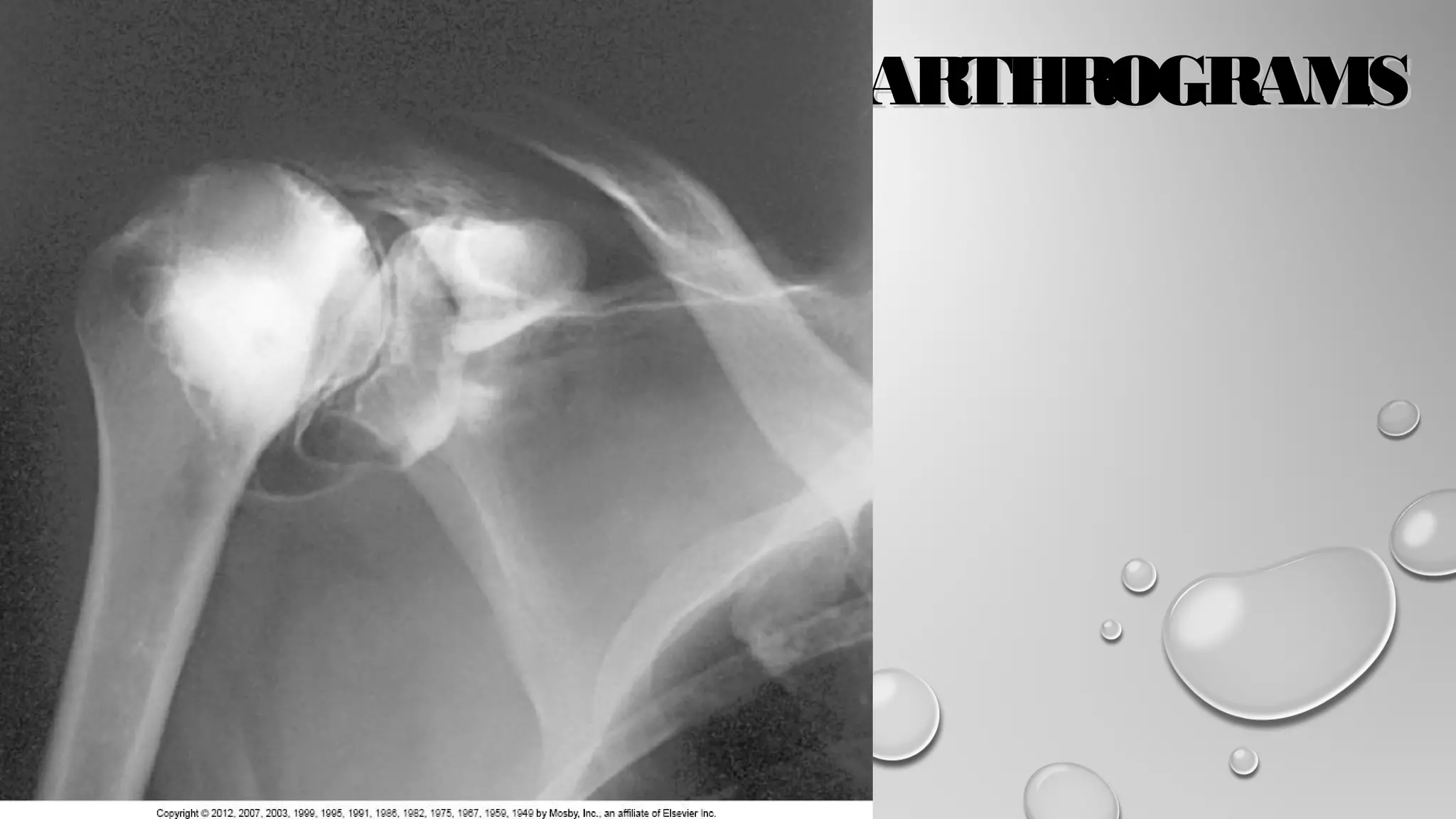
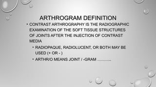


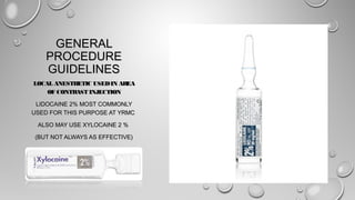

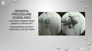
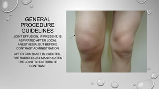
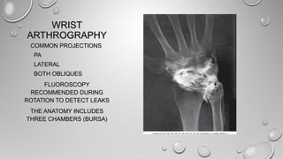
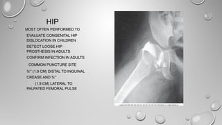
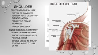

This document provides information on arthrograms, including definitions, terminology, procedures, and specifics on contrast arthrography of various joints like the knee, wrist, hip, shoulder, and TMJ. It explains that contrast arthrography involves injecting contrast media into a joint space to examine soft tissues under fluoroscopy. While MRI has replaced many arthrograms due to being noninvasive, contrast arthrography is still used to evaluate certain joints like the knee, wrist, hip and shoulder for conditions like trauma, pain, or prosthesis loosening.


