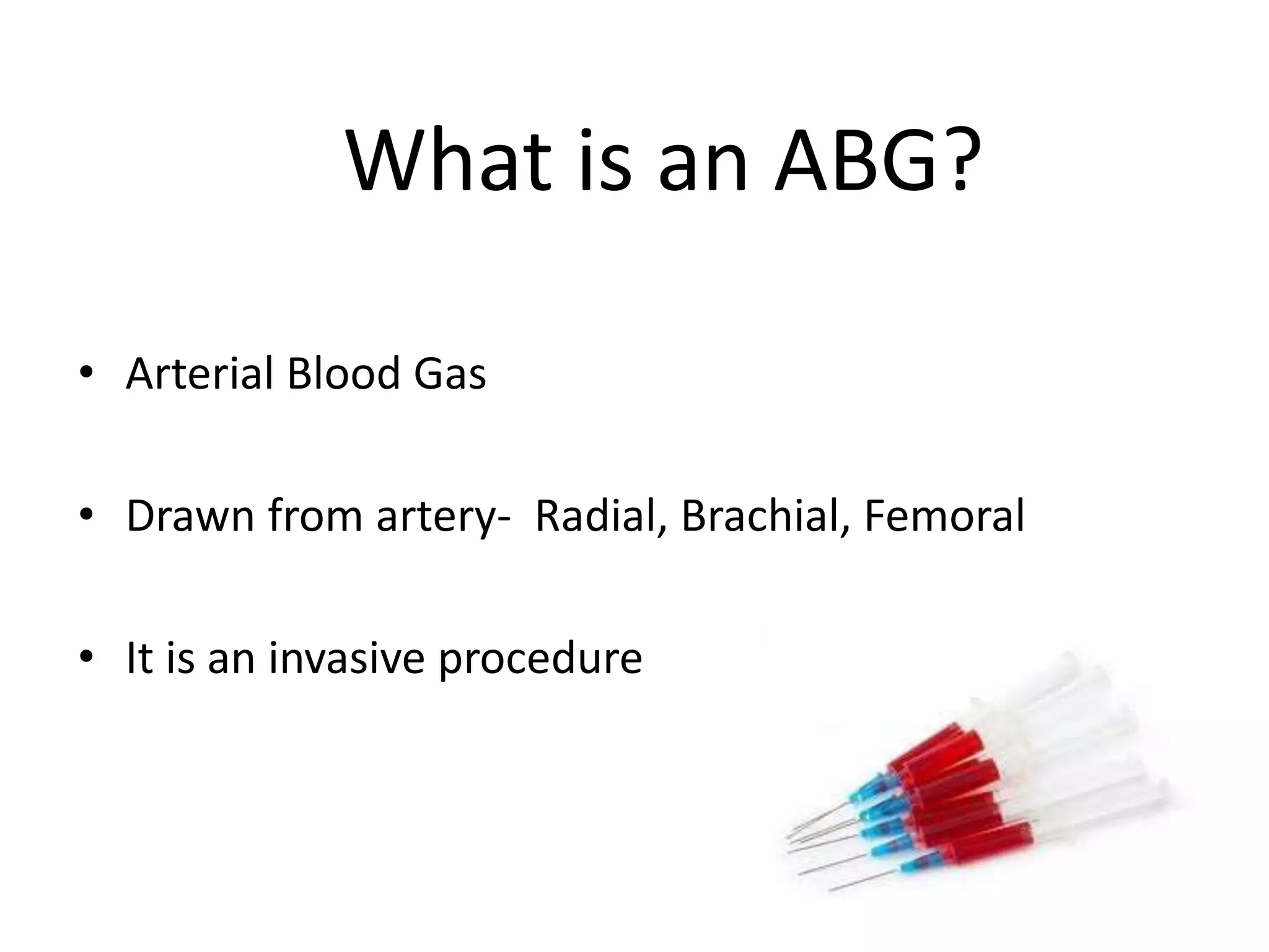
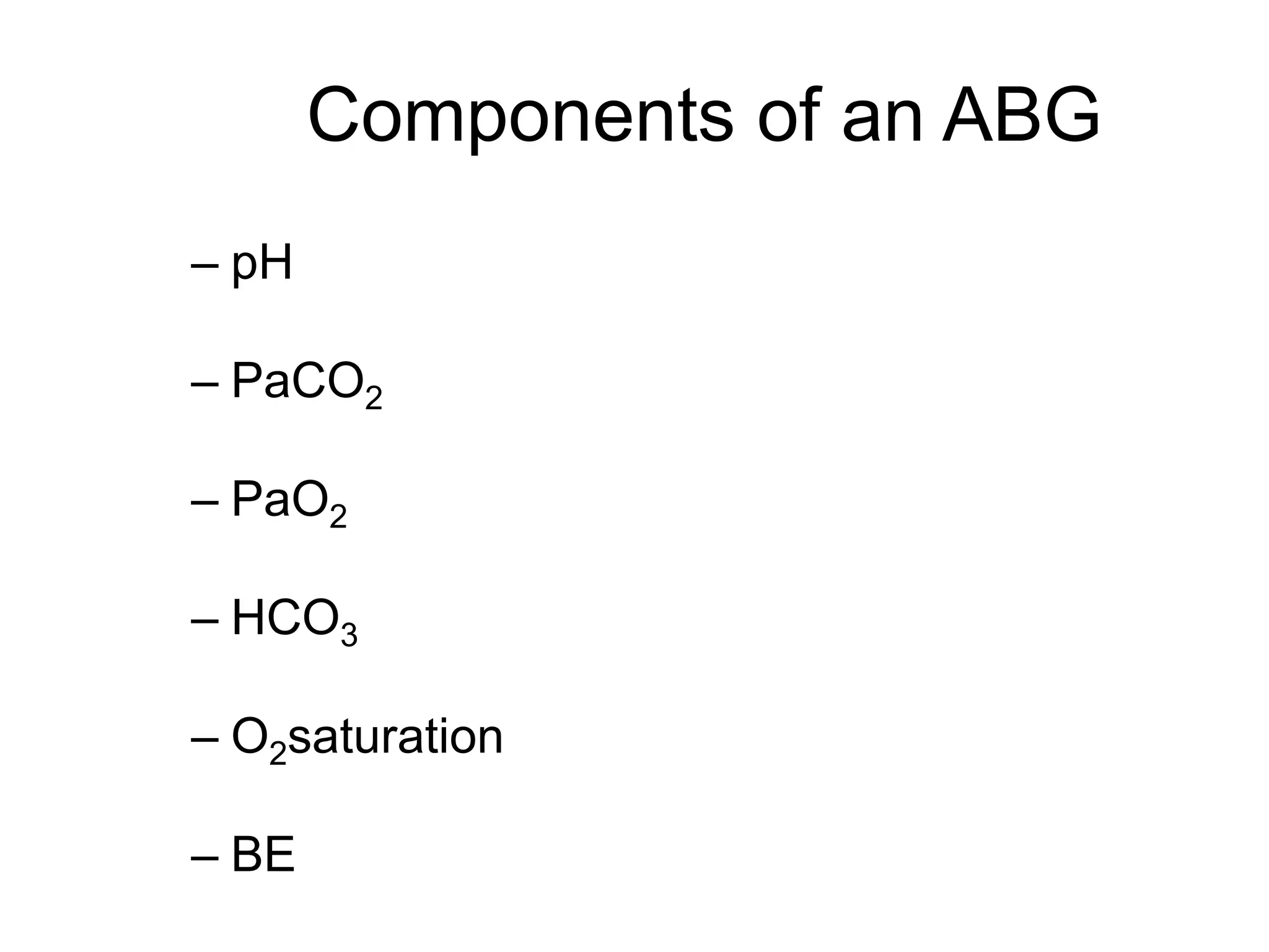
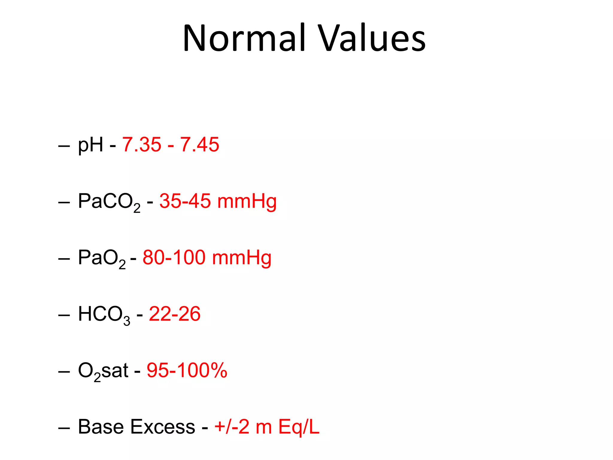
This document provides information about arterial blood gas (ABG) analysis. It defines an ABG as a blood sample drawn from an artery to assess acid-base balance, oxygenation, and ventilation. ABGs are ordered to diagnose and monitor respiratory failure and changes in acid-base homeostasis. The components of an ABG include pH, PaCO2, PaO2, HCO3, O2 saturation, and base excess. Normal values for these components are provided. The document discusses how to properly collect an ABG sample and handle it to avoid inaccurate results. It also covers acid-base physiology and the four primary acid-base disorders: metabolic acidosis, metabolic alkalosis, respiratory acidosis, and respiratory alk
ABGs or VBGs interpretation made simple straight forward easy to remember and easy to apply. The presentation is designed to help the residents and junior ER physicians. The second part will discuss the oxygenation and the third part will review the "Stewart Approach" while fourth and last part is meant for the Experts.
Acid base balance & ABG interpretation,Dept of anesthesiology,JJMMC,DavangereGopan Gopalakrisna Pillai
Acid base balance and ABG interpretation presented by Dr.Gopan.G,Post-Graduate student. Chairperson : Dr.Ravi.R,Professor, Department of Anaesthesiology & Critical care,JJMMC,Davangere.
This presentation discuss about acid-base-gas normal ratio and its indication in relation to varying abnormal level and how to manage it. This includes clinical analysis practice.
ABG test measures the blood gas tension values of the arterial partial pressure of oxygen, and the arterial partial pressure of carbon dioxide, and the blood's pH
Everolimus eluting stents or bypass surgery finalGOPAL GHOSH
Trials and registry studies have shown lower long-term mortality after CABG than after PCI among patients with multivessel disease.These previous analyses did not evaluate PCI with second-generation drug-eluting stents
Explore natural remedies for syphilis treatment in Singapore. Discover alternative therapies, herbal remedies, and lifestyle changes that may complement conventional treatments. Learn about holistic approaches to managing syphilis symptoms and supporting overall health.
Pulmonary Thromboembolism - etilogy, types, medical- Surgical and nursing man...VarunMahajani
Disruption of blood supply to lung alveoli due to blockage of one or more pulmonary blood vessels is called as Pulmonary thromboembolism. In this presentation we will discuss its causes, types and its management in depth.
The prostate is an exocrine gland of the male mammalian reproductive system
It is a walnut-sized gland that forms part of the male reproductive system and is located in front of the rectum and just below the urinary bladder
Function is to store and secrete a clear, slightly alkaline fluid that constitutes 10-30% of the volume of the seminal fluid that along with the spermatozoa, constitutes semen
A healthy human prostate measures (4cm-vertical, by 3cm-horizontal, 2cm ant-post ).
It surrounds the urethra just below the urinary bladder. It has anterior, median, posterior and two lateral lobes
It’s work is regulated by androgens which are responsible for male sex characteristics
Generalised disease of the prostate due to hormonal derangement which leads to non malignant enlargement of the gland (increase in the number of epithelial cells and stromal tissue)to cause compression of the urethra leading to symptoms (LUTS
Lung Cancer: Artificial Intelligence, Synergetics, Complex System Analysis, S...Oleg Kshivets
RESULTS: Overall life span (LS) was 2252.1±1742.5 days and cumulative 5-year survival (5YS) reached 73.2%, 10 years – 64.8%, 20 years – 42.5%. 513 LCP lived more than 5 years (LS=3124.6±1525.6 days), 148 LCP – more than 10 years (LS=5054.4±1504.1 days).199 LCP died because of LC (LS=562.7±374.5 days). 5YS of LCP after bi/lobectomies was significantly superior in comparison with LCP after pneumonectomies (78.1% vs.63.7%, P=0.00001 by log-rank test). AT significantly improved 5YS (66.3% vs. 34.8%) (P=0.00000 by log-rank test) only for LCP with N1-2. Cox modeling displayed that 5YS of LCP significantly depended on: phase transition (PT) early-invasive LC in terms of synergetics, PT N0—N12, cell ratio factors (ratio between cancer cells- CC and blood cells subpopulations), G1-3, histology, glucose, AT, blood cell circuit, prothrombin index, heparin tolerance, recalcification time (P=0.000-0.038). Neural networks, genetic algorithm selection and bootstrap simulation revealed relationships between 5YS and PT early-invasive LC (rank=1), PT N0—N12 (rank=2), thrombocytes/CC (3), erythrocytes/CC (4), eosinophils/CC (5), healthy cells/CC (6), lymphocytes/CC (7), segmented neutrophils/CC (8), stick neutrophils/CC (9), monocytes/CC (10); leucocytes/CC (11). Correct prediction of 5YS was 100% by neural networks computing (area under ROC curve=1.0; error=0.0).
CONCLUSIONS: 5YS of LCP after radical procedures significantly depended on: 1) PT early-invasive cancer; 2) PT N0--N12; 3) cell ratio factors; 4) blood cell circuit; 5) biochemical factors; 6) hemostasis system; 7) AT; 8) LC characteristics; 9) LC cell dynamics; 10) surgery type: lobectomy/pneumonectomy; 11) anthropometric data. Optimal diagnosis and treatment strategies for LC are: 1) screening and early detection of LC; 2) availability of experienced thoracic surgeons because of complexity of radical procedures; 3) aggressive en block surgery and adequate lymph node dissection for completeness; 4) precise prediction; 5) adjuvant chemoimmunoradiotherapy for LCP with unfavorable prognosis.
These lecture slides, by Dr Sidra Arshad, offer a quick overview of physiological basis of a normal electrocardiogram.
Learning objectives:
1. Define an electrocardiogram (ECG) and electrocardiography
2. Describe how dipoles generated by the heart produce the waveforms of the ECG
3. Describe the components of a normal electrocardiogram of a typical bipolar leads (limb II)
4. Differentiate between intervals and segments
5. Enlist some common indications for obtaining an ECG
Study Resources:
1. Chapter 11, Guyton and Hall Textbook of Medical Physiology, 14th edition
2. Chapter 9, Human Physiology - From Cells to Systems, Lauralee Sherwood, 9th edition
3. Chapter 29, Ganong’s Review of Medical Physiology, 26th edition
4. Electrocardiogram, StatPearls - https://www.ncbi.nlm.nih.gov/books/NBK549803/
5. ECG in Medical Practice by ABM Abdullah, 4th edition
6. ECG Basics, http://www.nataliescasebook.com/tag/e-c-g-basics
Ethanol (CH3CH2OH), or beverage alcohol, is a two-carbon alcohol
that is rapidly distributed in the body and brain. Ethanol alters many
neurochemical systems and has rewarding and addictive properties. It
is the oldest recreational drug and likely contributes to more morbidity,
mortality, and public health costs than all illicit drugs combined. The
5th edition of the Diagnostic and Statistical Manual of Mental Disorders
(DSM-5) integrates alcohol abuse and alcohol dependence into a single
disorder called alcohol use disorder (AUD), with mild, moderate,
and severe subclassifications (American Psychiatric Association, 2013).
In the DSM-5, all types of substance abuse and dependence have been
combined into a single substance use disorder (SUD) on a continuum
from mild to severe. A diagnosis of AUD requires that at least two of
the 11 DSM-5 behaviors be present within a 12-month period (mild
AUD: 2–3 criteria; moderate AUD: 4–5 criteria; severe AUD: 6–11 criteria).
The four main behavioral effects of AUD are impaired control over
drinking, negative social consequences, risky use, and altered physiological
effects (tolerance, withdrawal). This chapter presents an overview
of the prevalence and harmful consequences of AUD in the U.S.,
the systemic nature of the disease, neurocircuitry and stages of AUD,
comorbidities, fetal alcohol spectrum disorders, genetic risk factors, and
pharmacotherapies for AUD.
Prix Galien International 2024 Forum ProgramLevi Shapiro
June 20, 2024, Prix Galien International and Jerusalem Ethics Forum in ROME. Detailed agenda including panels:
- ADVANCES IN CARDIOLOGY: A NEW PARADIGM IS COMING
- WOMEN’S HEALTH: FERTILITY PRESERVATION
- WHAT’S NEW IN THE TREATMENT OF INFECTIOUS,
ONCOLOGICAL AND INFLAMMATORY SKIN DISEASES?
- ARTIFICIAL INTELLIGENCE AND ETHICS
- GENE THERAPY
- BEYOND BORDERS: GLOBAL INITIATIVES FOR DEMOCRATIZING LIFE SCIENCE TECHNOLOGIES AND PROMOTING ACCESS TO HEALTHCARE
- ETHICAL CHALLENGES IN LIFE SCIENCES
- Prix Galien International Awards Ceremony
Anti ulcer drugs and their Advance pharmacology ||
Anti-ulcer drugs are medications used to prevent and treat ulcers in the stomach and upper part of the small intestine (duodenal ulcers). These ulcers are often caused by an imbalance between stomach acid and the mucosal lining, which protects the stomach lining.
||Scope: Overview of various classes of anti-ulcer drugs, their mechanisms of action, indications, side effects, and clinical considerations.
2. What is an ABG?
• Arterial Blood Gas
• Drawn from artery- Radial, Brachial, Femoral
• It is an invasive procedure
3. Why to order an ABG?
• Assess adequacy of ventilation and oxygenation
• Aids in establishing a diagnosis and severity of respiratory
failure
• Assess changes in acid- base homeostasis
• Helps to guide treatment plan
• Helps in management of ICU patients.
4. Components of an ABG
– pH
– PaCO2
– PaO2
– HCO3
– O2saturation
– BE
5. Others include
• Haemoglobin
• Hematocrit
• Na
• K
• Glucose
• Standard bicarbonate
• Buffer base
• Standard base excess
• TCO2
6. • Base Excess: It indicates increase in the amount
of buffer base.
• It is the number of mmol of strong acid needed
to adjust pH to 7.4 at PCO2 40 mm Hg.
• Base Deficit: It indicates decrease in the
amount of buffer base.
7. • Standard bicarbonate
It is the bicarbonate conc. In plasma in a completely
oxygenated blood sample at Pco2 of 40 mm Hg at 370C .
SBC > 24 mmol/l indicates metabolic alkalosis
SBC<24 mmol/l indicates metabolic acidosis
Buffer base
It is the total equivalent conc. of all anion buffer
components of blood . Normal value =48 mmol/L
8. • Standard base excess
• It gives a view of the base excess of the entire extracellular
fluid.
• It indicates average Hb of the fluid space through which
bicarbonate distributes.
• standard base excess is the value when the hemoglobin is
at 5 g/dl.
Total plasma CO2 (T-CO2)
Total content of the CO2 normal value= 27 meq/L
10. HOW TO DRAW AN ABG..???
• Equipments
• Site Selection
• Contraindication
• Puncture procedure
• Post puncture procedure
• Sample handling
11. Equipments
1. One 1 cc to 5 cc vented, pre-heparinized, plastic syringe
2. One 20 - 25 gauge 1 –1 1/2” needle
Longer needles for brachial and femoral artery puncture
3. One Biohazard labelled plastic bag
4. . Two 1 x 1 inch sterile gauze
12. • 5. Alcohol prep pad
• 6. Specimen/Patient label
• 7. Iodine pad
• 8. One adhesive bandage
• 9. Lab Form
• 10. Ice
15. Contraindication
• No absolute contraindications
• Dialysis shunt – choose another site
• Mastectomy – use opposite side
• Patient on anticoagulant/aspirin therapy – may have to hold
pressure on puncture site longer than normal
17. Puncture procedure
1. Check for orders
2.Explain the patient about compression of puncture site
3. Make positive patient I.D.
4. Put on gloves
5. Assemble needle to syringe
a. keep needle sterile
b. eject excess heparin and air bubbles
c. pull back syringe plunger to at least 1 cc
18. 4 things that “hurt”
1. Needle through skin (sharp)
2. Needle through arterial wall (blunt)
3. Miss & catch nerve (shooting)
4. Miss & hit periostium (sharp)
19. Site selection
1. Radial artery is always the first choice because it provides
collateral circulation.
• A. Palpate the right and left radials arterial pulse and
visualize the course of the artery.
• B. Pick strongest pulse
i. if radial pulse weak on right, move to left
ii. if pulse on left weak, then try brachial
• 2. Brachial used as alternative site
• 3. Femoral is the last choice in normal situations
Highest complication rate
20.
21.
22.
23.
24.
25.
26.
27.
28. Check for:
• a. Bleeding
• b. movement of fingers and tingling sensation
• c. pulse distal to puncture
29. Post puncture procedure
1. Remove any air bubbles from sample and cap
syringe
• Dispose of needle in sharps container
2. Roll syringe to mix heparin with sample
3. Immerse in ice
4. On lab slip indicate:
a. FIO2
b. Patient temperature
c. Ventilator parameters
5. Deliver to lab
30. Sample handling
Sample should be analyzed as soon as possible
– If iced sample can be stored
» Glass syringe – 1 hour
» Plastic syringe – 15 minutes
Remember: Blood is living tissue that continues to
consume O2 and produce CO2
31. ABG Specimen Collection/Handling
• Transport specimen to laboratory in a biohazard container
• Analyze specimen on an instrument that has been recently
calibrated
• Temperature correction specimen in analyzer
• Increase in patient temp: PO2, PCO2, pH
• Decrease in patient temp: PO2, PCO2, pH
34. Technical Causes of Abnormal Results
• 1. Room air mixed with sample
• a. PaO2 will equilibrate to above 160
• 2. CO2 will be lower due to equilibration
• 3. Delay in running sample
• O2 consumption will continue as will CO2 production –
pH decreases
• Iced, sample will last an hour without a change in the result
• un-iced, ABG's can be significantly changed after 10 minutes
35. • 4. Venous sample drawn
• a. Usually this in patients with shock
• b. Should doubt when PO2 is significantly lower than expected
• i. draw venous blood to check comparison or
• ii. redraw sample
36. • 5. Heparin
• a. Sodium Heparin 1% solution should be used
• b. ammonium heparin will alter pH
• c. dry lithium heparin is OK
• • All unnecessary heparin should be ejected from syringe, excess
can effect results
37. • 6. Patient pain
• a. Can cause hyperventilation or breath holding
• b. An anaesthetic may be injected prior to stick for pain
• • Usually 2% lidocaine
• • CAUTION – some people allergic to “caines”
• 7. Machine errors
• a. Improper calibration
• b. Air bubbles in electrode
38. Acid-Base Physiology
• pH is the negative logarithm to the base 10 of the hydrogen ion
concentration in mmol/L
• pH = - log10[H+]
• An increase in pH indicates a proportionate decrease in the [H+] and
a decrease in the pH indicates a proportionate increase in the [H+].
• H2CO3 generates 12,500 mmol H+ per day.
• Normal metabolism of proteins and nucleotides generates about 100
mmol H+ per day in the form of sulphuric and phosphoric acids.
39. Calculation of pH
• pH is calculated from Henderson-Hasselbalch
equation .
• pH = pK + log acid/bas
• pH = 6.1 + log HCO3-
H2CO3
Kassirer and Bliech modified equation
• H+ = 24 x PCO2/HCO3-
40. Regulation of pH
pH is maintained in narrow range by
• 1) In seconds: buffer systems
• 2)In minutes: CO2 excretion by the lungs
• 3)In hours to days: renal excretion of H+,
reabsorption of HCO3
41. Regulation of arterial pH
• 1.BUFFERS –Buffer systems minimize the change in pH resulting from
production of acid .
• Main buffer system in humans is HCO3- in ECF and protein and
phosphate buffers in ICF.
2.ROLE OF THE RESPIRATORY SYSTEM–Elimination of volatile acid CO2.
• a. Respiratory centers in the brain respond to changes in pH of
CSF and blood to affect ventilatory rate.
• b. Ventilation directly controls the elimination of CO2.
42. ROLE OF KIDNEY
It retains and regenerate HCO3- thereby regenerating the
body buffer with the net effect of eliminating the non-
volatile acid load
a. H+ secretion
1. Free urinary H+ - minimal contribution
2. Ammonia
3. Phosphorus
b. HCO3- reabsorption
1. Proximal tubule – 90%
2. Distal tubule -10%
43. Carbon dioxide transport
• In Dissolved form : Carbon dioxide is twenty times more soluble in
water than oxygen.
• 100 ml of plasma with a PCO2 of 40 mmHg carries about 2.4 mlCO2
in solution. 5% of total CO2 carriage.
• As carbamino compounds : 5 -10% of CO2 carriage
• Bicarbonate: The remaining 85 -90% of carbon dioxide is carried by
blood in the form of bicarbonate ions.
44. Acid base disorders
• Acidemia –pH less than 7.35
• Acidosis – A process that would cause
acidemia, if not compensated
• Alkalemia–pH greater than 7.45
• Alkalosis – A process that would cause
alkalemia if not compensated
46. SIMPLE VS. MIXED ACID-BASE DISORDER
Simple acid-base disorder – a single primary
process of acidosis or alkalosis.
Mixed acid-base disorder – presence of more
than one acid base disorder simultaneously
48. Compensation
• In the presence of acidosis or alkalosis, regulatory
mechanisms occur which try to maintain arterial
pH.
• Disturbances in HCO3- result in respiratory
compensation
• Changes in CO2 are counteracted by renal
compensation
49. a. Renal compensation – kidneys adapt to alterations
in pH by changing the amount of HCO3-
generated/excreted.
Full renal compensation takes 2-5 days
b. Respiratory compensation – alteration in ventilation
allow immediate compensation for metabolic acid-
base disorders
51. Prediction of compensation
Metabolic acidosis PaCO2= (1.5 x HCO3
-) + 8 ± 2
Metabolic alkalosis
PaCO2 will↑ 0.75 mmHg per
mmol/L ↑ in [HCO3
-]
Respiratory
acidosis
Acute
[HCO3
-] will ↑ 0.1 mmol/L per
mmHg in PaCO2
Chronic
[HCO3
-] will ↑ 0.4 mmol/L per
mmHg in PaCO2
Respiratory
alkalosis
Acute
[HCO3
-] will ↑ 0.2 mmol/L per
mmHg in PaCO2
Chronic
[HCO3
-] will ↑ 0.4 mmol/L per
mmHg in PaCO2
52. Anion Gap
• Anion gap used to assess acid-base status in
D/D of meabolic acidosis
Anion gap based on principle of electro
neutrality:
• Total Serum Cations = Total Serum Anions
• Na – (HCO3 + Cl) = Anion gap
• Normal Anion gap – 10 +/- 2meq/L
53. • Albumin is the major unmeasured anion
• The anion gap should be corrected if there are
gross changes in serum albumin levels.
AG (CORRECTED) = AG + { (4 – [ALBUMIN]) × 2.5}
54. Delta gap
• Difference between
– Change in anion gap ( AG)
– Change in bicarbonate ( HCO3
- )
• Based on assumption that for each 1 meq/L
increase in AG, HCO3 will fall by 1 meq/L , to
maintain a stable anion content.
• Usual range: -6 to +6 meq/L ; should be 0
55. AG = HCO3
- Pure High AG Met Acidosis
AG > HCO3
- ( Gap >6) HCO3- does not
decrease as expected.
Associated Metabolic Alkalosis or respiratory acidosis
AG < HCO3
- ( Gap < -6) HCO3- does not
increase as expected
Associated N AG Met Acidosis or rarely respiratory
alkalosis
Delta ratio may also be calculated
57. URINARY ANION GAP
• Urinary NH4
+ levels can be estimated by calculating the
urine anion gap (UAG)
• UAG = [Na+ + K+]u – [Cl–]u
• [Cl–]u > [Na+ + K+], the urine gap is negative by definition
• Helps to distinguish GI from renal causes of loss of
HCO3 by estimating Urinary NH4+ (elevated in GI HCO3
loss but low in distal RTA).
• Hence a -ve UAG (av -20 meq/L) seen in former while
+ve value (av +23 meq/L) seen in latter.
58. Urine PH
• Non AG metabolic acidosis:
– If urine pH > 5.5 : Type 1 RTA
– If urine pH < 5.5 : Type 2 or Type 4 RTA
• Type 2 or Type 4 RTA can be later
differentiated using serum K+ level
60. Causes of High AG Met Acidosis
1. Ketoacidosis:
Diabetic
Alcoholic
Starvation
2. Lactic Acidosis:
Type A (Inadequate O2 Delivery to Cells)
Type B (Inability of Cells to utilise O2)
Type D (Abn bowel anatomy)
3. Toxicity:
Salicylates Paraldehyde
Methanol Toluene
Ethylene Glycol
4. Renal Failure
5. Rhabdomyolsis
61. CAUSES OF NORMAL ANION GAP
METABOLIC ACIDOSIS
1. HCO3 loss:
GIT Diarrhoea
Pancreatic or biliary drainage
Urinary diversions
(ureterosigmoidostomy)
Renal Proximal (type 2) RTA
Ketoacidosis (during therapy)
Post-chronic hypocapnia
62. 2. Impaired renal acid excretion:
Distal (type 1) RTA
Hyperkalemia (type 4) RTA
Hypoaldosteronism
3. Misc:
Acid Administration (NH4Cl)
Hyperalimentation
Cholestyramine Cl
HCl therapy (Rx of severe met alkalosis)
74. Step – 1 : Check for ERRORS
Have the required parameters been correctly
fed..???
Patient’s Temperature
Fi O₂ : specially if patient is in ventilator
Hemoglobin : some machines may not measure it
Barometric pressure : some machines may not measure it
79. pH is inversely related to [H+]; a pH change of 1.00
represents a 10-fold change in [H+]
pH [H+] in nanomoles/L
7.00 100
7.10 80
7.30 50
7.40 40
7.52 30
7.70 20
8.00 10
Relation b/w pH & H+ conc.
Assessment of validity of test results
80. Assessment of validity of test results
• H+ in nmol/L = 24 × PCO₂/HCO₃
• If there is a discripancy between the 2
results, the blood should be reanalyzed.
82. STEP -2 : Comprehensive history and physical
examination.
STEP -3 : Acidosis or alkalosis..???
See the pH (<7.35 or >7.45)
STEP -4 : Identify the primary disorder
See the change in PCo2 & HCO3
STEP -5 : Calculate the compensatory response
Is adequately compensated???
83. STEP -6 : Calculate anion gap
STEP -7 : Calculate the delta gap (unmask hidden
mixed disorders)
STEP -8 : Calculate the osmolar gap (for high AG
acidosis)
STEP -9 : Calculate the urinary anion gap (Non
AG metabolic acidosis)
STEP -10 : Formulate differential diagnosis
85. Case-1
• 60 years old M, presents to the ED with rapid
breathing and less responsive than usual. No
other history available.
ABG results
pH 7.31
PCO₂ 10
HCO₃ 5
Na 123
K 5
Cl 99
86. Stepwise interpretation
1. At pH 7.3 H+ conc. Should be ≈50nmol/L
– Calculated H+ = 24 × 10/5 = 24 × 2 = 48
– Both values corroborate, hence result is valid.
2. pH is 7.3 i.e Acidosis
3. HCO₃ value has gone down, primary process
is metabolic
4. Respiratory compensation:
– Calculated PCO₂ = (1.5 × 5)+8 ± 2 = 13.5 to 17.5
– Over compensated ; mixed disorder
– a/w respiratory alkalosis
87. 5. Anion gap: (123+5) – (99 + 5) = 19
– High anion gap metabolic acidosis
6. Delta gap :
– Change in AG = (19-10) = 9
– Change in HCO₃ = (24-5) = 19
– Delta gap = 9-19 = -10
Presence of non anion gap metabolic acidosis also.!!
7. Osmolar gap: data not provided.
88. Finally …
• Mixed acid base disorder, with presence of
both high AG & normal AG metabolic acidosis
and respiratory alkalosis.
89. Case-2
• A k/c/o COPD with cor pulmonale on
treatment presented with progressive
breathlessness.
ABG results
pH 7.42
PCO₂ 67
HCO₃ 42
Na 140
K 3.5
Cl 88
90. • pH is normal; but PCO₂ & HCO₃ both are
increased.
• Change in PCO₂ is 67-40 = 27
• Expected rise in HCO₃ should be 27 × 0.4 =
10.8
• Expected HCO₃ = 24+10.8 ≈ 35
• Actual HCO₃ = 42
• AG = 12 (N)
• Mixed disorder, both respiratory acidosis &
metabolic alkalosis.
91. Case -3
• A known case of case of chronic kidney disease,
discontinued MHD & presented to the emergency in
an altered state of sensorium. Attendants gave
history of repeated episodes of vomiting at home.
ABG results
pH 7.42
PCO₂ 40
HCO₃ 25
Na 140
K 3.0
Cl 95
92. • pH, PCO₂, HCO₃ all WNL
• AG = 23 (↑)
• Delta gap = 13 – 1 = 12 (↑)
• AG >> HCO3
–
• Mixed disorder with presence of both high AG
metabolic acidosis and metabolic alkalosis.
93. Case-4
• 65 yrs old M, past h/o AMI on medication, presented
with high grade fever with, cough & yellowish
expectoration for 5 days. Acute increase in shortness of
breath.
ABG results
pH 7.3
PCO₂ 38
HCO₃ 16
Na 136
K 4
Cl 102
96. • ABG is a very useful diagnostic tool for our day
to day practice.
• Approach to interpret should be step wise & in
a systematic manner.
• Any abnormal result should be analyzed
cautiously in light of clinical context.
• Appropriate use of this tool using clinical
judgment is of paramount importance