SlideShare a Scribd company logo
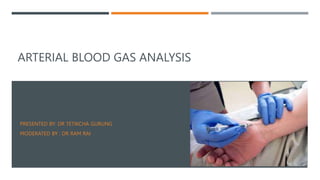
ARTERIAL BLOOD GAS ANALYSIS
PRESENTED BY: DR TETIKCHA GURUNG
MODERATED BY : DR RAM RAI
INTRODUCTION
 ABG provides rapid information on three physiological processes:
i. Ventilation( reflected by PaCO2)
ii. Oxygenation status( assessed primarily by PaO2 andSaO2
iii. Acid Base Balance
 ABG analysis  essential for diagnosing and managing the patient’s
oxygenation status , ventilation failure and acid base balance.
INDICATION
 Assess the ventilatory setting, oxygenation and acid base status.
 Assess the response to an intervention.
 Regulate electrolyte therapy.
 Establish preoperative baseline parameters.
CONTRAINDICATION
 An abnormal modified Allen’s test.
 Local infection or distorted anatomy at puncture site.
 Severe peripheral vascular disease of the artery.
 Active Raynaud’s Syndrome
SITE
 Radial artery ( most common )
 Brachial artery
 Femoral artery
 Radial is the most preferable site used
because:
i. It is easy to access
ii. It is not a deep artery which facilitate
palpation,
stabilization and puncturing
iii. The artery has a collateral blood
circulation
EQUIPMENTS
Blood gas kit OR
 1ml syringe
 23-26 gauge needle
 Stopper or cap
 Alcohol swab
 Disposable gloves
 Plastic bag & crushed
ice
 Lidocaine (optional)
 Vial of heparin
(1:1000)
 Bar code or label
METHODOLOGY
PREPARATORY PHASE:
 Record patient inspired oxygen concentration.
 Explain the procedure to the patient.
 Heparinize the needle.
• Donot leave excess heparin in the syringe
• ↑↑ heparin  ↑↑ dilutional effect  ↓↓ HCO3
−
and ↓↓pCO2
 Wait at least 20 minutes before drawing blood for ABG after changing
settings of mechanical ventilation, after suctioning the patient or after
extubation.
 After preparing the site, the artery is palpated for maximum pulsation
 In case of radial artery , Modified Allen test is done.
 Skin and subcutaneous tissue may be infiltrated with local anesthetic agent if
needed
 The needle is inserted at 45 in radial, 60 in brachial and 90 in femoral.
• Ensure no air bubble
Air Bubble has pO2 − 150 mm Hg and pCO2 −0 mmHg
Air Bubble + Blood = ↑↑ pO2 and ↓↓ pCO2
 Place the capped syringe in the container of ice immediately
 Maintain firm pressure on the puncture site for 5 minutes.
MODIFIED ALLEN’S TEST
 Test to determine collateral circulation is present from the ulnar
artery in case thrombosis occur in the radial artery.
 ABG syringe must be transported at the earliest to the laboratory for early
analysis via cold chain
 ABG sample should always be sent with relevant information regarding
O2, FiO2 status and Temperature.
COMPLICATION
 Arteriospasm
 Infection
 Hematoma
 Hemorrhage
 Distal Ischemia
 Gangrene
 AV Fistula
ACID BASE BALANCE
 Acid base balance is defined by the concentration of hydrogen ion
 The hydrogen ion concentration in aqueous solution is expressed by pH
which is defined as negative logarithm( base 10 ) of [H+
].
pH = log(1/ [H+
]) = -log [H+
].
The acid base equilibrium is described using Henderson Hasselbach Equation:
 BICARBONATE BUFFER
SYSTEM
 Acts within few seconds
 RESPIRATORY
REGULATION
 Acts within few minutes
 RENAL REGULATION
 Acts in hours to days
CHEMICAL BUFFER
 Buffer = base molecule and its weak conjugate acid.
 pKa= dissociation ionization constant  pH at which acid is 50 %
dissociated and 50% undissociated.
 pKA indicates strength of the acid
 There are 2 buffer system:
i. Extracellular buffer system
ii. Intracellular buffer system
EXTRACELLULAR BUFFER SYSTEM
 It includes : Bicarbonate buffer system(pKa=6.1) and Phosphate buffer
system(pKa=6.8).
1. Bicarbonate Buffer System(H2CO3/HCO3
−
)
The base = bicarbonate and its weak acid conjugate= carbonic acid
CO2 + H2O carbonic anhydrase H2CO3 H+ + HCO3
-
INTRACELLULAR BUFFER
 It includes :
i. Hemoglobin buffer(HbH/Hb)
ii. Other protein buffer(PrH/Pr−)
iii. Phosphate buffer(H2PO4 −/HPO4 2−),
HEMOGLOBIN BUFFER SYSTEM
RESPIRATORY REGULATION
RENAL REGULATION
 Occurs via 3 mechanism:
i. reabsorption of the filtered HCO3
−
ii. excretion of titratable acids,
iii. production of ammonia
ANALYTE Normal Value Units
pH 7.35 - 7.45
PCO2 35 - 45 mm Hg
PO2 72 – 104 mm Hg`
[HCO3] 22 – 30 meq/L
SaO2 95-100 %
Anion Gap 9 + 3 meq/L
B.E +2 to -2 meq/L
DEFINITIONS
 ACID: molecule that can act as a proton (H+) donor
 BASE: molecule that can act as a proton acceptor.
 ACIDEMIA:A blood pH less than 7.35
 ALKALKEMIA : a blood pH greater than 7.45
 ACIDOSIS – presence of a process which tends to  pH by virtue of gain of H
+ or loss of HCO3
-
 ALKALOSIS – presence of a process which tends to  pH by virtue of loss of H+
or gain of HCO3
-
 Simple Acid Base Disorder/ Primary Acid Base disorder – a single primary
process of acidosis or alkalosis due to an initial change in PCO2 and HCO3.
 Compensation - The normal response of the respiratory system or kidneys to
change in pH induced by a primary acid-base disorder
The Compensatory responses to a primary Acid Base disturbance are never
enough to correct the change in pH they only act to reduce the severity.
 Mixed Acid Base Disorder – Presence of more than one acid base disorder
simultaneously .
 Buffer Base:
 It is total quantity of buffers in blood including both volatile(Hco3) and non
volatile (as Hgb,albumin,Po4)
 Base Excess/Base Deficit:
 Amount of strong acid or base needed to restore plasma pH to 7.40 at a Pa
CO2 of 40 mm Hg,at 37*C.
 Calculated from pH, PaCO2 and HCT
 Negative BE also referred to as Base Deficit
 True reflection of non respiratory (metabolic) acid base status
 Normal value: -2 to +2mEq/L
 The H+ In extracellular fluid is determined by balance between the pCO2 and HCO3
- in the
fluid.
 This relationship is expressed as
H+ = 24 x (pCO2/ HCO3 )
STEPWISE APPROACH TO ACID BASE ANALYSIS
 STEP 1: Check for authenticity
 STEP 2: : Identify the primary Acid Base disorder
 STEP 3: Evaluate the Secondary Response
 STEP 4: Calculate Anion Gap
STEP 1 : CHECH FOR AUTHENTICITY
 [H+] neq/l = 24 X (PCO2 / HCO3)
Calculate it from the ABG report and if this value
is equal to H+ in the report,the ABG report is
authentic.
 Alternatively subtract the last two digits of the
pH(e.g 20 in Ph 7.20) from 80, this value is
approximately equal to the H+ concentration in
the ABG report.
 𝐻𝐶𝑂3
−
= 24 x
𝑝𝐶𝑂2
𝐻+ = ± 2 of 𝐻𝐶𝑂3
−
of venous
blood ; ifnot then the ABG is invalid and not
compatible
H+ ion pH
100 7.00
79 7.10
63 7.20
50 7.30
45 7.35
40 7.40
35 7.45
32 7.50
25 7.60
STEP 2 : IDENTIFY THE PRIMARY ACID BASE DISORDER.
 RULE 1 : If the 𝑃𝑎𝐶𝑂2 and /or pH is outside the normal range acid base
disorder
 RULE 2: if the 𝑃𝑎𝐶𝑂2 and pH are both abnormal, compare the directional
change
 2a: if 𝑡ℎ𝑒 ↑ 𝑃𝑎𝐶𝑂2 and ↑pH or ↓ 𝑃𝑎𝐶𝑂2 and ↓pH  primary metabolic acid
base disorder
 2b : if 𝑡ℎ𝑒 ↑ 𝑃𝑎𝐶𝑂2 and↓pH or ↑ pH and ↓ 𝑃𝑎𝐶𝑂2 primary respiratory acid
base disorder
 RULE 3: if the 𝑃𝑎𝐶𝑂2 or pH is abnormal, the condition is a mixed metabolic
and respiratory disorder.
 3a: if 𝑃𝑎𝐶𝑂2 is abnormal, directional change in 𝑃𝑎𝐶𝑂2  type of respiratory
disorder
 3b: if pH is abnormal , the directional change in pH metabolic disorder.
STEP 3 : EVALUATE THE SECONDARY RESPONSE
 RULE 4: For a primary metabolic acidosis , if
measured 𝑃𝑎𝐶𝑂2 is higher than expected secondary respiratory acidosis and
measured 𝑃𝑎𝐶𝑂2 is less than expected  secondary respiratory alkalosis.
 Metabolic Acidosis
 Winter’s formula: Expected pCO2 = 1.5[HCO3] + 8 ± 2
OR
 pCO2 = 1.2 ( HCO3)
 If serum pCO2 > expected pCO2 -> additional respiratory acidosis and vice versa
EXPECTED CHANGES IN ACID-BASE DISORDERS
Primary Disorder Expected Changes
Metabolic acidosis PCO2 = 1.5 × HCO3 + (8 ± 2)
Metabolic alkalosis PCO2 = 0.7 × HCO3 + (21 ± 2)
Acute respiratory acidosis delta pH = 0.008 × (PCO2 - 40)
Chronic respiratory acidosis delta pH = 0.003 × (PCO2 - 40)
Acute respiratory alkalosis delta pH = 0.008 × (40 - PCO2)
Chronic respiratory alkalosis delta pH = 0.003 × (40 - PCO2)
From: THE ICU BOOK - 2nd Ed. (1998) [Corrected]
 RULE 5: For a primary respiratory disorder,
a normal or near normal 𝐻𝐶𝑂3
−
 acute
 RULE 6: For a primary respiratory disorder where the 𝐻𝐶𝑂3
−
is abnormal , determine the
expected 𝐻𝐶𝑂3
−
for a chronic respiratory disorder
 6a : For a chronic respiratory acidosis, if
𝐻𝐶𝑂3
−
is lower than expected  incomplete renal response
𝐻𝐶𝑂3
−
is higher than expected secondary metabolic alkalosis
 6b : For a chronic respiratory alkalosis, if
𝐻𝐶𝑂3
−
is higher than expected incomplete renal response
𝐻𝐶𝑂3
−
is lower than expected  secondary metabolic alkalosis
 Respiratory Acidosis
 Acute (Uncompensated): for every 10 increase in pCO2 -> HCO3 increases by 1 and there is a decre
ase of 0.08 in pH
 Chronic (Compensated): for every 10 increase in pCO2 -> HCO3 increases by 4 and there is a decreas
e of 0.03 in pH
 Respiratory Alkalosis
 Acute (Uncompensated): for every 10 decrease in pCO2 -> HCO3 decreases by 2 and there is a increas
e of 0.08 in PH
 Chronic (Compensated): for every 10 decrease in pCO2 -> HCO3 decreases by 5 and there is a increas
e of 0.03 in PH
ANION GAP
 Normally, measured cation(MC) + unmeasured cation(UC) = measured anion(MA) + unmeasured
anion(UA).
 MC – MA = UA-UC
 Measured cation = 𝑁𝑎+
and Measured Anion = 𝐶𝑙−
and 𝐻𝐶𝑂3
−
 𝑁𝑎+ - (𝐶𝑙− + 𝐻𝐶𝑂3
−
) = UA –UC
 UA - UC = Anion Gap(AG)= 8-12mEq/L
 Corrected Anion Gap= Anion Gap + 2.5(4.5- albumin of patient)
 1g/dL of Albumin contribute to 3 mEq/L of Anion Gap
1. HAGMA( High Anion Gap Metabolic Acidosis)
 Anion gap is high because fall in 𝐻𝐶𝑂3
−
is not compensated by 𝐶𝑙−
 Causes
K- Ketoacidosis due to endogenous causes/ acid  Diabetes, Alcohol , Starvation
U-Uremic Acidosis
S-Salicylate/Paraldehyde
M-Methanol
E-Ethylene Glycol
L-Lactic Acidosi
2. NAGMA(Normal Anion Gap Metabolic Acidosis)
 A.k.a Hyperchloremic Metabolic Acidosis
 Fall in 𝐻𝐶𝑂3
−
is compensated by 𝐶𝑙−
 Causes:
Causes of nongap metabolic acidosis - DURHAM
Diarrhea, ileostomy, colostomy, enteric fistulas
Ureteral diversions or pancreatic fistulas
RTA type I or IV, early renal failure
Hyperailmentation, hydrochloric acid administration
Acetazolamide, Addison’s
Miscellaneous – post-hypocapnia, toulene, sevelamer, cholestyramine ingestion
CALCULATE ANION GAP
 𝑁𝑎+ - (𝐶𝑙− + 𝐻𝐶𝑂3
−
) = UA –UC; 𝑁𝑎+ = 140 and 𝐶𝑙− = 106
 UA - UC = Anion Gap(AG)= 8-12mEq/L
 In case of HAGMA, calculate Delta Ratio= 1-2
Delta Ratio =
∆𝐴𝐺
∆𝐻𝐶𝑂3
−
=
𝐴𝐺−12
24−𝐻𝐶𝑂3
−
 When Delta Ratio < 1 ; ∆ 𝐻𝐶𝑂3
−
increased disproportionately  HAGMA+NAGMA
 When Delta Ratio >2 ; ∆ 𝐻𝐶𝑂3
−
decreased disproportionately  HAGMA+ metabolic alkalosis
 When Delta Ratio 1-2  HAGMA
 If a patient has normal anion gap , cause may be
i. RTA
ii. GI loss of bicarbonate
 History to be noted
 If no history + , check for Urine Anion Gap
Urine Anion Gap( UAG) = Urine 𝑁𝑎+
+(Urine𝐾+
− 𝐶𝑙−
)
 In RTA UAG more positive
 In GI Loss of 𝐻𝐶𝑂3
−
 UAG negative.
33636222222222222222222879566

More Related Content

Similar to arterial blood gases , a guide to pg students of anesthesiology

Arterial Blood Gas (ABG) analysis
Arterial Blood Gas (ABG) analysisArterial Blood Gas (ABG) analysis
Arterial Blood Gas (ABG) analysisAbdullah Ansari
 
abg-151118185050-lva1-app68911111111.pdf
abg-151118185050-lva1-app68911111111.pdfabg-151118185050-lva1-app68911111111.pdf
abg-151118185050-lva1-app68911111111.pdfDivyanshJoshi39
 
abg-121230031055-phpapp02.pdf
abg-121230031055-phpapp02.pdfabg-121230031055-phpapp02.pdf
abg-121230031055-phpapp02.pdfDivyanshJoshi39
 
abg objetives.pptx
abg objetives.pptxabg objetives.pptx
abg objetives.pptxjavier
 
Arterial blood gas analysis in clinical practice (2)
Arterial blood gas analysis in clinical practice (2)Arterial blood gas analysis in clinical practice (2)
Arterial blood gas analysis in clinical practice (2)Mohit Aggarwal
 
Arterial blood gas analysis 1
Arterial blood gas analysis 1Arterial blood gas analysis 1
Arterial blood gas analysis 1Ajay Kurian
 
Pravin Pawal Abg
Pravin Pawal AbgPravin Pawal Abg
Pravin Pawal Abgpravinpawal
 
Abg clinical-interpretation navneet-2004
Abg clinical-interpretation navneet-2004Abg clinical-interpretation navneet-2004
Abg clinical-interpretation navneet-2004manoj kumar
 
Acid Base Balance
Acid Base BalanceAcid Base Balance
Acid Base Balancewashinca
 
Arterial blood gas analysis
Arterial blood gas analysisArterial blood gas analysis
Arterial blood gas analysisKrishna Yadarala
 
acid base and electrolye disorder in ICU.ppt
acid base and electrolye disorder in ICU.pptacid base and electrolye disorder in ICU.ppt
acid base and electrolye disorder in ICU.pptMisganawMengie
 
abg content.pptx
abg content.pptxabg content.pptx
abg content.pptxjavier
 
Blood gas analysis.pptx
Blood gas analysis.pptxBlood gas analysis.pptx
Blood gas analysis.pptxvivekraghavanm
 

Similar to arterial blood gases , a guide to pg students of anesthesiology (20)

Abg interpretation
Abg interpretationAbg interpretation
Abg interpretation
 
Presentation1
Presentation1Presentation1
Presentation1
 
Arterial Blood Gas (ABG) analysis
Arterial Blood Gas (ABG) analysisArterial Blood Gas (ABG) analysis
Arterial Blood Gas (ABG) analysis
 
abg-151118185050-lva1-app68911111111.pdf
abg-151118185050-lva1-app68911111111.pdfabg-151118185050-lva1-app68911111111.pdf
abg-151118185050-lva1-app68911111111.pdf
 
abg-121230031055-phpapp02.pdf
abg-121230031055-phpapp02.pdfabg-121230031055-phpapp02.pdf
abg-121230031055-phpapp02.pdf
 
abg objetives.pptx
abg objetives.pptxabg objetives.pptx
abg objetives.pptx
 
Arterial blood gas analysis in clinical practice (2)
Arterial blood gas analysis in clinical practice (2)Arterial blood gas analysis in clinical practice (2)
Arterial blood gas analysis in clinical practice (2)
 
Arterial blood gas analysis 1
Arterial blood gas analysis 1Arterial blood gas analysis 1
Arterial blood gas analysis 1
 
ABG Analysis
ABG Analysis ABG Analysis
ABG Analysis
 
Pravin Pawal Abg
Pravin Pawal AbgPravin Pawal Abg
Pravin Pawal Abg
 
Abg clinical-interpretation navneet-2004
Abg clinical-interpretation navneet-2004Abg clinical-interpretation navneet-2004
Abg clinical-interpretation navneet-2004
 
pediatrics.ppt
pediatrics.pptpediatrics.ppt
pediatrics.ppt
 
Acid Base Balance
Acid Base BalanceAcid Base Balance
Acid Base Balance
 
Arterial blood gas analysis
Arterial blood gas analysisArterial blood gas analysis
Arterial blood gas analysis
 
acid base and electrolye disorder in ICU.ppt
acid base and electrolye disorder in ICU.pptacid base and electrolye disorder in ICU.ppt
acid base and electrolye disorder in ICU.ppt
 
abg content.pptx
abg content.pptxabg content.pptx
abg content.pptx
 
Acid base balance
Acid base balanceAcid base balance
Acid base balance
 
Arterial blood gas analysis
Arterial blood gas analysisArterial blood gas analysis
Arterial blood gas analysis
 
The biochemical aspect of pH imbalance
The biochemical aspect of pH imbalanceThe biochemical aspect of pH imbalance
The biochemical aspect of pH imbalance
 
Blood gas analysis.pptx
Blood gas analysis.pptxBlood gas analysis.pptx
Blood gas analysis.pptx
 

Recently uploaded

Temporal, Infratemporal & Pterygopalatine BY Dr.RIG.pptx
Temporal, Infratemporal & Pterygopalatine BY Dr.RIG.pptxTemporal, Infratemporal & Pterygopalatine BY Dr.RIG.pptx
Temporal, Infratemporal & Pterygopalatine BY Dr.RIG.pptxDr. Rabia Inam Gandapore
 
TEST BANK For Advanced Practice Nursing in the Care of Older Adults, 2nd Edit...
TEST BANK For Advanced Practice Nursing in the Care of Older Adults, 2nd Edit...TEST BANK For Advanced Practice Nursing in the Care of Older Adults, 2nd Edit...
TEST BANK For Advanced Practice Nursing in the Care of Older Adults, 2nd Edit...kevinkariuki227
 
Alcohol_Dr. Jeenal Mistry MD Pharmacology.pdf
Alcohol_Dr. Jeenal Mistry MD Pharmacology.pdfAlcohol_Dr. Jeenal Mistry MD Pharmacology.pdf
Alcohol_Dr. Jeenal Mistry MD Pharmacology.pdfDr Jeenal Mistry
 
Ocular injury ppt Upendra pal optometrist upums saifai etawah
Ocular injury  ppt  Upendra pal  optometrist upums saifai etawahOcular injury  ppt  Upendra pal  optometrist upums saifai etawah
Ocular injury ppt Upendra pal optometrist upums saifai etawahpal078100
 
Non-Invasive assessment of arterial stiffness in advanced heart failure patie...
Non-Invasive assessment of arterial stiffness in advanced heart failure patie...Non-Invasive assessment of arterial stiffness in advanced heart failure patie...
Non-Invasive assessment of arterial stiffness in advanced heart failure patie...Catherine Liao
 
Couples presenting to the infertility clinic- Do they really have infertility...
Couples presenting to the infertility clinic- Do they really have infertility...Couples presenting to the infertility clinic- Do they really have infertility...
Couples presenting to the infertility clinic- Do they really have infertility...Sujoy Dasgupta
 
"Central Hypertension"‚ in China: Towards the nation-wide use of SphygmoCor t...
"Central Hypertension"‚ in China: Towards the nation-wide use of SphygmoCor t..."Central Hypertension"‚ in China: Towards the nation-wide use of SphygmoCor t...
"Central Hypertension"‚ in China: Towards the nation-wide use of SphygmoCor t...Catherine Liao
 
Young at heart: Cardiovascular health stations to empower healthy lifestyle b...
Young at heart: Cardiovascular health stations to empower healthy lifestyle b...Young at heart: Cardiovascular health stations to empower healthy lifestyle b...
Young at heart: Cardiovascular health stations to empower healthy lifestyle b...Catherine Liao
 
The Normal Electrocardiogram - Part I of II
The Normal Electrocardiogram - Part I of IIThe Normal Electrocardiogram - Part I of II
The Normal Electrocardiogram - Part I of IIMedicoseAcademics
 
The History of Diagnostic Medical imaging
The History of Diagnostic Medical imagingThe History of Diagnostic Medical imaging
The History of Diagnostic Medical imagingYahye Mohamed
 
Phone Us ❤85270-49040❤ #ℂall #gIRLS In Surat By Surat @ℂall @Girls Hotel With...
Phone Us ❤85270-49040❤ #ℂall #gIRLS In Surat By Surat @ℂall @Girls Hotel With...Phone Us ❤85270-49040❤ #ℂall #gIRLS In Surat By Surat @ℂall @Girls Hotel With...
Phone Us ❤85270-49040❤ #ℂall #gIRLS In Surat By Surat @ℂall @Girls Hotel With...Savita Shen $i11
 
Hemodialysis: Chapter 3, Dialysis Water Unit - Dr.Gawad
Hemodialysis: Chapter 3, Dialysis Water Unit - Dr.GawadHemodialysis: Chapter 3, Dialysis Water Unit - Dr.Gawad
Hemodialysis: Chapter 3, Dialysis Water Unit - Dr.GawadNephroTube - Dr.Gawad
 
How to Give Better Lectures: Some Tips for Doctors
How to Give Better Lectures: Some Tips for DoctorsHow to Give Better Lectures: Some Tips for Doctors
How to Give Better Lectures: Some Tips for DoctorsLanceCatedral
 
Final CAPNOCYTOPHAGA INFECTION by Gauri Gawande.pptx
Final CAPNOCYTOPHAGA INFECTION by Gauri Gawande.pptxFinal CAPNOCYTOPHAGA INFECTION by Gauri Gawande.pptx
Final CAPNOCYTOPHAGA INFECTION by Gauri Gawande.pptxgauripg8
 
Effects of vaping e-cigarettes on arterial health
Effects of vaping e-cigarettes on arterial healthEffects of vaping e-cigarettes on arterial health
Effects of vaping e-cigarettes on arterial healthCatherine Liao
 
EATING DISORDERS (Psychiatry-7)by dr Shivam sharma.pptx
EATING DISORDERS (Psychiatry-7)by dr Shivam sharma.pptxEATING DISORDERS (Psychiatry-7)by dr Shivam sharma.pptx
EATING DISORDERS (Psychiatry-7)by dr Shivam sharma.pptxShivam Sharma
 
US E-cigarette Summit: Taming the nicotine industrial complex
US E-cigarette Summit: Taming the nicotine industrial complexUS E-cigarette Summit: Taming the nicotine industrial complex
US E-cigarette Summit: Taming the nicotine industrial complexClive Bates
 
TEST BANK For Williams' Essentials of Nutrition and Diet Therapy, 13th Editio...
TEST BANK For Williams' Essentials of Nutrition and Diet Therapy, 13th Editio...TEST BANK For Williams' Essentials of Nutrition and Diet Therapy, 13th Editio...
TEST BANK For Williams' Essentials of Nutrition and Diet Therapy, 13th Editio...kevinkariuki227
 
Antiplatelets in IHD, Dose Duration, DAPT vs SAPT
Antiplatelets in IHD, Dose Duration, DAPT vs SAPTAntiplatelets in IHD, Dose Duration, DAPT vs SAPT
Antiplatelets in IHD, Dose Duration, DAPT vs SAPTAkashGanganePatil1
 

Recently uploaded (20)

Temporal, Infratemporal & Pterygopalatine BY Dr.RIG.pptx
Temporal, Infratemporal & Pterygopalatine BY Dr.RIG.pptxTemporal, Infratemporal & Pterygopalatine BY Dr.RIG.pptx
Temporal, Infratemporal & Pterygopalatine BY Dr.RIG.pptx
 
TEST BANK For Advanced Practice Nursing in the Care of Older Adults, 2nd Edit...
TEST BANK For Advanced Practice Nursing in the Care of Older Adults, 2nd Edit...TEST BANK For Advanced Practice Nursing in the Care of Older Adults, 2nd Edit...
TEST BANK For Advanced Practice Nursing in the Care of Older Adults, 2nd Edit...
 
Alcohol_Dr. Jeenal Mistry MD Pharmacology.pdf
Alcohol_Dr. Jeenal Mistry MD Pharmacology.pdfAlcohol_Dr. Jeenal Mistry MD Pharmacology.pdf
Alcohol_Dr. Jeenal Mistry MD Pharmacology.pdf
 
Ocular injury ppt Upendra pal optometrist upums saifai etawah
Ocular injury  ppt  Upendra pal  optometrist upums saifai etawahOcular injury  ppt  Upendra pal  optometrist upums saifai etawah
Ocular injury ppt Upendra pal optometrist upums saifai etawah
 
basicmodesofventilation2022-220313203758.pdf
basicmodesofventilation2022-220313203758.pdfbasicmodesofventilation2022-220313203758.pdf
basicmodesofventilation2022-220313203758.pdf
 
Non-Invasive assessment of arterial stiffness in advanced heart failure patie...
Non-Invasive assessment of arterial stiffness in advanced heart failure patie...Non-Invasive assessment of arterial stiffness in advanced heart failure patie...
Non-Invasive assessment of arterial stiffness in advanced heart failure patie...
 
Couples presenting to the infertility clinic- Do they really have infertility...
Couples presenting to the infertility clinic- Do they really have infertility...Couples presenting to the infertility clinic- Do they really have infertility...
Couples presenting to the infertility clinic- Do they really have infertility...
 
"Central Hypertension"‚ in China: Towards the nation-wide use of SphygmoCor t...
"Central Hypertension"‚ in China: Towards the nation-wide use of SphygmoCor t..."Central Hypertension"‚ in China: Towards the nation-wide use of SphygmoCor t...
"Central Hypertension"‚ in China: Towards the nation-wide use of SphygmoCor t...
 
Young at heart: Cardiovascular health stations to empower healthy lifestyle b...
Young at heart: Cardiovascular health stations to empower healthy lifestyle b...Young at heart: Cardiovascular health stations to empower healthy lifestyle b...
Young at heart: Cardiovascular health stations to empower healthy lifestyle b...
 
The Normal Electrocardiogram - Part I of II
The Normal Electrocardiogram - Part I of IIThe Normal Electrocardiogram - Part I of II
The Normal Electrocardiogram - Part I of II
 
The History of Diagnostic Medical imaging
The History of Diagnostic Medical imagingThe History of Diagnostic Medical imaging
The History of Diagnostic Medical imaging
 
Phone Us ❤85270-49040❤ #ℂall #gIRLS In Surat By Surat @ℂall @Girls Hotel With...
Phone Us ❤85270-49040❤ #ℂall #gIRLS In Surat By Surat @ℂall @Girls Hotel With...Phone Us ❤85270-49040❤ #ℂall #gIRLS In Surat By Surat @ℂall @Girls Hotel With...
Phone Us ❤85270-49040❤ #ℂall #gIRLS In Surat By Surat @ℂall @Girls Hotel With...
 
Hemodialysis: Chapter 3, Dialysis Water Unit - Dr.Gawad
Hemodialysis: Chapter 3, Dialysis Water Unit - Dr.GawadHemodialysis: Chapter 3, Dialysis Water Unit - Dr.Gawad
Hemodialysis: Chapter 3, Dialysis Water Unit - Dr.Gawad
 
How to Give Better Lectures: Some Tips for Doctors
How to Give Better Lectures: Some Tips for DoctorsHow to Give Better Lectures: Some Tips for Doctors
How to Give Better Lectures: Some Tips for Doctors
 
Final CAPNOCYTOPHAGA INFECTION by Gauri Gawande.pptx
Final CAPNOCYTOPHAGA INFECTION by Gauri Gawande.pptxFinal CAPNOCYTOPHAGA INFECTION by Gauri Gawande.pptx
Final CAPNOCYTOPHAGA INFECTION by Gauri Gawande.pptx
 
Effects of vaping e-cigarettes on arterial health
Effects of vaping e-cigarettes on arterial healthEffects of vaping e-cigarettes on arterial health
Effects of vaping e-cigarettes on arterial health
 
EATING DISORDERS (Psychiatry-7)by dr Shivam sharma.pptx
EATING DISORDERS (Psychiatry-7)by dr Shivam sharma.pptxEATING DISORDERS (Psychiatry-7)by dr Shivam sharma.pptx
EATING DISORDERS (Psychiatry-7)by dr Shivam sharma.pptx
 
US E-cigarette Summit: Taming the nicotine industrial complex
US E-cigarette Summit: Taming the nicotine industrial complexUS E-cigarette Summit: Taming the nicotine industrial complex
US E-cigarette Summit: Taming the nicotine industrial complex
 
TEST BANK For Williams' Essentials of Nutrition and Diet Therapy, 13th Editio...
TEST BANK For Williams' Essentials of Nutrition and Diet Therapy, 13th Editio...TEST BANK For Williams' Essentials of Nutrition and Diet Therapy, 13th Editio...
TEST BANK For Williams' Essentials of Nutrition and Diet Therapy, 13th Editio...
 
Antiplatelets in IHD, Dose Duration, DAPT vs SAPT
Antiplatelets in IHD, Dose Duration, DAPT vs SAPTAntiplatelets in IHD, Dose Duration, DAPT vs SAPT
Antiplatelets in IHD, Dose Duration, DAPT vs SAPT
 

arterial blood gases , a guide to pg students of anesthesiology

  • 1. ARTERIAL BLOOD GAS ANALYSIS PRESENTED BY: DR TETIKCHA GURUNG MODERATED BY : DR RAM RAI
  • 2. INTRODUCTION  ABG provides rapid information on three physiological processes: i. Ventilation( reflected by PaCO2) ii. Oxygenation status( assessed primarily by PaO2 andSaO2 iii. Acid Base Balance  ABG analysis  essential for diagnosing and managing the patient’s oxygenation status , ventilation failure and acid base balance.
  • 3. INDICATION  Assess the ventilatory setting, oxygenation and acid base status.  Assess the response to an intervention.  Regulate electrolyte therapy.  Establish preoperative baseline parameters.
  • 4. CONTRAINDICATION  An abnormal modified Allen’s test.  Local infection or distorted anatomy at puncture site.  Severe peripheral vascular disease of the artery.  Active Raynaud’s Syndrome
  • 5. SITE  Radial artery ( most common )  Brachial artery  Femoral artery  Radial is the most preferable site used because: i. It is easy to access ii. It is not a deep artery which facilitate palpation, stabilization and puncturing iii. The artery has a collateral blood circulation
  • 6. EQUIPMENTS Blood gas kit OR  1ml syringe  23-26 gauge needle  Stopper or cap  Alcohol swab  Disposable gloves  Plastic bag & crushed ice  Lidocaine (optional)  Vial of heparin (1:1000)  Bar code or label
  • 7. METHODOLOGY PREPARATORY PHASE:  Record patient inspired oxygen concentration.  Explain the procedure to the patient.  Heparinize the needle. • Donot leave excess heparin in the syringe • ↑↑ heparin  ↑↑ dilutional effect  ↓↓ HCO3 − and ↓↓pCO2  Wait at least 20 minutes before drawing blood for ABG after changing settings of mechanical ventilation, after suctioning the patient or after extubation.
  • 8.  After preparing the site, the artery is palpated for maximum pulsation  In case of radial artery , Modified Allen test is done.  Skin and subcutaneous tissue may be infiltrated with local anesthetic agent if needed  The needle is inserted at 45 in radial, 60 in brachial and 90 in femoral. • Ensure no air bubble Air Bubble has pO2 − 150 mm Hg and pCO2 −0 mmHg Air Bubble + Blood = ↑↑ pO2 and ↓↓ pCO2  Place the capped syringe in the container of ice immediately  Maintain firm pressure on the puncture site for 5 minutes.
  • 9. MODIFIED ALLEN’S TEST  Test to determine collateral circulation is present from the ulnar artery in case thrombosis occur in the radial artery.
  • 10.  ABG syringe must be transported at the earliest to the laboratory for early analysis via cold chain  ABG sample should always be sent with relevant information regarding O2, FiO2 status and Temperature.
  • 11. COMPLICATION  Arteriospasm  Infection  Hematoma  Hemorrhage  Distal Ischemia  Gangrene  AV Fistula
  • 12. ACID BASE BALANCE  Acid base balance is defined by the concentration of hydrogen ion  The hydrogen ion concentration in aqueous solution is expressed by pH which is defined as negative logarithm( base 10 ) of [H+ ]. pH = log(1/ [H+ ]) = -log [H+ ].
  • 13. The acid base equilibrium is described using Henderson Hasselbach Equation:
  • 14.  BICARBONATE BUFFER SYSTEM  Acts within few seconds  RESPIRATORY REGULATION  Acts within few minutes  RENAL REGULATION  Acts in hours to days
  • 15. CHEMICAL BUFFER  Buffer = base molecule and its weak conjugate acid.  pKa= dissociation ionization constant  pH at which acid is 50 % dissociated and 50% undissociated.  pKA indicates strength of the acid  There are 2 buffer system: i. Extracellular buffer system ii. Intracellular buffer system
  • 16. EXTRACELLULAR BUFFER SYSTEM  It includes : Bicarbonate buffer system(pKa=6.1) and Phosphate buffer system(pKa=6.8). 1. Bicarbonate Buffer System(H2CO3/HCO3 − ) The base = bicarbonate and its weak acid conjugate= carbonic acid CO2 + H2O carbonic anhydrase H2CO3 H+ + HCO3 -
  • 17. INTRACELLULAR BUFFER  It includes : i. Hemoglobin buffer(HbH/Hb) ii. Other protein buffer(PrH/Pr−) iii. Phosphate buffer(H2PO4 −/HPO4 2−),
  • 20.
  • 21. RENAL REGULATION  Occurs via 3 mechanism: i. reabsorption of the filtered HCO3 − ii. excretion of titratable acids, iii. production of ammonia
  • 22.
  • 23. ANALYTE Normal Value Units pH 7.35 - 7.45 PCO2 35 - 45 mm Hg PO2 72 – 104 mm Hg` [HCO3] 22 – 30 meq/L SaO2 95-100 % Anion Gap 9 + 3 meq/L B.E +2 to -2 meq/L
  • 24. DEFINITIONS  ACID: molecule that can act as a proton (H+) donor  BASE: molecule that can act as a proton acceptor.  ACIDEMIA:A blood pH less than 7.35  ALKALKEMIA : a blood pH greater than 7.45  ACIDOSIS – presence of a process which tends to  pH by virtue of gain of H + or loss of HCO3 -  ALKALOSIS – presence of a process which tends to  pH by virtue of loss of H+ or gain of HCO3 -
  • 25.  Simple Acid Base Disorder/ Primary Acid Base disorder – a single primary process of acidosis or alkalosis due to an initial change in PCO2 and HCO3.  Compensation - The normal response of the respiratory system or kidneys to change in pH induced by a primary acid-base disorder The Compensatory responses to a primary Acid Base disturbance are never enough to correct the change in pH they only act to reduce the severity.  Mixed Acid Base Disorder – Presence of more than one acid base disorder simultaneously .
  • 26.  Buffer Base:  It is total quantity of buffers in blood including both volatile(Hco3) and non volatile (as Hgb,albumin,Po4)  Base Excess/Base Deficit:  Amount of strong acid or base needed to restore plasma pH to 7.40 at a Pa CO2 of 40 mm Hg,at 37*C.  Calculated from pH, PaCO2 and HCT  Negative BE also referred to as Base Deficit  True reflection of non respiratory (metabolic) acid base status  Normal value: -2 to +2mEq/L
  • 27.  The H+ In extracellular fluid is determined by balance between the pCO2 and HCO3 - in the fluid.  This relationship is expressed as H+ = 24 x (pCO2/ HCO3 )
  • 28. STEPWISE APPROACH TO ACID BASE ANALYSIS
  • 29.  STEP 1: Check for authenticity  STEP 2: : Identify the primary Acid Base disorder  STEP 3: Evaluate the Secondary Response  STEP 4: Calculate Anion Gap
  • 30. STEP 1 : CHECH FOR AUTHENTICITY  [H+] neq/l = 24 X (PCO2 / HCO3) Calculate it from the ABG report and if this value is equal to H+ in the report,the ABG report is authentic.  Alternatively subtract the last two digits of the pH(e.g 20 in Ph 7.20) from 80, this value is approximately equal to the H+ concentration in the ABG report.  𝐻𝐶𝑂3 − = 24 x 𝑝𝐶𝑂2 𝐻+ = ± 2 of 𝐻𝐶𝑂3 − of venous blood ; ifnot then the ABG is invalid and not compatible H+ ion pH 100 7.00 79 7.10 63 7.20 50 7.30 45 7.35 40 7.40 35 7.45 32 7.50 25 7.60
  • 31. STEP 2 : IDENTIFY THE PRIMARY ACID BASE DISORDER.  RULE 1 : If the 𝑃𝑎𝐶𝑂2 and /or pH is outside the normal range acid base disorder  RULE 2: if the 𝑃𝑎𝐶𝑂2 and pH are both abnormal, compare the directional change  2a: if 𝑡ℎ𝑒 ↑ 𝑃𝑎𝐶𝑂2 and ↑pH or ↓ 𝑃𝑎𝐶𝑂2 and ↓pH  primary metabolic acid base disorder  2b : if 𝑡ℎ𝑒 ↑ 𝑃𝑎𝐶𝑂2 and↓pH or ↑ pH and ↓ 𝑃𝑎𝐶𝑂2 primary respiratory acid base disorder
  • 32.  RULE 3: if the 𝑃𝑎𝐶𝑂2 or pH is abnormal, the condition is a mixed metabolic and respiratory disorder.  3a: if 𝑃𝑎𝐶𝑂2 is abnormal, directional change in 𝑃𝑎𝐶𝑂2  type of respiratory disorder  3b: if pH is abnormal , the directional change in pH metabolic disorder.
  • 33. STEP 3 : EVALUATE THE SECONDARY RESPONSE
  • 34.  RULE 4: For a primary metabolic acidosis , if measured 𝑃𝑎𝐶𝑂2 is higher than expected secondary respiratory acidosis and measured 𝑃𝑎𝐶𝑂2 is less than expected  secondary respiratory alkalosis.
  • 35.  Metabolic Acidosis  Winter’s formula: Expected pCO2 = 1.5[HCO3] + 8 ± 2 OR  pCO2 = 1.2 ( HCO3)  If serum pCO2 > expected pCO2 -> additional respiratory acidosis and vice versa
  • 36. EXPECTED CHANGES IN ACID-BASE DISORDERS Primary Disorder Expected Changes Metabolic acidosis PCO2 = 1.5 × HCO3 + (8 ± 2) Metabolic alkalosis PCO2 = 0.7 × HCO3 + (21 ± 2) Acute respiratory acidosis delta pH = 0.008 × (PCO2 - 40) Chronic respiratory acidosis delta pH = 0.003 × (PCO2 - 40) Acute respiratory alkalosis delta pH = 0.008 × (40 - PCO2) Chronic respiratory alkalosis delta pH = 0.003 × (40 - PCO2) From: THE ICU BOOK - 2nd Ed. (1998) [Corrected]
  • 37.  RULE 5: For a primary respiratory disorder, a normal or near normal 𝐻𝐶𝑂3 −  acute  RULE 6: For a primary respiratory disorder where the 𝐻𝐶𝑂3 − is abnormal , determine the expected 𝐻𝐶𝑂3 − for a chronic respiratory disorder  6a : For a chronic respiratory acidosis, if 𝐻𝐶𝑂3 − is lower than expected  incomplete renal response 𝐻𝐶𝑂3 − is higher than expected secondary metabolic alkalosis  6b : For a chronic respiratory alkalosis, if 𝐻𝐶𝑂3 − is higher than expected incomplete renal response 𝐻𝐶𝑂3 − is lower than expected  secondary metabolic alkalosis
  • 38.  Respiratory Acidosis  Acute (Uncompensated): for every 10 increase in pCO2 -> HCO3 increases by 1 and there is a decre ase of 0.08 in pH  Chronic (Compensated): for every 10 increase in pCO2 -> HCO3 increases by 4 and there is a decreas e of 0.03 in pH  Respiratory Alkalosis  Acute (Uncompensated): for every 10 decrease in pCO2 -> HCO3 decreases by 2 and there is a increas e of 0.08 in PH  Chronic (Compensated): for every 10 decrease in pCO2 -> HCO3 decreases by 5 and there is a increas e of 0.03 in PH
  • 39.
  • 40. ANION GAP  Normally, measured cation(MC) + unmeasured cation(UC) = measured anion(MA) + unmeasured anion(UA).  MC – MA = UA-UC  Measured cation = 𝑁𝑎+ and Measured Anion = 𝐶𝑙− and 𝐻𝐶𝑂3 −  𝑁𝑎+ - (𝐶𝑙− + 𝐻𝐶𝑂3 − ) = UA –UC  UA - UC = Anion Gap(AG)= 8-12mEq/L  Corrected Anion Gap= Anion Gap + 2.5(4.5- albumin of patient)  1g/dL of Albumin contribute to 3 mEq/L of Anion Gap
  • 41. 1. HAGMA( High Anion Gap Metabolic Acidosis)  Anion gap is high because fall in 𝐻𝐶𝑂3 − is not compensated by 𝐶𝑙−  Causes K- Ketoacidosis due to endogenous causes/ acid  Diabetes, Alcohol , Starvation U-Uremic Acidosis S-Salicylate/Paraldehyde M-Methanol E-Ethylene Glycol L-Lactic Acidosi
  • 42. 2. NAGMA(Normal Anion Gap Metabolic Acidosis)  A.k.a Hyperchloremic Metabolic Acidosis  Fall in 𝐻𝐶𝑂3 − is compensated by 𝐶𝑙−  Causes: Causes of nongap metabolic acidosis - DURHAM Diarrhea, ileostomy, colostomy, enteric fistulas Ureteral diversions or pancreatic fistulas RTA type I or IV, early renal failure Hyperailmentation, hydrochloric acid administration Acetazolamide, Addison’s Miscellaneous – post-hypocapnia, toulene, sevelamer, cholestyramine ingestion
  • 43. CALCULATE ANION GAP  𝑁𝑎+ - (𝐶𝑙− + 𝐻𝐶𝑂3 − ) = UA –UC; 𝑁𝑎+ = 140 and 𝐶𝑙− = 106  UA - UC = Anion Gap(AG)= 8-12mEq/L  In case of HAGMA, calculate Delta Ratio= 1-2 Delta Ratio = ∆𝐴𝐺 ∆𝐻𝐶𝑂3 − = 𝐴𝐺−12 24−𝐻𝐶𝑂3 −  When Delta Ratio < 1 ; ∆ 𝐻𝐶𝑂3 − increased disproportionately  HAGMA+NAGMA  When Delta Ratio >2 ; ∆ 𝐻𝐶𝑂3 − decreased disproportionately  HAGMA+ metabolic alkalosis  When Delta Ratio 1-2  HAGMA
  • 44.  If a patient has normal anion gap , cause may be i. RTA ii. GI loss of bicarbonate  History to be noted  If no history + , check for Urine Anion Gap Urine Anion Gap( UAG) = Urine 𝑁𝑎+ +(Urine𝐾+ − 𝐶𝑙− )  In RTA UAG more positive  In GI Loss of 𝐻𝐶𝑂3 −  UAG negative.