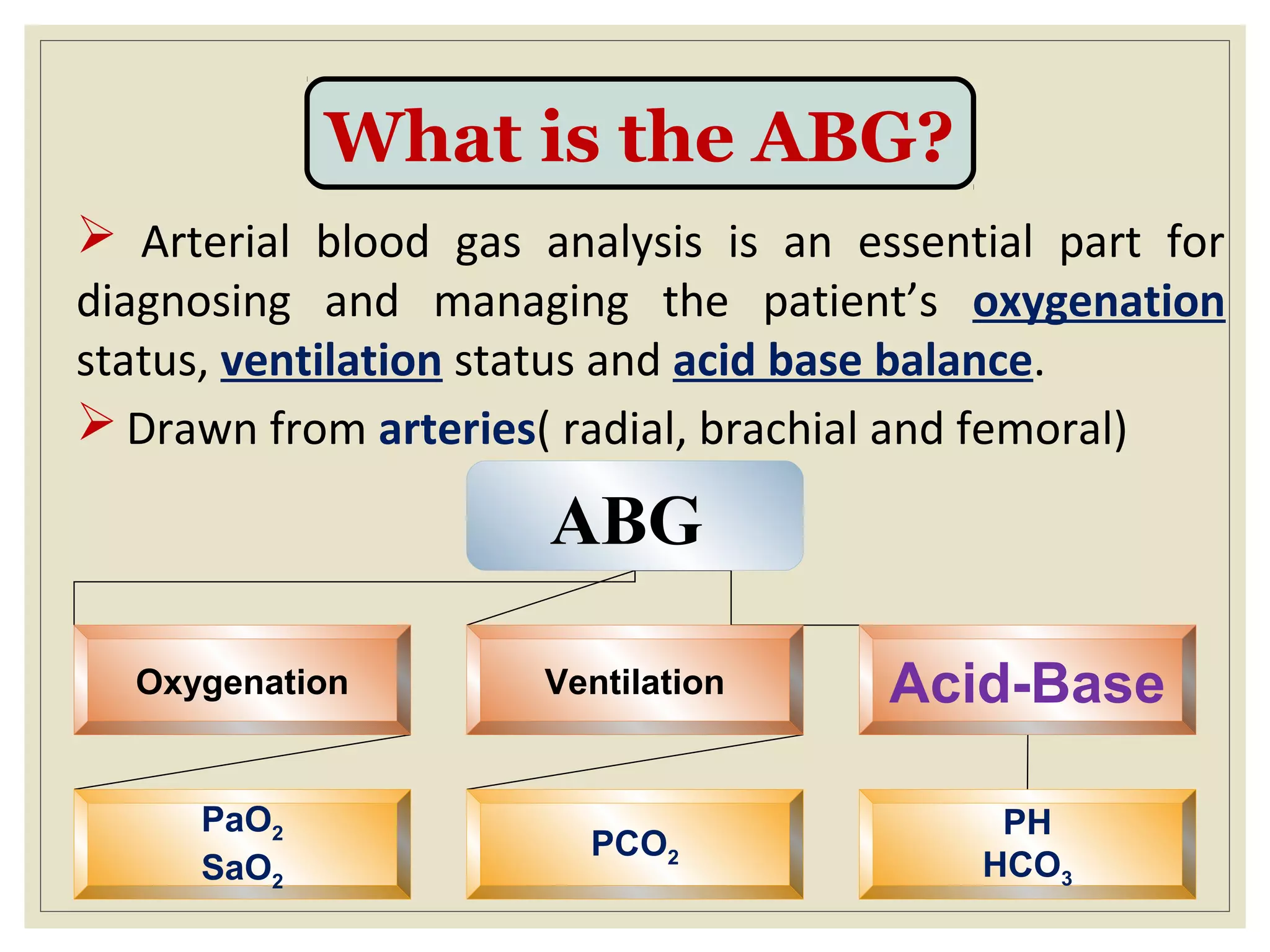
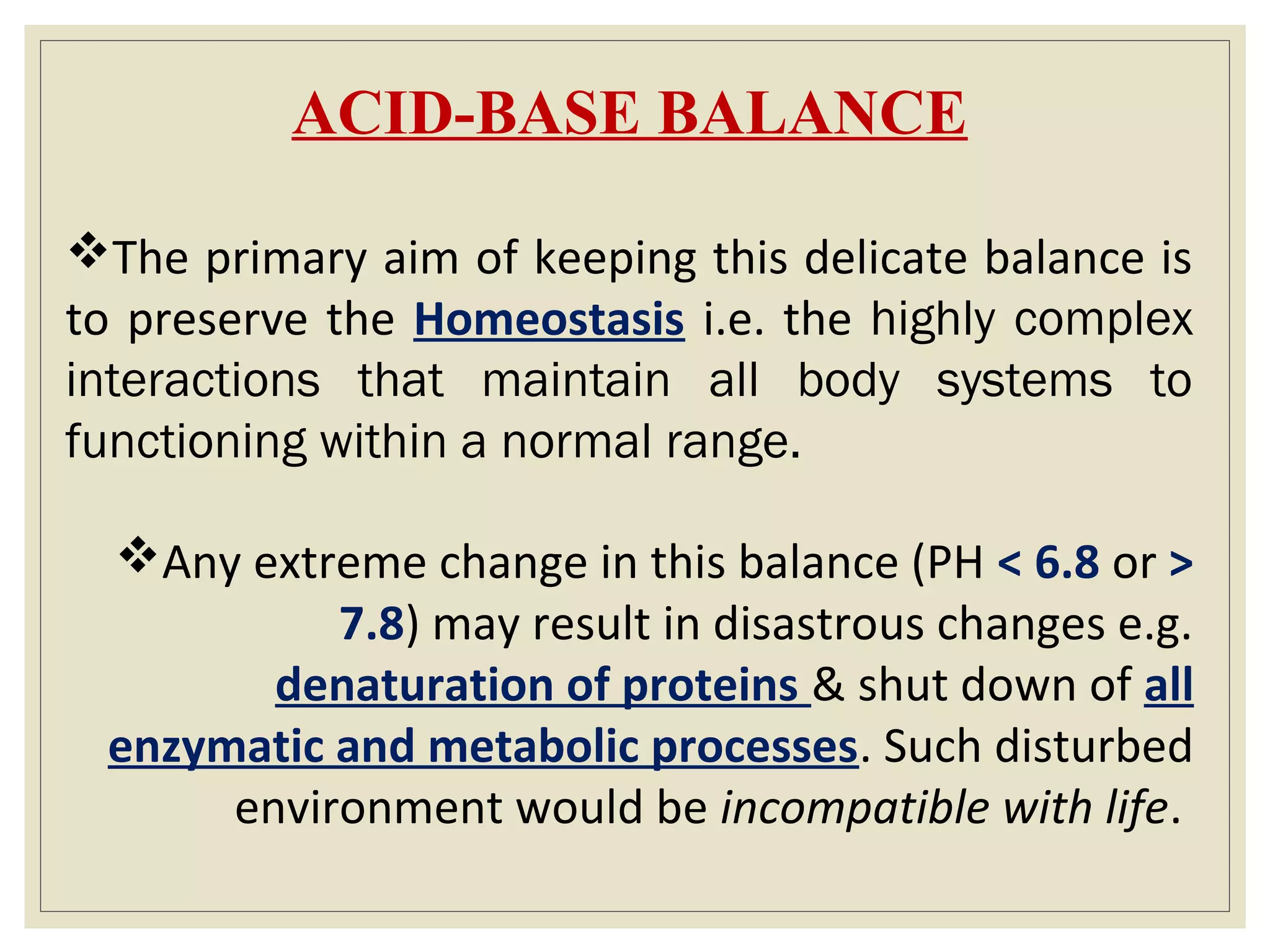
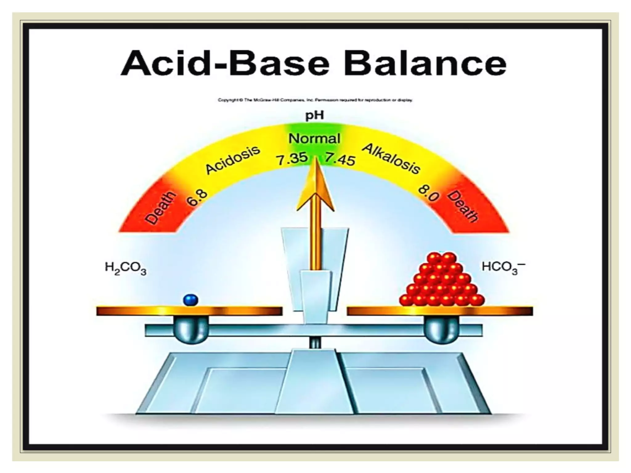
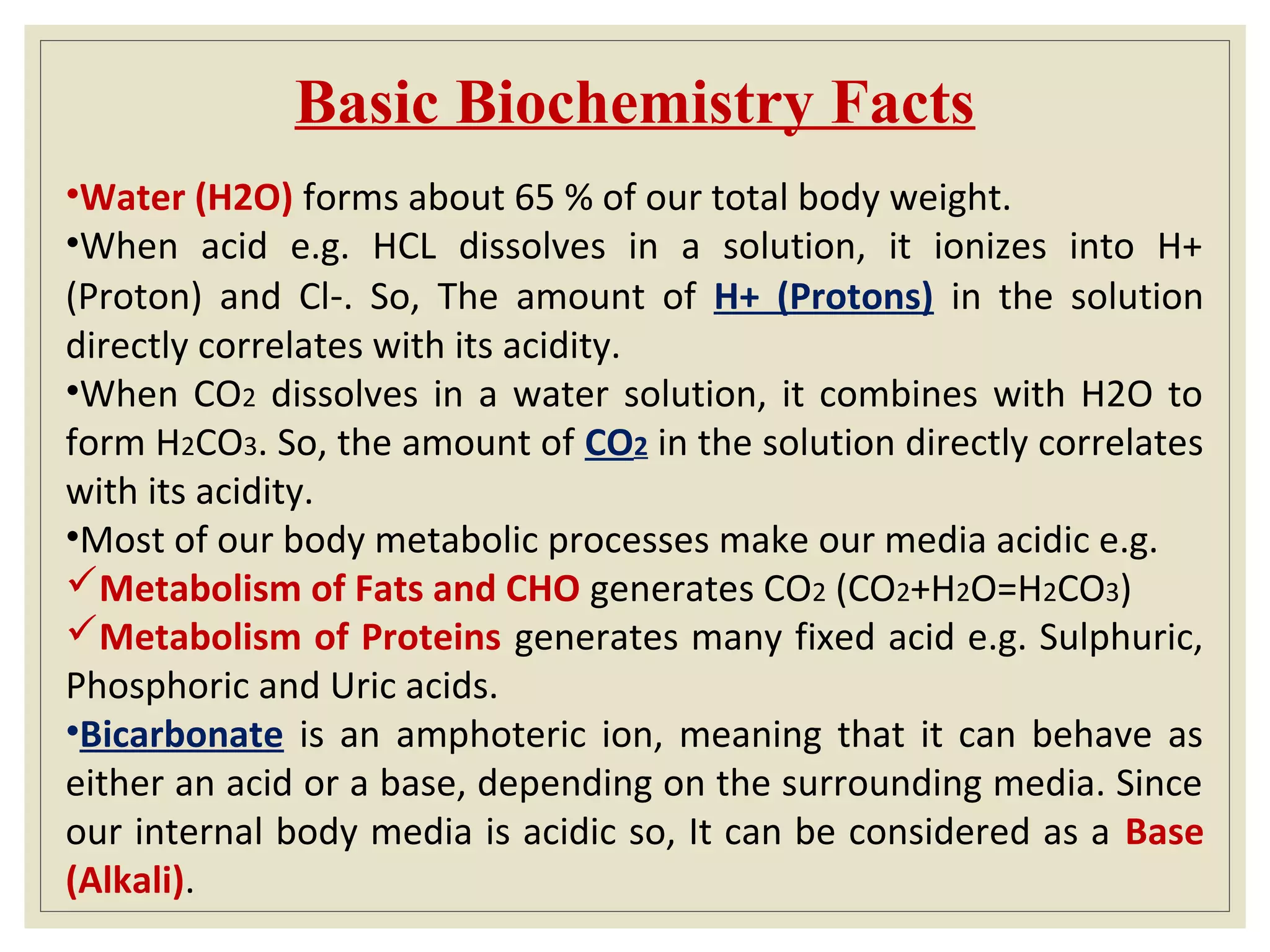
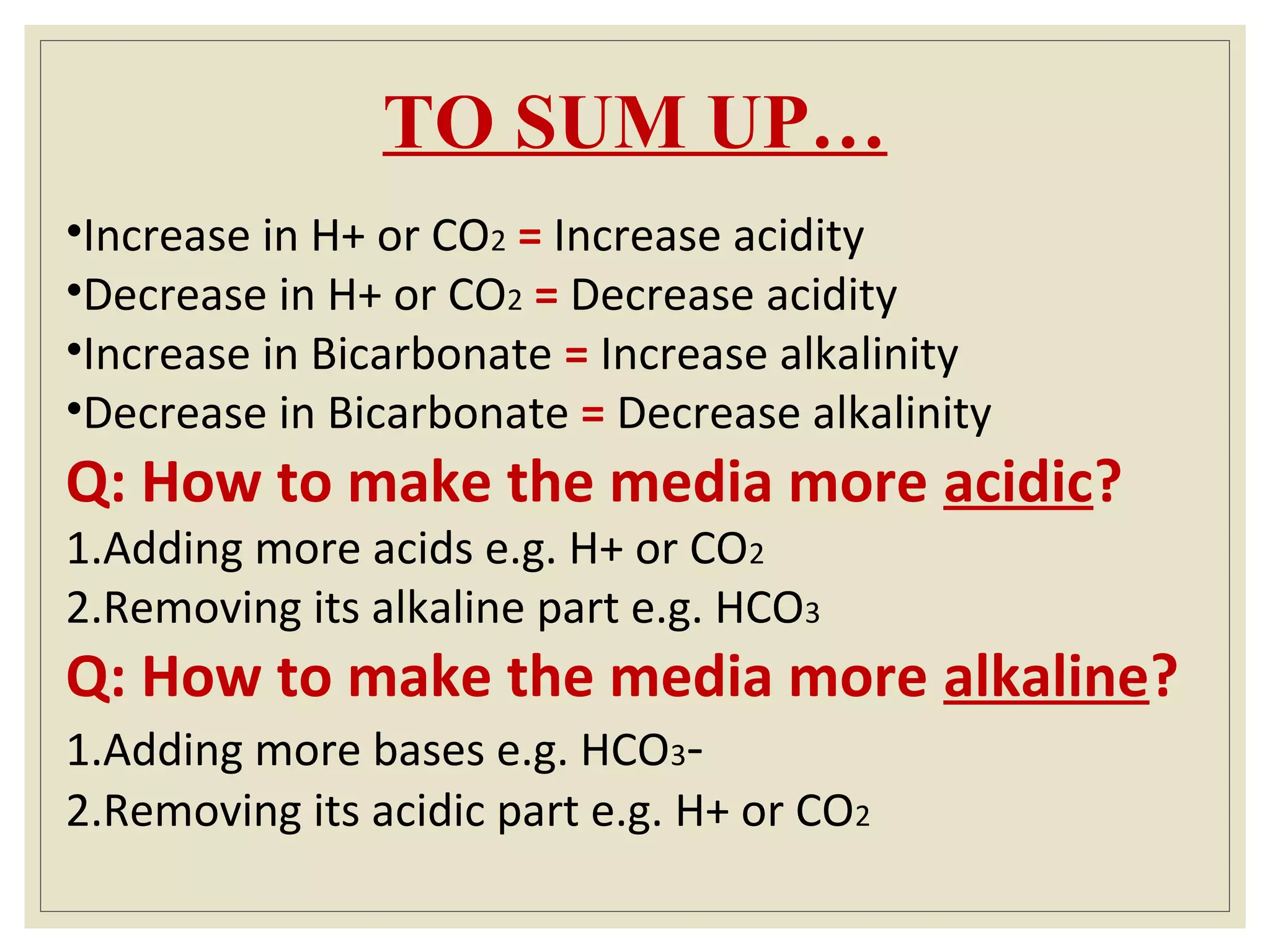
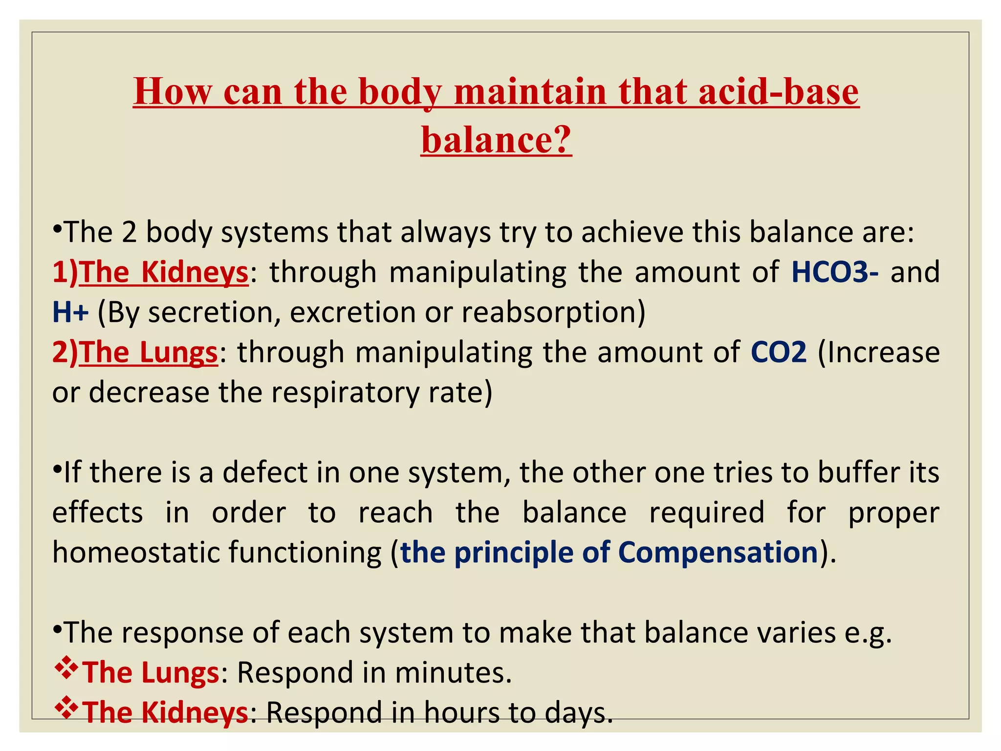
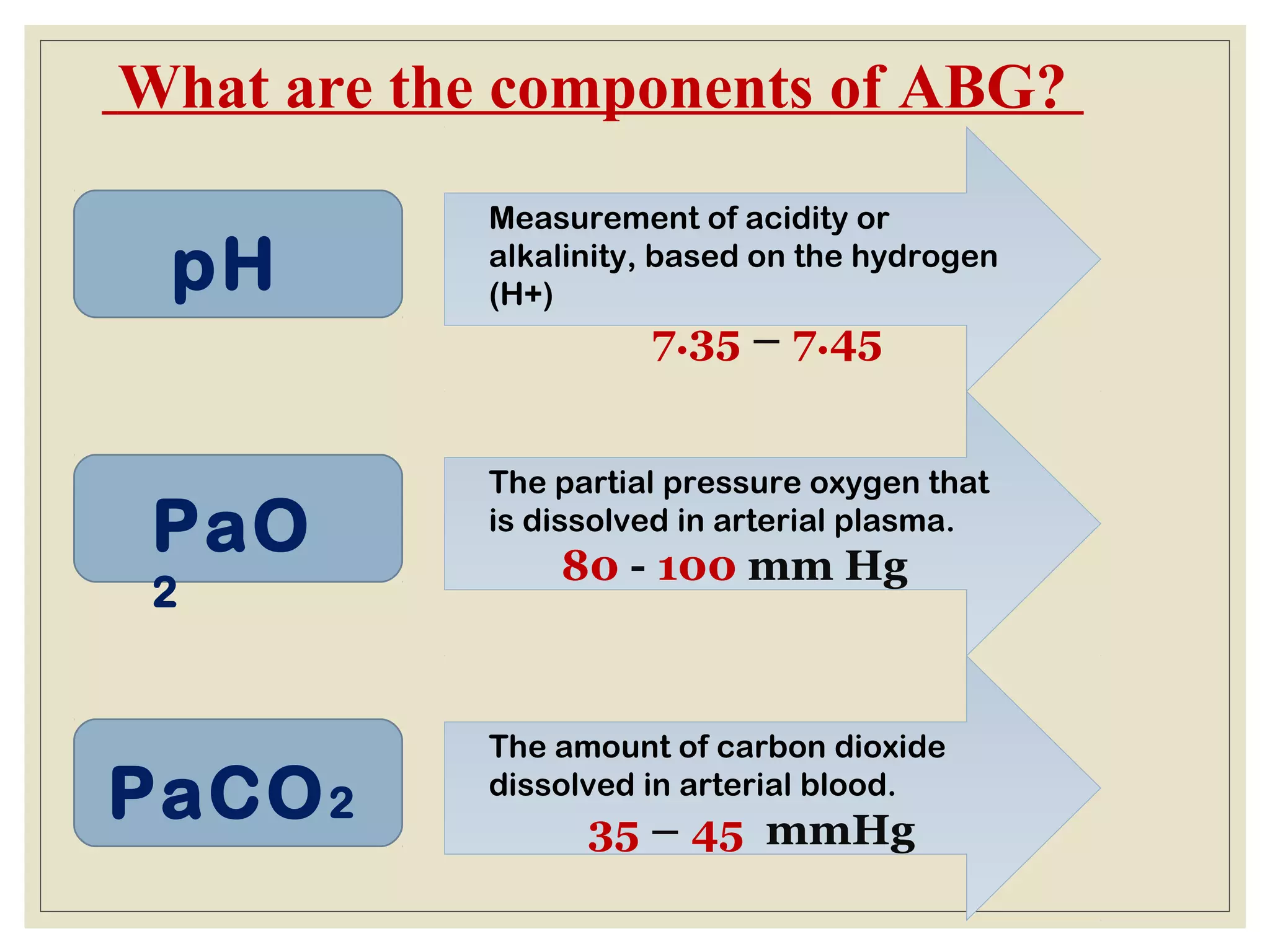
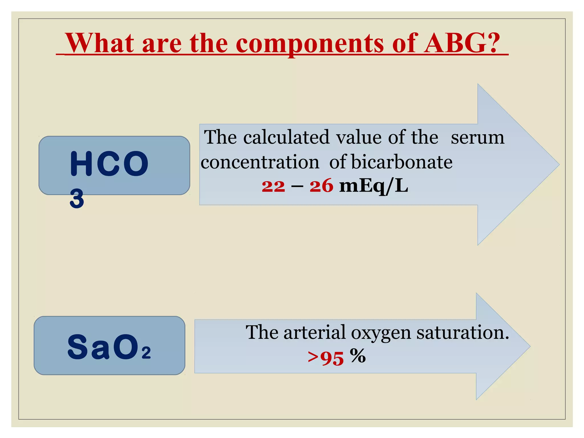
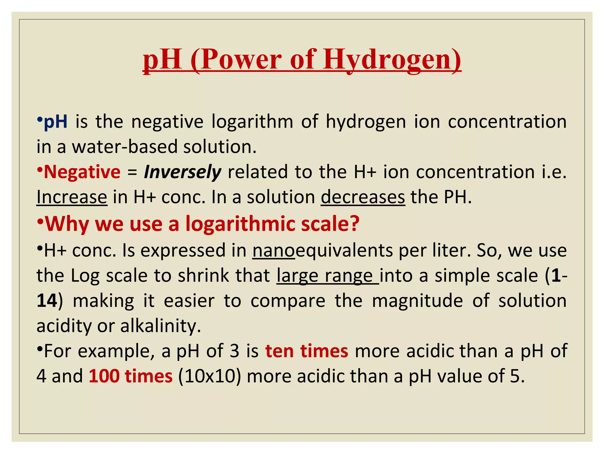
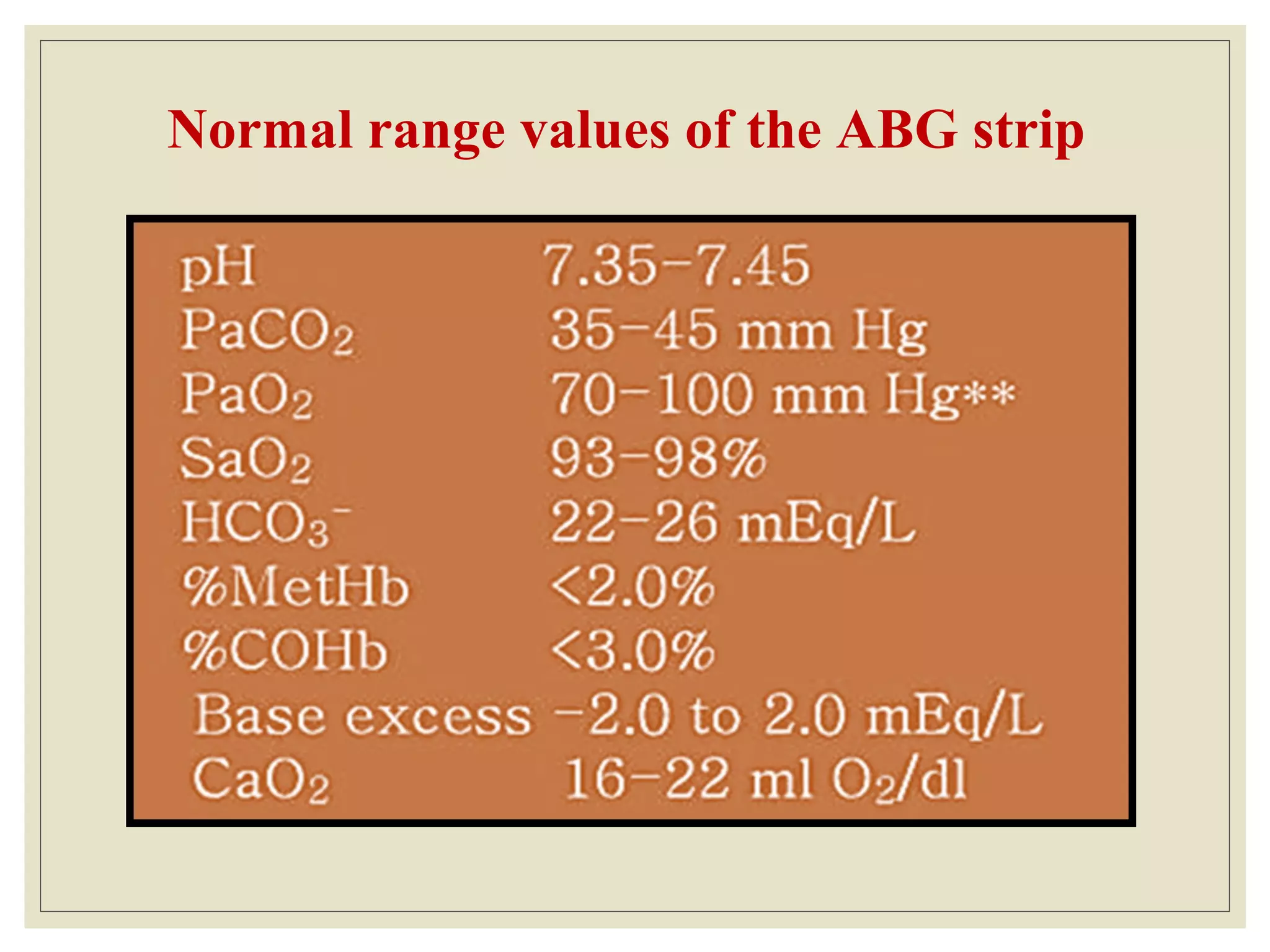
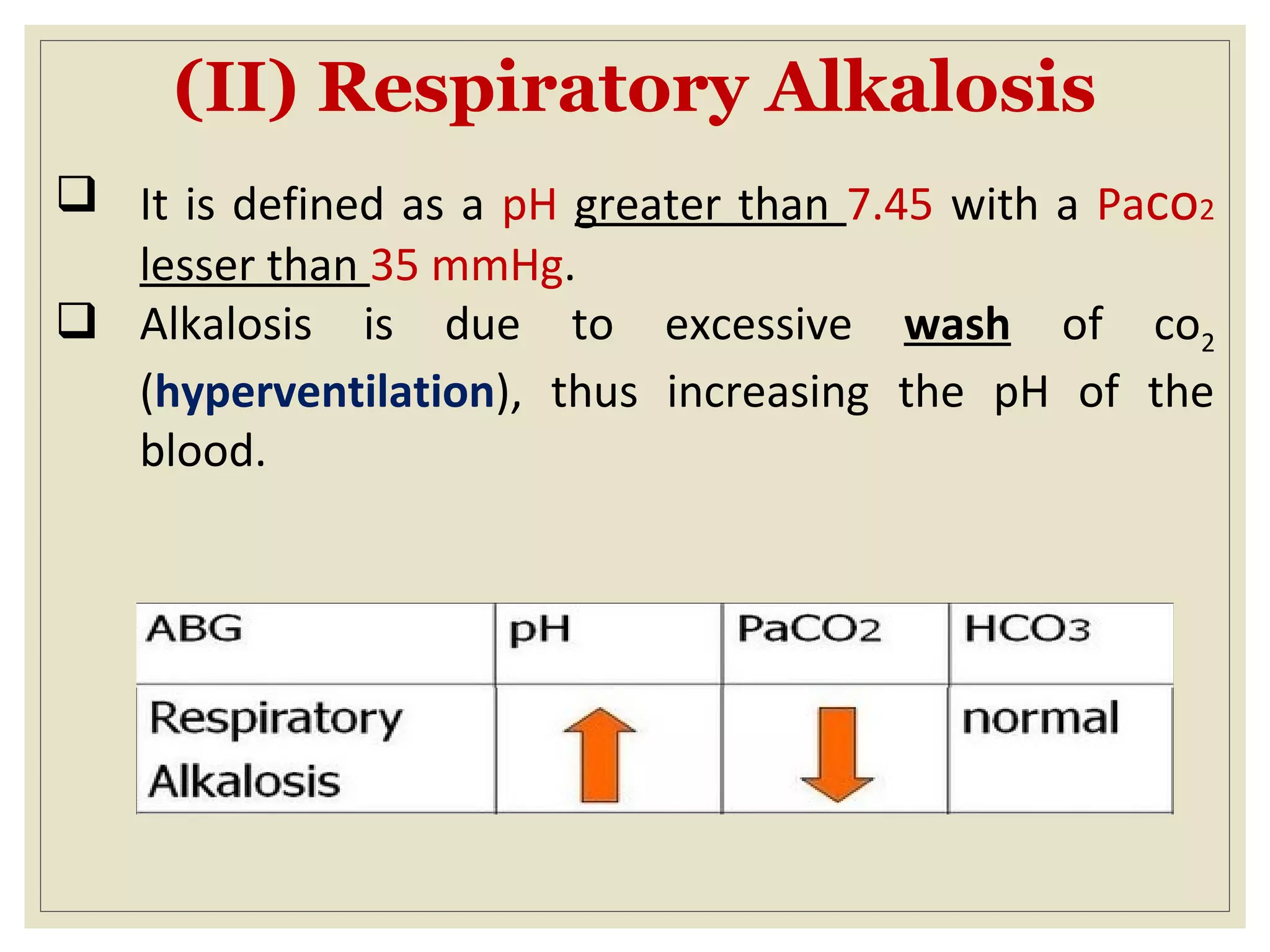
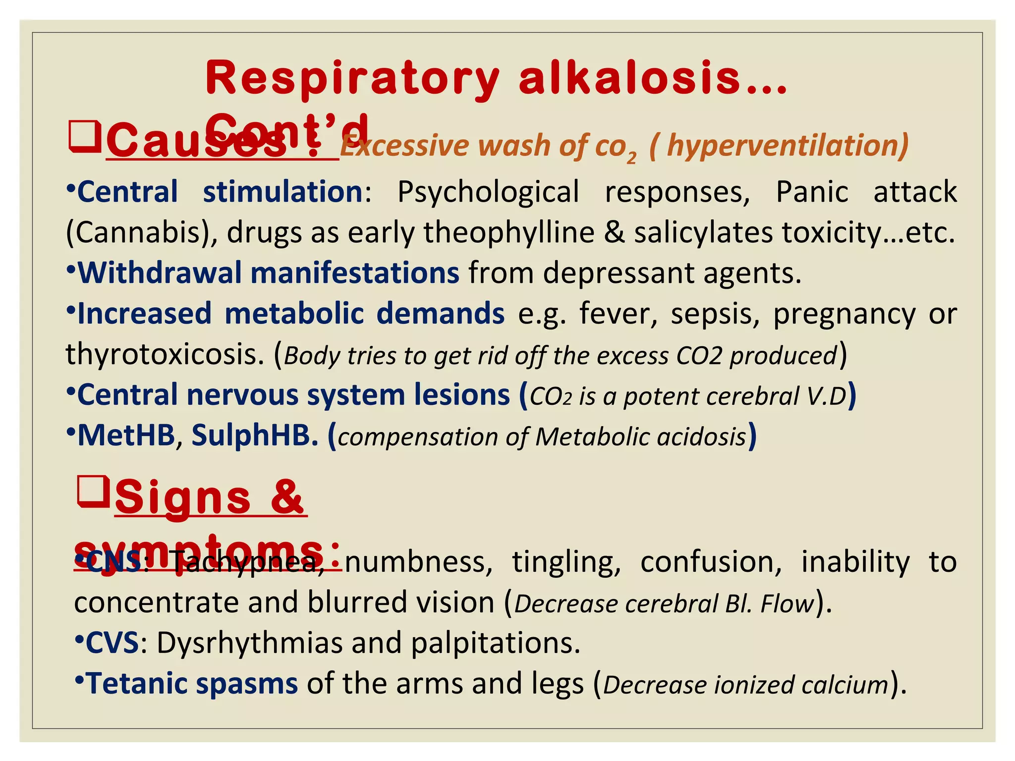
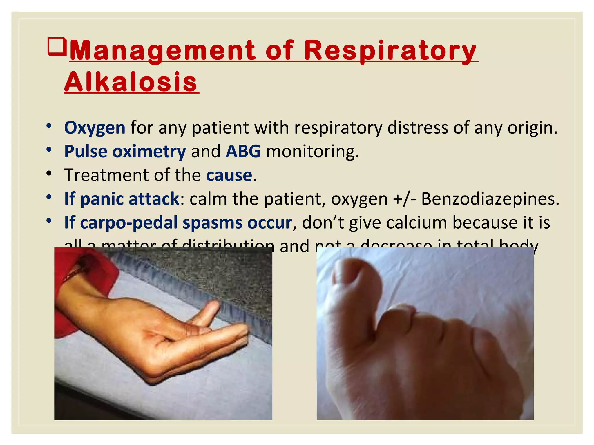
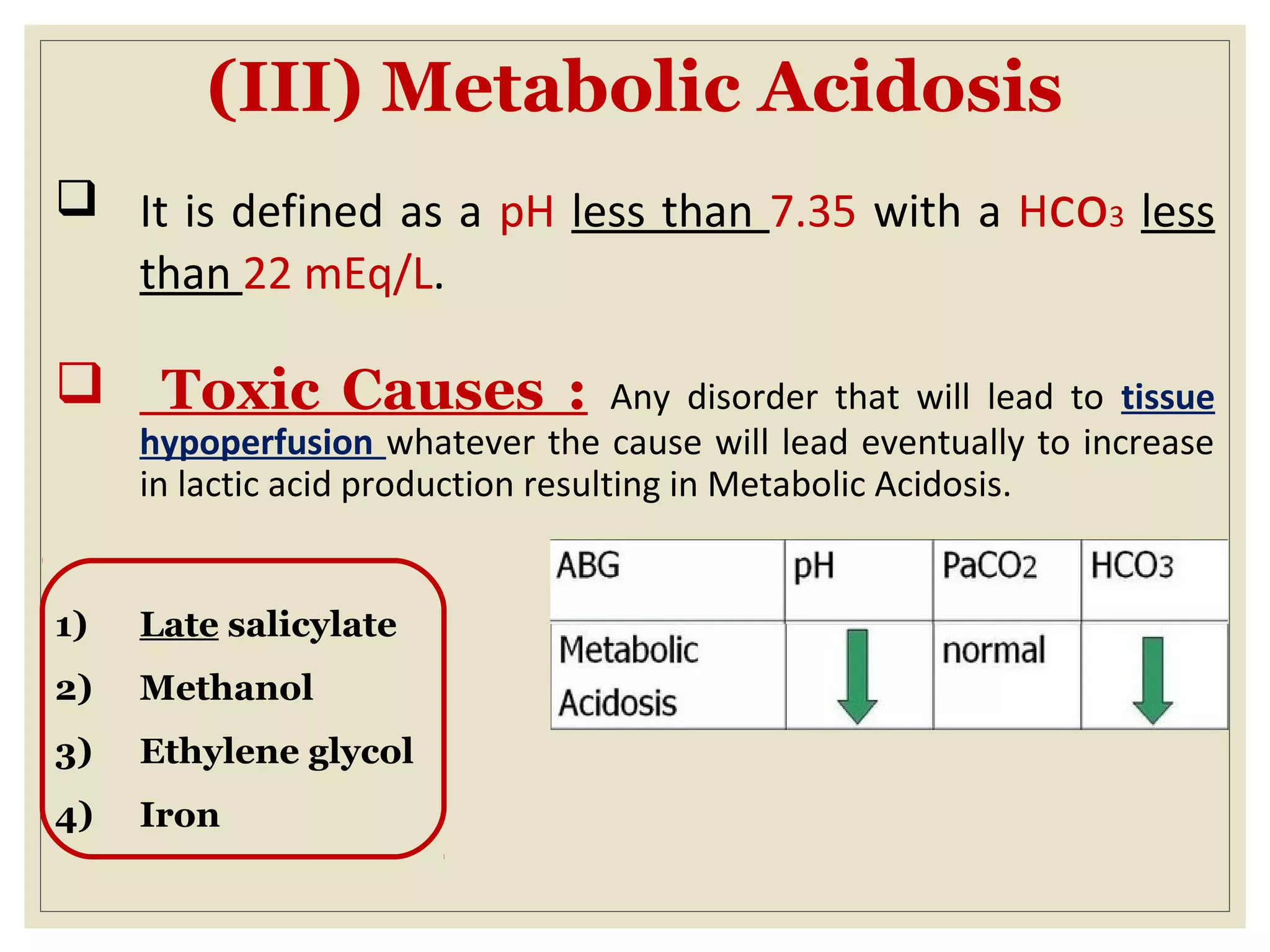
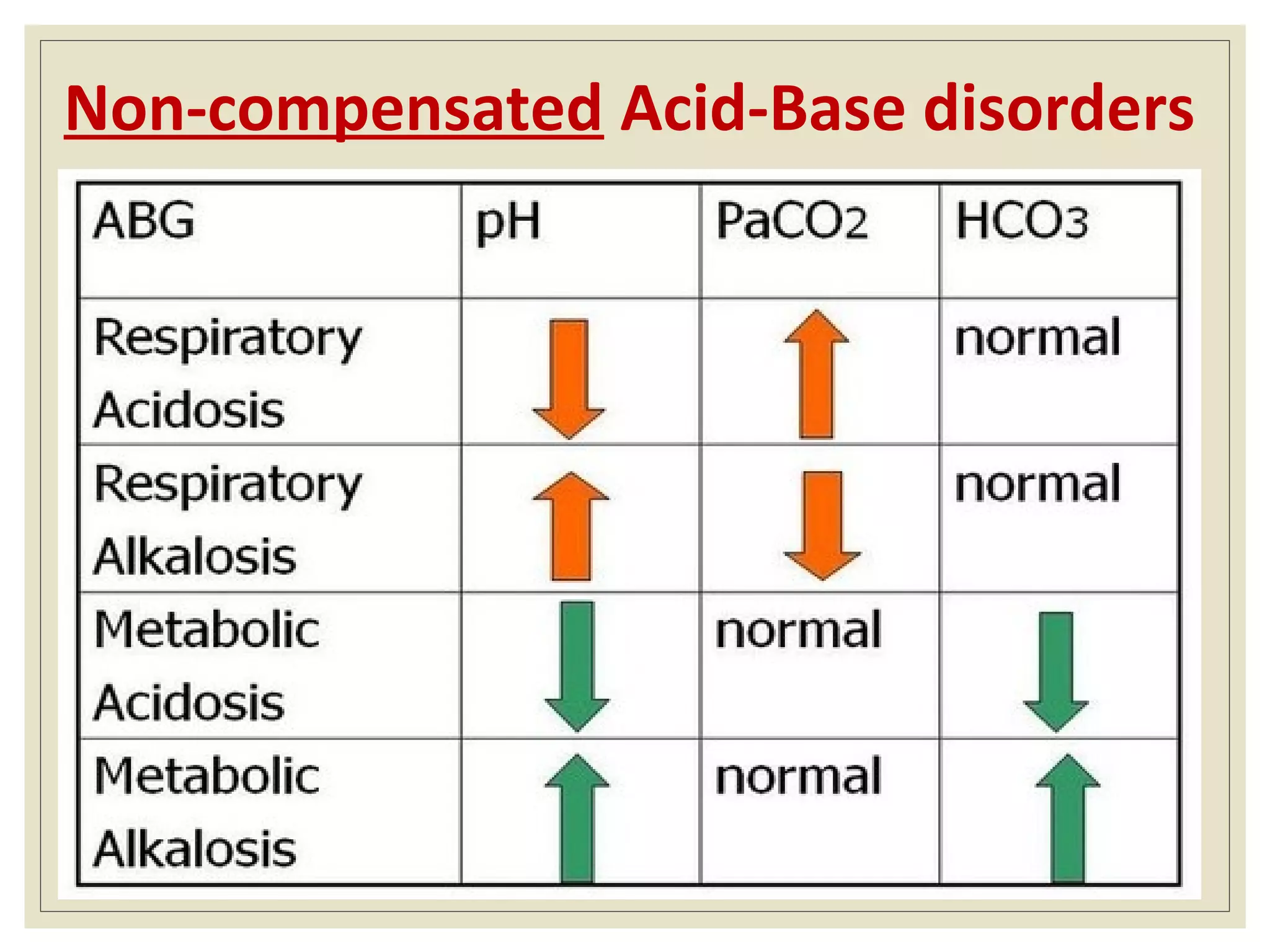
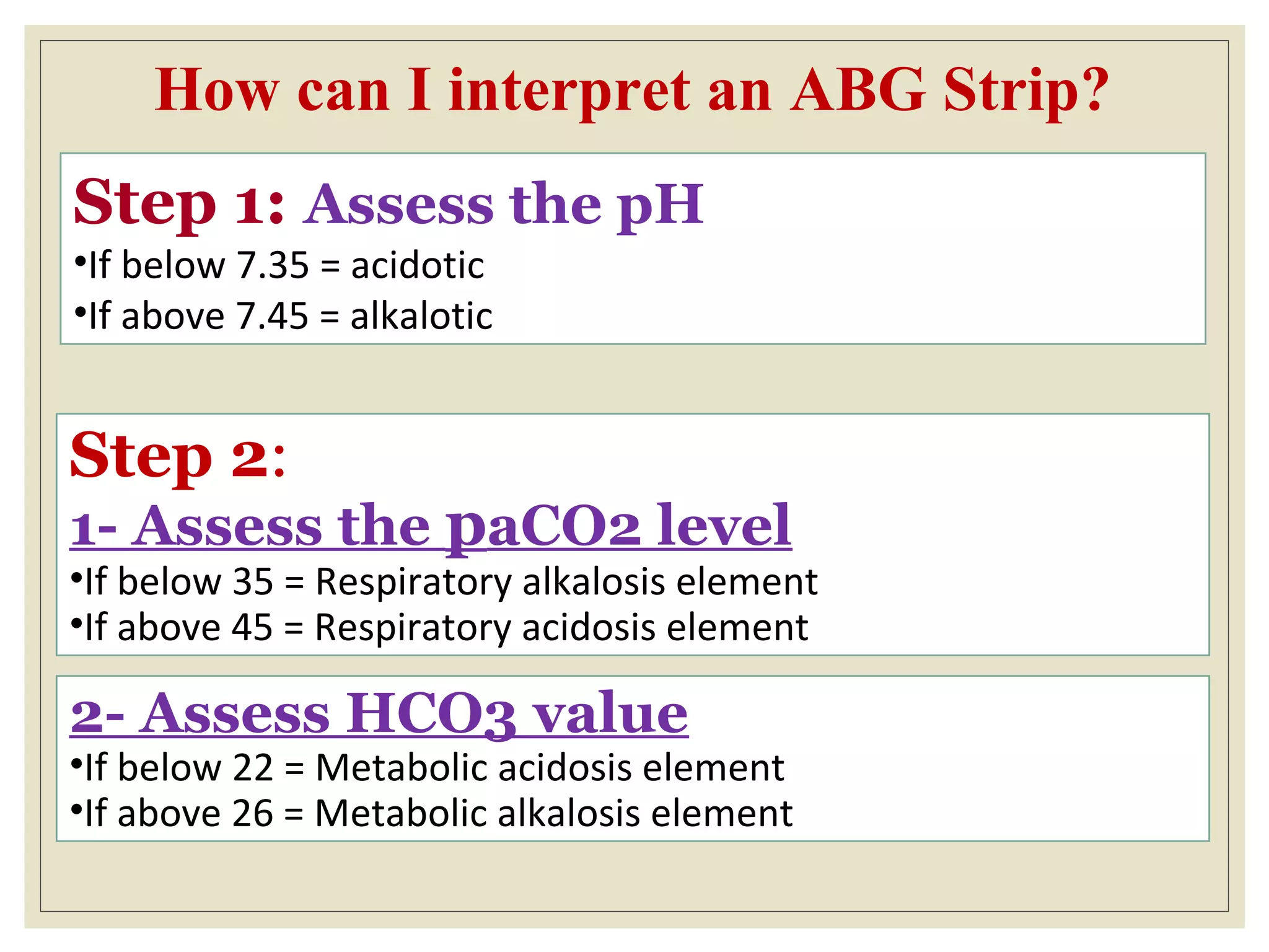
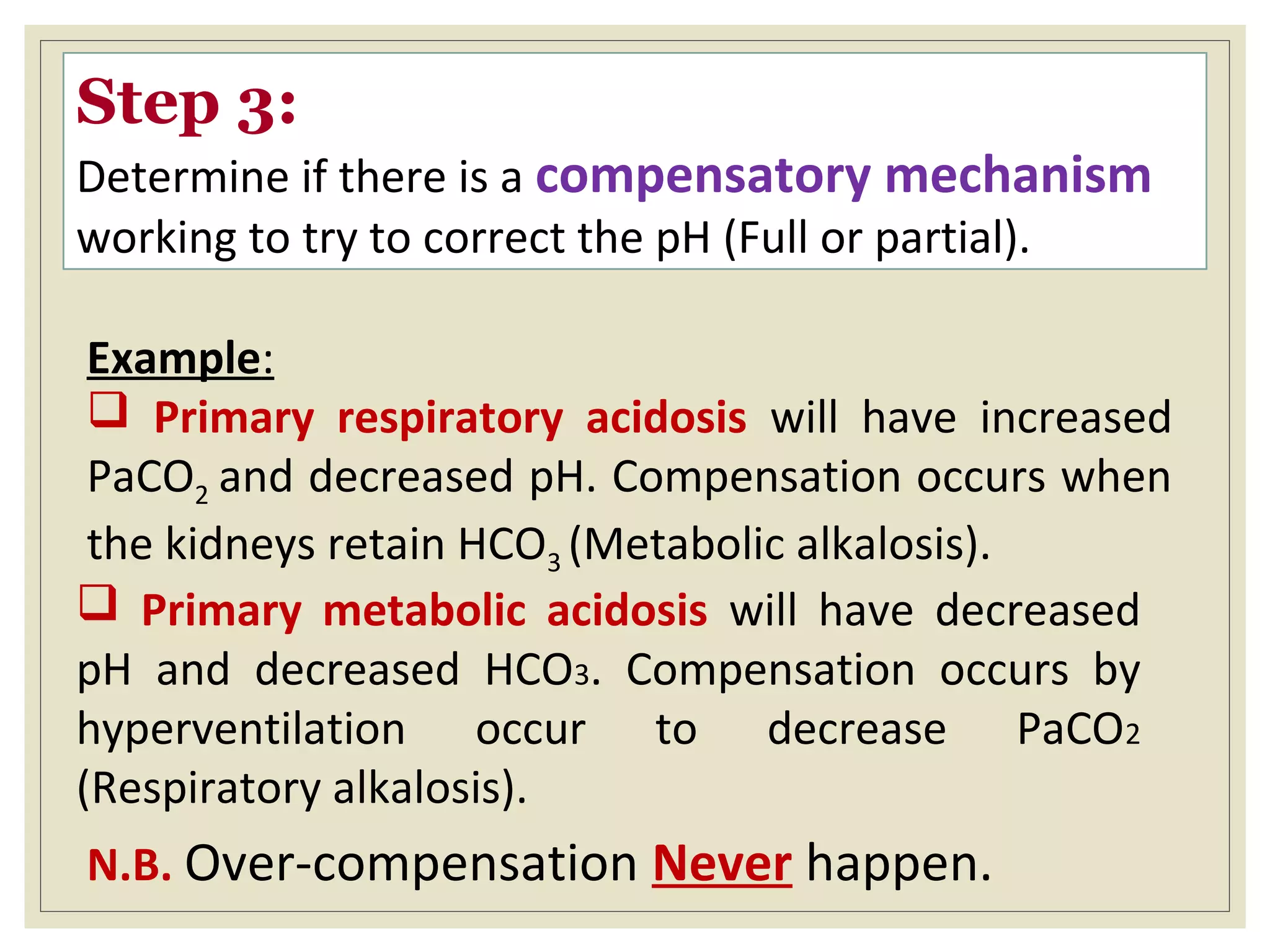
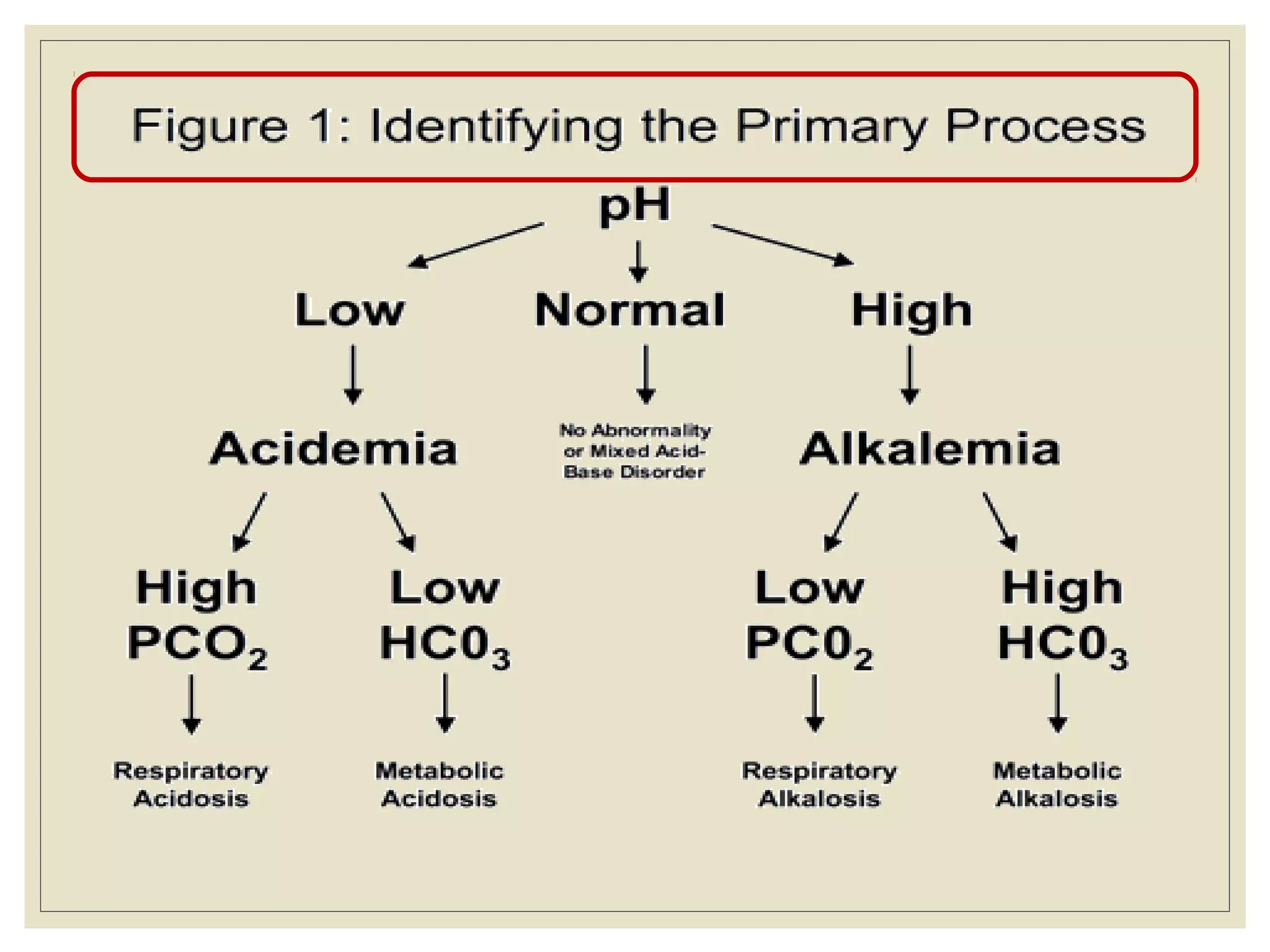
This document defines an arterial blood gas (ABG) analysis and its components. It discusses the normal acid-base balance and how acid-base imbalances present as respiratory or metabolic acidosis or alkalosis. Specific examples of each type of acid-base disorder are provided along with their typical causes, signs, symptoms, and management approaches. Common toxins that can cause acid-base disturbances are also listed. The document aims to equip readers to interpret ABG results in the clinical toxicology setting.
this slide focuses on all the acid base disorder pertaining to the respiratory system. it focus on the compensatory mechanism, causes, clinical features and treatment.
one can learn the step by step approach of ABG interpritation and its analysis from basics with the help of different case scenarios,Ref-NEJM article regarding physiological approach to acid base disbalance
Pulmonary edema is often caused by congestive heart failure. When the heart is not able to pump efficiently, blood can back up into the veins that take blood through the lungs. As the pressure in these blood vessels increases, fluid is pushed into the air spaces (alveoli) in the lungs.
Acute respiratory distress syndrome (ARDS) occurs when fluid builds up in the tiny, elastic air sacs (alveoli) in your lungs. The fluid keeps your lungs from filling with enough air, which means less oxygen reaches your bloodstream. This deprives your organs of the oxygen they need to function.
The CVP catheter is an important tool used to assess right ventricular function and systemic fluid status. Normal CVP is 2-6 mm Hg. CVP is elevated by : overhydration which increases venous return.
This article was originally a blog posted on Veritas Collaborative's website found here: http://veritascollaborative.com/blog/2016/07/building-a-life-worth-living-part-3
At Veritas Collaborative, we promise to drive a new standard of care in eating disorders treatment so individuals and families can thrive. The multidisciplinary treatment team members of Veritas Collaborative share a passion and a mission inspired by a collaborative community of care. Veritas Collaborative has three treatment center locations in Atlanta, GA, Durham, NC, and Richmond, VA. http://veritascollaborative.com/
An arterial-blood gas test measures the amounts of arterial gases, such as oxygen and carbon dioxide. An ABG test requires that a small volume of blood be drawn from the radial artery with a syringe and a thin needle, but sometimes the femoral artery in the groin or another site is used.
The common indications for ABGs are:
Respiratory compromise, which leads to hypoxia or diminished ventilation.
Peri- or postcardiopulmonary arrest or collapse.
Medical conditions that cause significant metabolic derangement, such as sepsis, diabetic ketoacidosis, renal failure, heart failure, toxic substance ingestion, drug overdose, trauma, or burns.
Evaluating the effectiveness of therapies, monitoring the patient's clinical status, and determining treatment needs. For instance, clinicians often titrate oxygenation therapy, adjust the level of ventilator support, and make decisions about fluid and electrolyte therapy based on ABG results.
During the perioperative phase of major surgeries, which includes the preoperative, intraoperative, and postoperative care of the patient.
The components of an ABG analysis are PaO2, SaO2, hydrogen ion concentration (pH), PaCO2, HCO3-, base excess, and serum levels of hemoglobin, lactate, glucose, and electrolytes (sodium, potassium, calcium, and chloride).
First aid course focusing on management of burns, wounds of different types, disturbed conscious level and chemical intoxication whether by inhalation, ingestion or skin exposure.
Provides a simple organized way for ABG analysis with special emphasis on Acid-base balance interpretation & its crucial rule in clinical toxicology practice.
MANAGEMENT OF ATRIOVENTRICULAR CONDUCTION BLOCK.pdfJim Jacob Roy
Cardiac conduction defects can occur due to various causes.
Atrioventricular conduction blocks ( AV blocks ) are classified into 3 types.
This document describes the acute management of AV block.
These lecture slides, by Dr Sidra Arshad, offer a quick overview of physiological basis of a normal electrocardiogram.
Learning objectives:
1. Define an electrocardiogram (ECG) and electrocardiography
2. Describe how dipoles generated by the heart produce the waveforms of the ECG
3. Describe the components of a normal electrocardiogram of a typical bipolar leads (limb II)
4. Differentiate between intervals and segments
5. Enlist some common indications for obtaining an ECG
Study Resources:
1. Chapter 11, Guyton and Hall Textbook of Medical Physiology, 14th edition
2. Chapter 9, Human Physiology - From Cells to Systems, Lauralee Sherwood, 9th edition
3. Chapter 29, Ganong’s Review of Medical Physiology, 26th edition
4. Electrocardiogram, StatPearls - https://www.ncbi.nlm.nih.gov/books/NBK549803/
5. ECG in Medical Practice by ABM Abdullah, 4th edition
6. ECG Basics, http://www.nataliescasebook.com/tag/e-c-g-basics
- Video recording of this lecture in English language: https://youtu.be/lK81BzxMqdo
- Video recording of this lecture in Arabic language: https://youtu.be/Ve4P0COk9OI
- Link to download the book free: https://nephrotube.blogspot.com/p/nephrotube-nephrology-books.html
- Link to NephroTube website: www.NephroTube.com
- Link to NephroTube social media accounts: https://nephrotube.blogspot.com/p/join-nephrotube-on-social-media.html
micro teaching on communication m.sc nursing.pdfAnurag Sharma
Microteaching is a unique model of practice teaching. It is a viable instrument for the. desired change in the teaching behavior or the behavior potential which, in specified types of real. classroom situations, tends to facilitate the achievement of specified types of objectives.
Ethanol (CH3CH2OH), or beverage alcohol, is a two-carbon alcohol
that is rapidly distributed in the body and brain. Ethanol alters many
neurochemical systems and has rewarding and addictive properties. It
is the oldest recreational drug and likely contributes to more morbidity,
mortality, and public health costs than all illicit drugs combined. The
5th edition of the Diagnostic and Statistical Manual of Mental Disorders
(DSM-5) integrates alcohol abuse and alcohol dependence into a single
disorder called alcohol use disorder (AUD), with mild, moderate,
and severe subclassifications (American Psychiatric Association, 2013).
In the DSM-5, all types of substance abuse and dependence have been
combined into a single substance use disorder (SUD) on a continuum
from mild to severe. A diagnosis of AUD requires that at least two of
the 11 DSM-5 behaviors be present within a 12-month period (mild
AUD: 2–3 criteria; moderate AUD: 4–5 criteria; severe AUD: 6–11 criteria).
The four main behavioral effects of AUD are impaired control over
drinking, negative social consequences, risky use, and altered physiological
effects (tolerance, withdrawal). This chapter presents an overview
of the prevalence and harmful consequences of AUD in the U.S.,
the systemic nature of the disease, neurocircuitry and stages of AUD,
comorbidities, fetal alcohol spectrum disorders, genetic risk factors, and
pharmacotherapies for AUD.
The prostate is an exocrine gland of the male mammalian reproductive system
It is a walnut-sized gland that forms part of the male reproductive system and is located in front of the rectum and just below the urinary bladder
Function is to store and secrete a clear, slightly alkaline fluid that constitutes 10-30% of the volume of the seminal fluid that along with the spermatozoa, constitutes semen
A healthy human prostate measures (4cm-vertical, by 3cm-horizontal, 2cm ant-post ).
It surrounds the urethra just below the urinary bladder. It has anterior, median, posterior and two lateral lobes
It’s work is regulated by androgens which are responsible for male sex characteristics
Generalised disease of the prostate due to hormonal derangement which leads to non malignant enlargement of the gland (increase in the number of epithelial cells and stromal tissue)to cause compression of the urethra leading to symptoms (LUTS
Tom Selleck Health: A Comprehensive Look at the Iconic Actor’s Wellness Journeygreendigital
Tom Selleck, an enduring figure in Hollywood. has captivated audiences for decades with his rugged charm, iconic moustache. and memorable roles in television and film. From his breakout role as Thomas Magnum in Magnum P.I. to his current portrayal of Frank Reagan in Blue Bloods. Selleck's career has spanned over 50 years. But beyond his professional achievements. fans have often been curious about Tom Selleck Health. especially as he has aged in the public eye.
Follow us on: Pinterest
Introduction
Many have been interested in Tom Selleck health. not only because of his enduring presence on screen but also because of the challenges. and lifestyle choices he has faced and made over the years. This article delves into the various aspects of Tom Selleck health. exploring his fitness regimen, diet, mental health. and the challenges he has encountered as he ages. We'll look at how he maintains his well-being. the health issues he has faced, and his approach to ageing .
Early Life and Career
Childhood and Athletic Beginnings
Tom Selleck was born on January 29, 1945, in Detroit, Michigan, and grew up in Sherman Oaks, California. From an early age, he was involved in sports, particularly basketball. which played a significant role in his physical development. His athletic pursuits continued into college. where he attended the University of Southern California (USC) on a basketball scholarship. This early involvement in sports laid a strong foundation for his physical health and disciplined lifestyle.
Transition to Acting
Selleck's transition from an athlete to an actor came with its physical demands. His first significant role in "Magnum P.I." required him to perform various stunts and maintain a fit appearance. This role, which he played from 1980 to 1988. necessitated a rigorous fitness routine to meet the show's demands. setting the stage for his long-term commitment to health and wellness.
Fitness Regimen
Workout Routine
Tom Selleck health and fitness regimen has evolved. adapting to his changing roles and age. During his "Magnum, P.I." days. Selleck's workouts were intense and focused on building and maintaining muscle mass. His routine included weightlifting, cardiovascular exercises. and specific training for the stunts he performed on the show.
Selleck adjusted his fitness routine as he aged to suit his body's needs. Today, his workouts focus on maintaining flexibility, strength, and cardiovascular health. He incorporates low-impact exercises such as swimming, walking, and light weightlifting. This balanced approach helps him stay fit without putting undue strain on his joints and muscles.
Importance of Flexibility and Mobility
In recent years, Selleck has emphasized the importance of flexibility and mobility in his fitness regimen. Understanding the natural decline in muscle mass and joint flexibility with age. he includes stretching and yoga in his routine. These practices help prevent injuries, improve posture, and maintain mobilit
Title: Sense of Taste
Presenter: Dr. Faiza, Assistant Professor of Physiology
Qualifications:
MBBS (Best Graduate, AIMC Lahore)
FCPS Physiology
ICMT, CHPE, DHPE (STMU)
MPH (GC University, Faisalabad)
MBA (Virtual University of Pakistan)
Learning Objectives:
Describe the structure and function of taste buds.
Describe the relationship between the taste threshold and taste index of common substances.
Explain the chemical basis and signal transduction of taste perception for each type of primary taste sensation.
Recognize different abnormalities of taste perception and their causes.
Key Topics:
Significance of Taste Sensation:
Differentiation between pleasant and harmful food
Influence on behavior
Selection of food based on metabolic needs
Receptors of Taste:
Taste buds on the tongue
Influence of sense of smell, texture of food, and pain stimulation (e.g., by pepper)
Primary and Secondary Taste Sensations:
Primary taste sensations: Sweet, Sour, Salty, Bitter, Umami
Chemical basis and signal transduction mechanisms for each taste
Taste Threshold and Index:
Taste threshold values for Sweet (sucrose), Salty (NaCl), Sour (HCl), and Bitter (Quinine)
Taste index relationship: Inversely proportional to taste threshold
Taste Blindness:
Inability to taste certain substances, particularly thiourea compounds
Example: Phenylthiocarbamide
Structure and Function of Taste Buds:
Composition: Epithelial cells, Sustentacular/Supporting cells, Taste cells, Basal cells
Features: Taste pores, Taste hairs/microvilli, and Taste nerve fibers
Location of Taste Buds:
Found in papillae of the tongue (Fungiform, Circumvallate, Foliate)
Also present on the palate, tonsillar pillars, epiglottis, and proximal esophagus
Mechanism of Taste Stimulation:
Interaction of taste substances with receptors on microvilli
Signal transduction pathways for Umami, Sweet, Bitter, Sour, and Salty tastes
Taste Sensitivity and Adaptation:
Decrease in sensitivity with age
Rapid adaptation of taste sensation
Role of Saliva in Taste:
Dissolution of tastants to reach receptors
Washing away the stimulus
Taste Preferences and Aversions:
Mechanisms behind taste preference and aversion
Influence of receptors and neural pathways
Impact of Sensory Nerve Damage:
Degeneration of taste buds if the sensory nerve fiber is cut
Abnormalities of Taste Detection:
Conditions: Ageusia, Hypogeusia, Dysgeusia (parageusia)
Causes: Nerve damage, neurological disorders, infections, poor oral hygiene, adverse drug effects, deficiencies, aging, tobacco use, altered neurotransmitter levels
Neurotransmitters and Taste Threshold:
Effects of serotonin (5-HT) and norepinephrine (NE) on taste sensitivity
Supertasters:
25% of the population with heightened sensitivity to taste, especially bitterness
Increased number of fungiform papillae
2. Objectives
1)Define ABG & its indications.
2)Describe components of ABG & their normal
values.
3)Acid-Base imbalance.
4)Interpret ABG changes in clinical toxicology
practice.
3. What is the ABG?
Arterial blood gas analysis is an essential part for
diagnosing and managing the patient’s oxygenation
status, ventilation status and acid base balance.
Drawn from arteries( radial, brachial and femoral)
ABG
Oxygenation Ventilation Acid-Base
PaO2
SaO2
PCO2
PH
HCO3
4. ACID-BASE BALANCE
The primary aim of keeping this delicate balance is
to preserve the Homeostasis i.e. the highly complex
interactions that maintain all body systems to
functioning within a normal range.
Any extreme change in this balance (PH < 6.8 or >
7.8) may result in disastrous changes e.g.
denaturation of proteins & shut down of all
enzymatic and metabolic processes. Such disturbed
environment would be incompatible with life.
5.
6. Basic Biochemistry Facts
•Water (H2O) forms about 65 % of our total body weight.
•When acid e.g. HCL dissolves in a solution, it ionizes into H+
(Proton) and Cl-. So, The amount of H+ (Protons) in the solution
directly correlates with its acidity.
•When CO2 dissolves in a water solution, it combines with H2O to
form H2CO3. So, the amount of CO2 in the solution directly correlates
with its acidity.
•Most of our body metabolic processes make our media acidic e.g.
Metabolism of Fats and CHO generates CO2 (CO2+H2O=H2CO3)
Metabolism of Proteins generates many fixed acid e.g. Sulphuric,
Phosphoric and Uric acids.
•Bicarbonate is an amphoteric ion, meaning that it can behave as
either an acid or a base, depending on the surrounding media. Since
our internal body media is acidic so, It can be considered as a Base
(Alkali).
7. TO SUM UP…
•Increase in H+ or CO2 = Increase acidity
•Decrease in H+ or CO2 = Decrease acidity
•Increase in Bicarbonate = Increase alkalinity
•Decrease in Bicarbonate = Decrease alkalinity
Q: How to make the media more acidic?
1.Adding more acids e.g. H+ or CO2
2.Removing its alkaline part e.g. HCO3
Q: How to make the media more alkaline?
1.Adding more bases e.g. HCO3-
2.Removing its acidic part e.g. H+ or CO2
8. How can the body maintain that acid-base
balance?
•The 2 body systems that always try to achieve this balance are:
1)The Kidneys: through manipulating the amount of HCO3- and
H+ (By secretion, excretion or reabsorption)
2)The Lungs: through manipulating the amount of CO2 (Increase
or decrease the respiratory rate)
•If there is a defect in one system, the other one tries to buffer its
effects in order to reach the balance required for proper
homeostatic functioning (the principle of Compensation).
•The response of each system to make that balance varies e.g.
The Lungs: Respond in minutes.
The Kidneys: Respond in hours to days.
9. What are the components of ABG?
pH
Measurement of acidity or
alkalinity, based on the hydrogen
(H+)
7.35 – 7.45
PaO
2
The partial pressure oxygen that
is dissolved in arterial plasma.
80 - 100 mm Hg
PaCO2
The amount of carbon dioxide
dissolved in arterial blood.
35 – 45 mmHg
10. What are the components of ABG?
HCO
3
The calculated value of the serum
concentration of bicarbonate
22 – 26 mEq/L
SaO2
The arterial oxygen saturation.
>95 %
11. pH (Power of Hydrogen)
•pH is the negative logarithm of hydrogen ion concentration
in a water-based solution.
•Negative = Inversely related to the H+ ion concentration i.e.
Increase in H+ conc. In a solution decreases the PH.
•Why we use a logarithmic scale?
•H+ conc. Is expressed in nanoequivalents per liter. So, we use
the Log scale to shrink that large range into a simple scale (1-
14) making it easier to compare the magnitude of solution
acidity or alkalinity.
•For example, a pH of 3 is ten times more acidic than a pH of
4 and 100 times (10x10) more acidic than a pH value of 5.
13. ACID BASE DISORDERSACID BASE DISORDERS
BASIC CONCEPTS
•ABG shouldn’t be used alone in the diagnosis. Correlate the
clinical findings with the other lab and imaging studies to get a
panoramic assessment of the patient’s condition.
•ABG findings can assist not only in reaching a diagnosis, but
also in determining the prognosis of a patient.
14. (I) Respiratory Acidosis
It is defined as a pH less than 7.35 with a Paco2
greater than 45 mmHg.
Acidosis is the accumulation of co2 which
combines with water in the body to produce
carbonic acid, thus lowering the pH of the
blood.
15. Toxic Causes :
•Any condition that results in hypoventilation can cause
respiratory acidosis.
(a)
Central
(b)
Peripheral
CNS depression
opiates, sedatives,
anesthesia, methanol,
ethylene glycol...etc.
1-Respiratory muscle
paralysis
e.g. botulism
2- lung disease
e.g. pulmonary edema
3- respiratory passage
obstruction
e.g. organophosphorus
16. Signs & symptoms of Respiratory
Acidosis:
•Respiratory: Respiratory distress & shallow respiration.
•Nervous: (CO2 Narcosis) Headache, restlessness and
confusion. If co2 is extremely high, drowsiness and
unresponsiveness may be noted.
•CVS: Tachycardia and Dysrhythmias due to myocardial hypoxia.
Management:
•Oxygen & suctioning as needed.
•Pulse oximetry & ABG follow up.
•Treatment of the cause e.g. pneumothorax, severe pain (Rib
fracture) and CNS depressants toxicity.
•If the cause can not be readily resolved, mechanical ventilation.
17. (II) Respiratory Alkalosis
It is defined as a pH greater than 7.45 with a Paco2
lesser than 35 mmHg.
Alkalosis is due to excessive wash of co2
(hyperventilation), thus increasing the pH of the
blood.
18. Respiratory alkalosis…
Cont’dCauses : Excessive wash of co2 ( hyperventilation)
•Central stimulation: Psychological responses, Panic attack
(Cannabis), drugs as early theophylline & salicylates toxicity…etc.
•Withdrawal manifestations from depressant agents.
•Increased metabolic demands e.g. fever, sepsis, pregnancy or
thyrotoxicosis. (Body tries to get rid off the excess CO2 produced)
•Central nervous system lesions (CO2 is a potent cerebral V.D)
•MetHB, SulphHB. (compensation of Metabolic acidosis)
Signs &
symptoms:•CNS: Tachypnea, numbness, tingling, confusion, inability to
concentrate and blurred vision (Decrease cerebral Bl. Flow).
•CVS: Dysrhythmias and palpitations.
•Tetanic spasms of the arms and legs (Decrease ionized calcium).
19. Management of Respiratory
Alkalosis
• Oxygen for any patient with respiratory distress of any origin.
• Pulse oximetry and ABG monitoring.
• Treatment of the cause.
• If panic attack: calm the patient, oxygen +/- Benzodiazepines.
• If carpo-pedal spasms occur, don’t give calcium because it is
all a matter of distribution and not a decrease in total body
calcium.
20. (III) Metabolic Acidosis
It is defined as a pH less than 7.35 with a Hco3 less
than 22 mEq/L.
Toxic Causes : Any disorder that will lead to tissue
hypoperfusion whatever the cause will lead eventually to increase
in lactic acid production resulting in Metabolic Acidosis.
1) Late salicylate
2) Methanol
3) Ethylene glycol
4) Iron
21. Bicarbonate less than 22mEq/L with a pH of less than 7.35
Causes:
• Renal failure (Sulphuric, phosphoric, uric acids…etc.)
•Diabetic Ketoacidosis (Ketoacids)
•Anaerobic metabolism (Lactic acid)
•Starvation (Ketoacids)
•Convulsions (Lactic acid)
•Drugs: Salicylates, methanol, ethylene glycol, metformin intoxication.
•Diarrhea & ATN…normal anion gap metabolic acidosis.
Metabolic Acidosis…
Cont’d
Sign & symptoms
•CNS: Headache, confusion and restlessness progressing to lethargy,
then stupor or coma.
•Respiratory: Acidotic (Kussmaul) breathing: Rapid and shallow
•CVS: Tachycardia and Dysrhythmias
22. Management of Metabolic Acidosis
• Treatment of the cause should be our primary aim.
• Maintain adequate tissue oxygenation & Hemodynamic
stability.
• In severe cases, we can use Sodium Bicarbonate as a buffer
to maintain a pH value that is compatible with a proper
homeostatic functioning.
• N.B. Correction with NaHCO3 should proceed in a cautious
and non-aggressive way because pouring too much base
into the circulation would cause a left shift in the O2
dissociation curve (Less release of O2 from HB into the
tissues) causing more tissue hypoxia and may worsen the
patient’s condition.
• So, NaHCO3 correction should be guided by the
Hemodynamic status of the patient and ABG monitoring to
make a proper adjustment of the milliequivalents needed.
23. (IV) Metabolic Alkalosis
It is defined as a pH greater than 7.45 with Hco3 greater than
28 mEq/L
Causes
It is due to excessive acid loss (repeated vomiting and
nasogastric suction) OR bicarbonate retention e.g.
overuse of sodium bicarbonate .
24. Metabolic alkalosis…
Cont’dBicarbonate more than 26:28 mEq/L with a pH more than 7.45
Causes:
Excess of base OR loss of acid.
•Ingestion of excess antacids, excess use of bicarbonate, or use of
lactate in dialysis.
•Sever repeated vomiting, gastric suction, excess use of diuretics
(Furosemide & HCTZ), or high levels of aldosterone.
•Excess Corticosteroids use.
Signs/symptoms:
•CNS: Dizziness, lethargy disorientation, seizures & coma.
•M/S: weakness, muscle twitching, muscle cramps and tetanic spasms.
•GIT: Nausea, vomiting
•Respiratory depression (Compensation).
Treatment of the cause and stop the offending
agent.
26. Step 1: Assess the pH
•If below 7.35 = acidotic
•If above 7.45 = alkalotic
Step 2:
1- Assess the paCO2 level
•If below 35 = Respiratory alkalosis element
•If above 45 = Respiratory acidosis element
How can I interpret an ABG Strip?
2- Assess HCO3 value
•If below 22 = Metabolic acidosis element
•If above 26 = Metabolic alkalosis element
27. Step 3:
Determine if there is a compensatory mechanism
working to try to correct the pH (Full or partial).
Primary metabolic acidosis will have decreased
pH and decreased HCO3. Compensation occurs by
hyperventilation occur to decrease PaCO2
(Respiratory alkalosis).
Example:
Primary respiratory acidosis will have increased
PaCO2 and decreased pH. Compensation occurs when
the kidneys retain HCO3 (Metabolic alkalosis).
N.B. Over-compensation Never happen.
28.
29.
30. Case (1)
45 years old female patient admitted with a severe
attack of asthma. She has been experiencing increasing
shortness of breath since admission three hours ago.
Her arterial blood gas result is as follows:
pH: 7.22
PaCO2: 55 mmHg
HCO3: 25 mEq/L
Q1: What is the primary acid-base disorder in this patient?
Q2: Name 3 toxins that would give a similar ABG findings.
31. Comment:
•PH is low = Acidosis.
•PaCO2 is high = Respiratory acidosis element.
•Hco3 is Normal = Normal Metabolic element.
“Respiratory Acidosis, Not compensated”
Some toxins that would result in respiratory
acidosis:
1. Opiates & Opioids toxicity.
2. Methanol & Ethylene glycol toxicity.
3. Barbiturates & Clonidine Toxicity.
4. Botulinum toxin.
5. Paralytic snake venom.
32. Case ( 2)
55 years old male patient admitted with recurring
bowel obstruction. He has been experiencing
intractable vomiting for the last several hours.
His ABG findings are:
pH: 7.50
PaCO2: 42 mmHg
HCO3: 33 mEq/L
Q1: What is the primary acid-base disorder in this patient?
Q2: What do you expect serum level of K+ and CL- to be in this patient?
Q3: Name 3 toxins that would lead to intractable severe vomiting.
33. Comment:
PH: Increases = Alkalosis
PaCO2: Normal = Normal Respiratory element.
HCO3: Increased = Metabolic Alkalosis element.
“Metabolic alkalosis, Non Compensated”
Serum K+ & Cl- would decrease in the setting of repeated vomiting.
Some toxins that lead to severe intractable
vomiting:
1. Theophylline intoxication.
2. Organophosphorus intoxication.
3. Acetylcholinesterase inhibitors medications (TTT of
Myasthenia gravis) e.g. Neostigmine and Pyridostigmine.
4. Acute Digitalis toxicity.
34. Case (3)
A 65 year old kidney dialysis patient who has missed his
last 2 sessions at the dialysis center.
The ABG findings:
PH: 7.24
PaCO2: 31 mmHg
HCO3: 17 mEq/L
Q1: What is the primary acid-base disorder in this patient?
Q2: What do you expect regarding his breathing pattern?
Q3: Name 3 toxins that may lead to a similar ABG findings.
35. Comment:
PH: Decreased = Acidosis
PaCO2: Slightly decreases = Respiratory Alkalosis element
HCO3: Decreased = Metabolic acidosis element
“Metabolic Acidosis with mild compensatory
respiratory alkalosis”
Since the primary disorder is Metabolic acidosis, the respiratory system tries to
compensate by increasing the R.R. to get rid off CO2 (Acid) so, respiration will be
rapid and shallow acidotic (Kussmaul breathing).
Some toxins that may lead to Metabolic acidosis:
1. Metformin (Lactic acid).
2. Carbon Monoxide.
3. Iron & INH.
4. Any toxin that lead to tissue hypoperfusion & tissue hypoxia (Directly or indirectly)
36. Case (4)
23 year old female presents with dyspnea 2 hours after
ingestion of a preserved red meat. She has blue lips and
nails beds. She denies any drug intake for any reason.
Her ABG findings:
PH: 7.31
PaCO2: 24 mmHg
HCO3: 18 mEq/L
Q1: What is the primary acid-base disorder in this patient?
Q2: Name 2 differential diagnoses.
Q3: How to differentiate between these 2 differentials?
Q4: What do you expect the PaO2 and SaO2 to be if the condition was toxin-induced?
Q5: What is your management plan?
37. Comment:
PH: Decreased = Acidosis
PaCO2: Decreased = Respiratory alkalosis element
HCO3: Decreased = Metabolic acidosis element
“Metabolic acidosis partially compensated by
Respiratory alkalosis”
Differential diagnosis:
1.anxiety or panic attack
2.MetHB
How to Differentiate:
1.Presence or absence of metabolic acidosis.
2.MetHB level in the blood.
•In MetHB, SulphHB or CarboxyHB, the PaO2 & SaO2 are NORMAL.
Management:
1.Check vital signs.
2.Oxygen & Pulse oximeter.
3.Clinical, ABG and ECG monitoring
4.If no improvement, Methylene blue can be used to oxidize Fe+3 to normal ferrous HB.
Editor's Notes
Kidney impairment must be present to maintain the metabolic alkalosis.