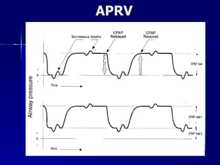
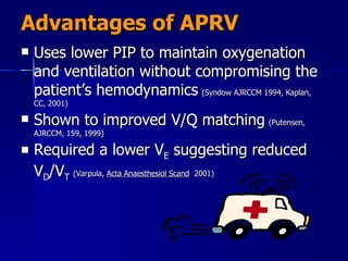
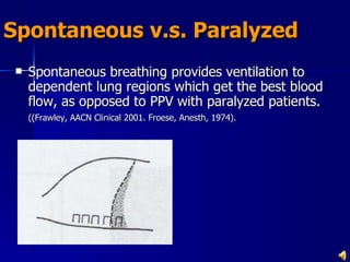
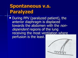
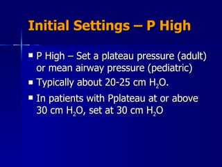
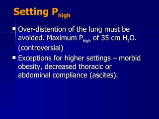
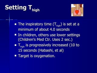
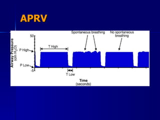
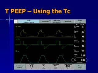
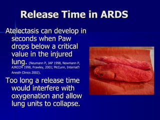
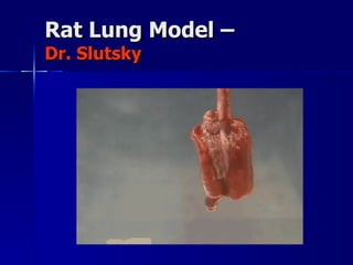
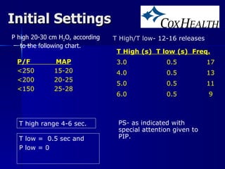
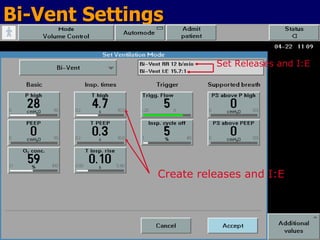
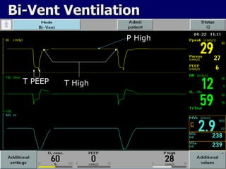
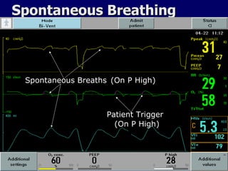
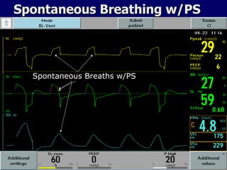
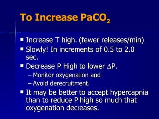
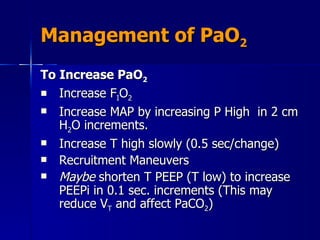
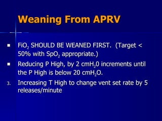
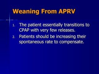
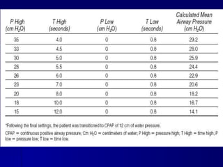
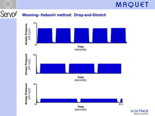
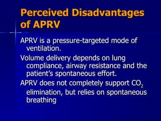
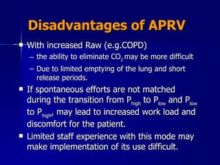
The “How To” of BiVent (APRV)  Created by: David Pitts II, RRT Clinical Applications Specialist, Maquet Birmingham, Alabama Sponsored by Maquet, Inc – Servo Ventilators
Objectives Provide the definition and names for APRV Explain the four set parameters. Identify recruitment in APRV using exhaled CO 2 . Recommend appropriate initial settings for APRV Make adjustments based on arterial blood gas results Discontinue ventilation with APRV
Lung Protective Strategies Acute lung injury (ALI) and acute respiratory distress syndrome (ARDS) Keep plateau pressures < 30 cm H 2 O Use low tidal volume ventilation (4-6 mL/kg IBW) Use PEEP to restore the functional residual capacity (FRC)
Keeping Plateau Pressure < 30 cm H 2 0 What do you do if CO 2  is rising and the plateau pressure is at 30 cm H 2 O?
Alternative Techniques Increase the ventilator rate Permissive Hypercapnia  Airway Pressure Release Ventilation  High Frequency Ventilation Extracorporeal Life Support
Indications Primarily used as an alternative ventilation technique in patients with ARDS. Used to help protect against ventilator induced lung injury.
Goal To provide the lung protective ventilation supported by the ARDSnet research.  Use an “Open lung” approach. Minimize alveolar overdistension. Avoid repeated alveolar collapse and reexpansion. Restore FRC through recruitment and, Maintain FRC by creating intrinsic PEEP.
APRV Description A mode of ventilation along with spontaneous ventilation to promote lung recruitment of collapsed and poorly ventilated alveoli. The CPAP is released periodically for a brief period. The short release along with spontaneous breathing promote CO 2  elimination. Release time is short to prevent the peak expiratory flow from returning to a zero baseline.
Ventilation With APRV The short release along with spontaneous breathing promote CO 2  elimination. Release time is short to prevent the peak expiratory flow from returning to a zero baseline.
APRV
AKA BiVent – Servo APRV – Drager BiLevel – Puritan Bennett APRV – Hamilton Etc.
Consider APRV when the Patient Has -- Bilateral Infiltrates PaO 2 /F I O 2  ratio < 300 and falling Plateau pressures greater than 30 cm H 2 O No evidence of left heart failure (e.g. PAOP of 18 mm Hg or greater) In other words, persistent ARDS
Possible Contraindications Unmanaged increases in intracraneal pressure. Large bronchopleural fistulas. Possibly obstructive lung disease. Technically, it may be possible to ventilate nearly any disorder.
Terminology P High   – the upper CPAP level. Analogous to MAP (mean airway pressure) and thus affects oxygenation PEEP   (Also called Plow) is the lower pressure setting. T  High -   is the inspiratory time IT(s) phase for the high CPAP level (P High). T PEEP or T low -  is the release time allowing CO 2  elimination
Terminology T High plus T PEEP (T low) is the total time of one cycle. I:E ratio becomes irrelevant because APRV is really best thought of as CPAP  With occasional releases
Bi-Vent Set-up Screen
Advantages of APRV Uses lower PIP to maintain oxygenation and ventilation without compromising the patient’s hemodynamics   (Syndow AJRCCM 1994, Kaplan, CC, 2001) Shown to improved V/Q matching   (Putensen, AJRCCM, 159, 1999) Required a lower V E  suggesting reduced V D /V T   (Varpula,  Acta Anaesthesiol Scand   2001)
Compared to PCIRV – Advantages of APRV APRV uses lower peak and mean airway pressures,  Increases cardiac index,  Decreases central venous pressure,
Additional Advantages - Compared to PCIRV APRV increases oxygen delivery and  Reduces the need for sedation and paralysis   APRV also improves renal perfusion and urine output when spontaneous breathing is maintained.  (Kaplan, Crit Care, 2001; Hering, Crit Care Med 2002)
New Water Coolers are Being Installed in the ICU Waiting Rooms
Advantages of Spontaneous Breathing The benefits of APRV may be related to the preservation of spontaneous breathing. Maintaining the normal cyclic decrease in pleural pressure, augmenting venous return and improving cardiac output.  (Putensen, AJRCCM, 1999) The need for sedation is decreased.
Preserve Spontaneous Breathing The dashed line in each figure represents the normal position of the diaphragm.  The shaded area represents the movement of the diaphragm.  (Froese, 1974)
Spontaneous v.s. Paralyzed Spontaneous breathing provides ventilation to dependent lung regions which get the best blood flow, as opposed to PPV with paralyzed patients.  ((Frawley, AACN Clinical 2001. Froese, Anesth, 1974).
Spontaneous v.s. Paralyzed During PPV (paralyzed patient), the anterior diaphragm is displaced towards the abdomen with the  non-dependent  regions of the lung receiving the most ventilation where perfusion is the least.
Try as we might.  We can’t get away from it?
Other Advantages of Spontaneous Breathing Reduces atrophy of the muscles of ventilation  associated with the use of PPV and paralytic agents.  (Neuman, ICM,2002)
Another Advantage During PPV atelectasis formation can occur near the diaphragm, when activity of this muscle is absent. (paralysis) However, if spontaneous breathing is preserved, the formation of atelectasis is offset by the activity of the diaphragm.  (Hedenstierna, Anesth, 1994)
Initial Settings – P High P High – Set a plateau pressure (adult) or mean airway pressure (pediatric) Typically about 20-25 cm H 2 O.  In patients with Pplateau at or above 30 cm H 2 O, set at 30 cm H 2 O
Setting P high Over-distention of the lung must be avoided. Maximum P high  of 35 cm H 2 O. (controversial) Exceptions for higher settings – morbid obesity, decreased thoracic or abdominal compliance (ascites).
Setting T high The inspiratory time (T high ) is set at a minimum of about 4.0 seconds  In children, others use lower settings (Children’s Med Ctr. Uses 2 sec.) T high  is progressively increased (10 to 15 seconds (Habashi, et al) Target is oxygenation.
Setting T high Progress slowly. For example, 5 sec T high  to 0.5 sec T low , a 10:1 ratio.  Increasing to 5.5 sec to 0.5 sec is an 11:1 ratio; not a big change.  Old patients may be fragile.
APRV
Release Time - T PEEP Currently, with ARDS thinking is not to let exhalation go to complete emptying, i.e. do not let expiratory flow returning to zero. ( McCunn, Crit Care 2002) Thus, regional auto-PEEP a desirable outcome with APRV FLOW
Setting PEEP or P low  in APRV Set PEEP at zero cm H 2 O. This provides a rapid drop in pressure, and a maximum   P for unimpeded expiratory gas flow.  (Frawley, AACN Clin Issues 2001) Avoid lung collapse during T low .
Establishing T PEEP  (Time at low pressure) Set T PEEP (T low) so that expiratory flow from patient ends at about 50 to 75% of peak expiratory flow. This can be determine saving a screen and calculating peak expiratory flow. Or, it can be estimated
Expiratory Flow
T PEEP – Setting The Time Adults 0.5 to 0.8 seconds Pediatric/neonatal settings 0.2 to 0.6 seconds. Or one time constant. (TC = C x R)
T PEEP – Using the Tc
Release Time in ARDS Atelectasis can develop in seconds when Paw drops below a critical value in the injured lung.   (Neumann P, JAP 1998, Newmann P, AJRCCM 1998, Frawley, 2001; McCunn, Internat’l Anesth Clinics 2002).   Too long a release time would interfere with oxygenation and allow lung units to collapse.
Rat Lung Model –  Dr. Slutsky
Initial Settings P high 20-30 cm H 2 O, according to the following chart. T High/T low - 12-16 releases T low =  0.5 sec  and  P low = 0 P/F  MAP <250  15-20 <200  20-25 <150  25-28 T High (s)  T low (s)  Freq. 3.0  0.5 17 4.0  0.5 13 5.0  0.5 11 6.0  0.5  9 T high range 4-6 sec. PS- as indicated with special attention given to PIP.
Bi-Vent Settings Set Releases and I:E Create releases and I:E
Bi-Vent Ventilation P High T High T PEEP
Spontaneous Breathing Spontaneous Breaths Patient Trigger (On P High) (On P High)
Spontaneous Breathing w/PS Spontaneous Breaths w/PS
Identifying Lung Recruitment – CO 2  Monitoring
Making Changes in APRV Settings Based on ABGs
Control Settings for CO 2    P (P high  – P low ) determines flow out of the lungs and volume exchange (V T  and PaCO 2 ). Some clinicians suggest a target minute ventilation of 2 to 3 L/min. (Frawley, 2001).  Optimize spontaneous ventilation.
CO 2  Elimination To Decrease PaCO 2 : Decrease T High. Shorter T High means more release/min. No shorter than 3 seconds  Example: T High 5 sec. = 12 releases/min T High 4 sec = 15 releases/min Increase P High to increase   P and volume exchange. (2-3 cm H 2 O/change) Monitor Vt PIP (best below 30 cm H 2 O) Check T low.  If possible increase T low to allow more time for “exhalation.”
To Increase PaCO 2 Increase T high. (fewer releases/min) Slowly! In increments of 0.5 to 2.0 sec. Decrease P High to lower   P.  Monitor oxygenation and Avoid derecruitment. It may be better to accept hypercapnia than to reduce P high so much that oxygenation decreases.
Management of PaO 2 To Increase PaO 2 Increase F I O 2 Increase MAP by increasing P High  in 2 cm H 2 O increments. Increase T high slowly (0.5 sec/change) Recruitment Maneuvers Maybe  shorten T PEEP (T low) to increase PEEPi in 0.1 sec. increments (This may reduce V T  and affect PaCO 2 )
Going Too Fast
Weaning From APRV FiO 2  SHOULD BE WEANED FIRST.  (Target < 50% with SpO 2  appropriate.) Reducing P High, by 2 cmH 2 0 increments until the P High is below 20 cmH 2 O. Increasing T High to change vent set rate by 5 releases/minute
Weaning From APRV The patient essentially transitions to CPAP with very few releases.  Patients should be increasing their spontaneous rate to compensate.
During Weaning Add Pressure Support judiciously.  Add Pressure Support to P High in order to decrease WOB  while avoiding over-distention,  P High + PS  <  30 cmH 2 O.
Pressure Support with APRV
Weaning Bi-Vent Lower Rate Longer T High Lower P High Add PS
 
 
Perceived Disadvantages of APRV APRV is a pressure-targeted mode of ventilation. Volume delivery depends on lung compliance, airway resistance and the patient’s spontaneous effort.  APRV does not completely support CO 2  elimination, but relies on spontaneous breathing
Disadvantages of APRV With increased Raw (e.g.COPD)  the ability to eliminate CO 2  may be more difficult Due to limited emptying of the lung and short release periods. If spontaneous efforts are not matched during the transition from P high  to P low  and P low  to P high , may lead to increased work load and discomfort for the patient.  Limited staff experience with this mode may make implementation of its use difficult.
The End  Thank You!

APRV

  • 1.
    The “How To”of BiVent (APRV) Created by: David Pitts II, RRT Clinical Applications Specialist, Maquet Birmingham, Alabama Sponsored by Maquet, Inc – Servo Ventilators
  • 2.
    Objectives Provide thedefinition and names for APRV Explain the four set parameters. Identify recruitment in APRV using exhaled CO 2 . Recommend appropriate initial settings for APRV Make adjustments based on arterial blood gas results Discontinue ventilation with APRV
  • 3.
    Lung Protective StrategiesAcute lung injury (ALI) and acute respiratory distress syndrome (ARDS) Keep plateau pressures < 30 cm H 2 O Use low tidal volume ventilation (4-6 mL/kg IBW) Use PEEP to restore the functional residual capacity (FRC)
  • 4.
    Keeping Plateau Pressure< 30 cm H 2 0 What do you do if CO 2 is rising and the plateau pressure is at 30 cm H 2 O?
  • 5.
    Alternative Techniques Increasethe ventilator rate Permissive Hypercapnia Airway Pressure Release Ventilation High Frequency Ventilation Extracorporeal Life Support
  • 6.
    Indications Primarily usedas an alternative ventilation technique in patients with ARDS. Used to help protect against ventilator induced lung injury.
  • 7.
    Goal To providethe lung protective ventilation supported by the ARDSnet research. Use an “Open lung” approach. Minimize alveolar overdistension. Avoid repeated alveolar collapse and reexpansion. Restore FRC through recruitment and, Maintain FRC by creating intrinsic PEEP.
  • 8.
    APRV Description Amode of ventilation along with spontaneous ventilation to promote lung recruitment of collapsed and poorly ventilated alveoli. The CPAP is released periodically for a brief period. The short release along with spontaneous breathing promote CO 2 elimination. Release time is short to prevent the peak expiratory flow from returning to a zero baseline.
  • 9.
    Ventilation With APRVThe short release along with spontaneous breathing promote CO 2 elimination. Release time is short to prevent the peak expiratory flow from returning to a zero baseline.
  • 10.
  • 11.
    AKA BiVent –Servo APRV – Drager BiLevel – Puritan Bennett APRV – Hamilton Etc.
  • 12.
    Consider APRV whenthe Patient Has -- Bilateral Infiltrates PaO 2 /F I O 2 ratio < 300 and falling Plateau pressures greater than 30 cm H 2 O No evidence of left heart failure (e.g. PAOP of 18 mm Hg or greater) In other words, persistent ARDS
  • 13.
    Possible Contraindications Unmanagedincreases in intracraneal pressure. Large bronchopleural fistulas. Possibly obstructive lung disease. Technically, it may be possible to ventilate nearly any disorder.
  • 14.
    Terminology P High – the upper CPAP level. Analogous to MAP (mean airway pressure) and thus affects oxygenation PEEP (Also called Plow) is the lower pressure setting. T High - is the inspiratory time IT(s) phase for the high CPAP level (P High). T PEEP or T low - is the release time allowing CO 2 elimination
  • 15.
    Terminology T Highplus T PEEP (T low) is the total time of one cycle. I:E ratio becomes irrelevant because APRV is really best thought of as CPAP With occasional releases
  • 16.
  • 17.
    Advantages of APRVUses lower PIP to maintain oxygenation and ventilation without compromising the patient’s hemodynamics (Syndow AJRCCM 1994, Kaplan, CC, 2001) Shown to improved V/Q matching (Putensen, AJRCCM, 159, 1999) Required a lower V E suggesting reduced V D /V T (Varpula, Acta Anaesthesiol Scand 2001)
  • 18.
    Compared to PCIRV– Advantages of APRV APRV uses lower peak and mean airway pressures, Increases cardiac index, Decreases central venous pressure,
  • 19.
    Additional Advantages -Compared to PCIRV APRV increases oxygen delivery and Reduces the need for sedation and paralysis APRV also improves renal perfusion and urine output when spontaneous breathing is maintained. (Kaplan, Crit Care, 2001; Hering, Crit Care Med 2002)
  • 20.
    New Water Coolersare Being Installed in the ICU Waiting Rooms
  • 21.
    Advantages of SpontaneousBreathing The benefits of APRV may be related to the preservation of spontaneous breathing. Maintaining the normal cyclic decrease in pleural pressure, augmenting venous return and improving cardiac output. (Putensen, AJRCCM, 1999) The need for sedation is decreased.
  • 22.
    Preserve Spontaneous BreathingThe dashed line in each figure represents the normal position of the diaphragm. The shaded area represents the movement of the diaphragm. (Froese, 1974)
  • 23.
    Spontaneous v.s. ParalyzedSpontaneous breathing provides ventilation to dependent lung regions which get the best blood flow, as opposed to PPV with paralyzed patients. ((Frawley, AACN Clinical 2001. Froese, Anesth, 1974).
  • 24.
    Spontaneous v.s. ParalyzedDuring PPV (paralyzed patient), the anterior diaphragm is displaced towards the abdomen with the non-dependent regions of the lung receiving the most ventilation where perfusion is the least.
  • 25.
    Try as wemight. We can’t get away from it?
  • 26.
    Other Advantages ofSpontaneous Breathing Reduces atrophy of the muscles of ventilation associated with the use of PPV and paralytic agents. (Neuman, ICM,2002)
  • 27.
    Another Advantage DuringPPV atelectasis formation can occur near the diaphragm, when activity of this muscle is absent. (paralysis) However, if spontaneous breathing is preserved, the formation of atelectasis is offset by the activity of the diaphragm. (Hedenstierna, Anesth, 1994)
  • 28.
    Initial Settings –P High P High – Set a plateau pressure (adult) or mean airway pressure (pediatric) Typically about 20-25 cm H 2 O. In patients with Pplateau at or above 30 cm H 2 O, set at 30 cm H 2 O
  • 29.
    Setting P highOver-distention of the lung must be avoided. Maximum P high of 35 cm H 2 O. (controversial) Exceptions for higher settings – morbid obesity, decreased thoracic or abdominal compliance (ascites).
  • 30.
    Setting T highThe inspiratory time (T high ) is set at a minimum of about 4.0 seconds In children, others use lower settings (Children’s Med Ctr. Uses 2 sec.) T high is progressively increased (10 to 15 seconds (Habashi, et al) Target is oxygenation.
  • 31.
    Setting T highProgress slowly. For example, 5 sec T high to 0.5 sec T low , a 10:1 ratio. Increasing to 5.5 sec to 0.5 sec is an 11:1 ratio; not a big change. Old patients may be fragile.
  • 32.
  • 33.
    Release Time -T PEEP Currently, with ARDS thinking is not to let exhalation go to complete emptying, i.e. do not let expiratory flow returning to zero. ( McCunn, Crit Care 2002) Thus, regional auto-PEEP a desirable outcome with APRV FLOW
  • 34.
    Setting PEEP orP low in APRV Set PEEP at zero cm H 2 O. This provides a rapid drop in pressure, and a maximum  P for unimpeded expiratory gas flow. (Frawley, AACN Clin Issues 2001) Avoid lung collapse during T low .
  • 35.
    Establishing T PEEP (Time at low pressure) Set T PEEP (T low) so that expiratory flow from patient ends at about 50 to 75% of peak expiratory flow. This can be determine saving a screen and calculating peak expiratory flow. Or, it can be estimated
  • 36.
  • 37.
    T PEEP –Setting The Time Adults 0.5 to 0.8 seconds Pediatric/neonatal settings 0.2 to 0.6 seconds. Or one time constant. (TC = C x R)
  • 38.
    T PEEP –Using the Tc
  • 39.
    Release Time inARDS Atelectasis can develop in seconds when Paw drops below a critical value in the injured lung. (Neumann P, JAP 1998, Newmann P, AJRCCM 1998, Frawley, 2001; McCunn, Internat’l Anesth Clinics 2002). Too long a release time would interfere with oxygenation and allow lung units to collapse.
  • 40.
    Rat Lung Model– Dr. Slutsky
  • 41.
    Initial Settings Phigh 20-30 cm H 2 O, according to the following chart. T High/T low - 12-16 releases T low = 0.5 sec and P low = 0 P/F MAP <250 15-20 <200 20-25 <150 25-28 T High (s) T low (s) Freq. 3.0 0.5 17 4.0 0.5 13 5.0 0.5 11 6.0 0.5 9 T high range 4-6 sec. PS- as indicated with special attention given to PIP.
  • 42.
    Bi-Vent Settings SetReleases and I:E Create releases and I:E
  • 43.
    Bi-Vent Ventilation PHigh T High T PEEP
  • 44.
    Spontaneous Breathing SpontaneousBreaths Patient Trigger (On P High) (On P High)
  • 45.
    Spontaneous Breathing w/PSSpontaneous Breaths w/PS
  • 46.
    Identifying Lung Recruitment– CO 2 Monitoring
  • 47.
    Making Changes inAPRV Settings Based on ABGs
  • 48.
    Control Settings forCO 2  P (P high – P low ) determines flow out of the lungs and volume exchange (V T and PaCO 2 ). Some clinicians suggest a target minute ventilation of 2 to 3 L/min. (Frawley, 2001). Optimize spontaneous ventilation.
  • 49.
    CO 2 Elimination To Decrease PaCO 2 : Decrease T High. Shorter T High means more release/min. No shorter than 3 seconds Example: T High 5 sec. = 12 releases/min T High 4 sec = 15 releases/min Increase P High to increase  P and volume exchange. (2-3 cm H 2 O/change) Monitor Vt PIP (best below 30 cm H 2 O) Check T low. If possible increase T low to allow more time for “exhalation.”
  • 50.
    To Increase PaCO2 Increase T high. (fewer releases/min) Slowly! In increments of 0.5 to 2.0 sec. Decrease P High to lower  P. Monitor oxygenation and Avoid derecruitment. It may be better to accept hypercapnia than to reduce P high so much that oxygenation decreases.
  • 51.
    Management of PaO2 To Increase PaO 2 Increase F I O 2 Increase MAP by increasing P High in 2 cm H 2 O increments. Increase T high slowly (0.5 sec/change) Recruitment Maneuvers Maybe shorten T PEEP (T low) to increase PEEPi in 0.1 sec. increments (This may reduce V T and affect PaCO 2 )
  • 52.
  • 53.
    Weaning From APRVFiO 2 SHOULD BE WEANED FIRST. (Target < 50% with SpO 2 appropriate.) Reducing P High, by 2 cmH 2 0 increments until the P High is below 20 cmH 2 O. Increasing T High to change vent set rate by 5 releases/minute
  • 54.
    Weaning From APRVThe patient essentially transitions to CPAP with very few releases. Patients should be increasing their spontaneous rate to compensate.
  • 55.
    During Weaning AddPressure Support judiciously. Add Pressure Support to P High in order to decrease WOB while avoiding over-distention, P High + PS < 30 cmH 2 O.
  • 56.
  • 57.
    Weaning Bi-Vent LowerRate Longer T High Lower P High Add PS
  • 58.
    Weaning Bi-Vent LowerRate Longer T High Lower P High Add PS
  • 59.
  • 60.
  • 61.
    Perceived Disadvantages ofAPRV APRV is a pressure-targeted mode of ventilation. Volume delivery depends on lung compliance, airway resistance and the patient’s spontaneous effort. APRV does not completely support CO 2 elimination, but relies on spontaneous breathing
  • 62.
    Disadvantages of APRVWith increased Raw (e.g.COPD) the ability to eliminate CO 2 may be more difficult Due to limited emptying of the lung and short release periods. If spontaneous efforts are not matched during the transition from P high to P low and P low to P high , may lead to increased work load and discomfort for the patient. Limited staff experience with this mode may make implementation of its use difficult.
  • 63.
    The End Thank You!

Editor's Notes

  • #24 Maintaining spontaneous ventilation tends to improve ventilation-perfusion matching by preferentially providing ventilation to dependent lung regions that receive the best blood flow.