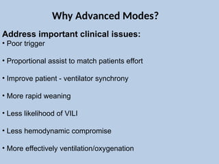
Address important clinicalissues:
• Poor trigger
• Proportional assist to match patients effort
• Improve patient - ventilator synchrony
• More rapid weaning
• Less likelihood of VILI
• Less hemodynamic compromise
• More effectively ventilation/oxygenation
Why Advanced Modes?
3.
Striving for betteroutcomes
The three S :
• Spontaneous breathing (Girard 2008; MacIntyre 2000, Levine 2008)
• Synchrony (Chao 1997;Thille 2006; De Wit 2009)
• Sedation management (Kress 2000, Girard 2008, De Wit 2009)
4.
Lung Protective StrategiesARDS
•Keep plateau pressures < 30 cm H 2 O
•Use low tidal volume ventilation (4-6 mL/kg IBW)
•Titration of PEEP to restore the functional residual capacity (FRC)
•Permissive hypoxia
•Permissive hypercapnia
6.
Classification of advancedmodes
Dual modes :
Which combine Volume mode + Pressure modes-
VS, MMV, VAPS, PRVC etc…
Modes which adapt to lung characteristics :
( Resistance & Compliance) PAV, ASV
Spontaneous breathing + higher FRC : APRV/ BIPAP
Knowledge based Weaning modes : Smartcare, ATC,
PAV, ASV, NAVA
Better trigger mechanism : NAVA
Airway pressure releaseventilation
•Time triggered, pressure-controlled, time-cycled mode
•Allows unsupported spontaneous respiration throughout
the respiratory cycle
•High-level CPAP with short pressure drops or ‘releases’ to
facilitate ventilation and CO2 clearance
•Considered to be an almost continuous, mild recruitment
maneuver
9.
Basics
• Hypoxaemia inALI or ARDS is largely due to V/Q mismatch and
intrapulmonary shunting in areas of consolidation and alveolar
collapse which occurs predominantly in the postero-basal portions
of the lung
• Recruitment of collapsed lung may be achieved by:
• CPAP with moderate to high levels of airway pressure
• Spontaneous breathing—contraction of the posterior part of
the diaphragm assists in recruiting the postero-basal lung
Airway pressure release ventilation
11.
Advantages of SpontaneousBreathing :
•Maintaining the normal respiratory cycle, decrease in pleural
pressure, augmenting venous return and improving cardiac output.
• Need for sedation is decreased.
•Spontaneous breathing provides ventilation to dependent lung
regions which get the best blood flow, as opposed to PPV with
paralyzed patients.
•Reduces atrophy of the muscles of ventilation
•Reduced PPV atelectasis formation near the diaphragm
Airway pressure release ventilation
13.
Airway pressure releaseventilation
Mechanisms for CO2 clearance :
• Collapsed alveoli are recruited, and ventilation to previously well
perfused alveoli is improved.
• As lung volume increases, pulmonary vascular resistance decreases
and blood flow to previously hypoperfused alveoli increases, reducing
physiological dead space.
• Unsupported spontaneous breathing increases cardiac output,
which will also improve V/Q matching
It may take up to 16h to achieve these effects on gas exchange. There
is little additional gain after 24h.
15.
Initial settings :
Phigh = high pressure level
• Initial setting 25–30 cm H2O.
• Consider up to 35 cmH 2 O if reduced chest wall/abdominal
compliance.
P low = low pressure level
• 0 cm H2O. Allows maximum Delta P and therefore maximum
flow during expiration.
• Lung collapse is avoided by manipulating T low rather than P low
Airway pressure release ventilation
16.
Initial settings :
Thigh = time spent at high pressure
• Initial setting 4–6 sec .
• Progressively increase for target oxygenation
T low = time spent at low pressure
• Initial setting 0.4 - 0.6s.
• Restrictive lung disease: 0.2–0.8s.
• Obstructive lung disease: 0.8–1.5s.
Airway pressure release ventilation
17.
Initial settings :
Methodto set T low :
1. The most common method described for setting T low uses
the expiratory flow waveform. T low should end when
expiratory flow falls to 50–75% of PEFR.
2. It can also be set at one time constant.
Airway pressure release ventilation
18.
Weaning APRV :The ‘drop and stretch’ method.
• FiO 2 should be < 50% before attempting any reduction in
airway pressure.
• P low remains at 0 for as long as the patient remains on APRV.
• Only adjust T low in response to changes in lung compliance.
• Reduce P high in 2cm H 2 O increments, guided by oxygenation.
Eventual target 8–10cmH 2 O.
• Simultaneously, increase T high in 2s increments. Monitor PaCO2
This effectively reduces the release rate and CO 2 clearance is
achieved by spontaneous ventilation.
Airway pressure release ventilation
19.
Weaning APRV :The ‘drop and stretch’ method.
• At CPAP 8–10cm H2O , an assessment of suitability for
tracheal extubation may be appropriate
• If the patient’s condition deteriorate at any stage during
this process :
• Increase P high (for deterioration of oxygenation) or
• Reduce T high (for unacceptable rise in CO 2
Airway pressure release ventilation
20.
Advantages :
• ImprovedV/Q matching
• Lower peak airway pressure for a given mean airway
pressure
• CO2 clearance maintained with lower MV
• Cardiovascular stability
• Reduced vasopressor requirements
• Preserved renal and splanchnic blood flow
• Reduced sedation and paralysis requirements.
Airway pressure release ventilation
21.
Advantages of APRVas compared to IRV :
•APRV uses lower peak and mean airway pressures
•Increases cardiac index
•Decreases central venous pressure
•APRV increases oxygen delivery and
•Reduces the need for sedation and paralysis
•APRV also improves renal perfusion and urine output
Airway pressure release ventilation
Pressure-regulated volume control
Breathto breath dual-control ventilation
Form of assist-control ventilation.
Breaths can be: ventilator initiated (control breath) or patient
initiated (assist breath)
Constant pressure applied throughout inspiration (like pressure
control), regardless of whether breath is a control breath or an
assist breath
Improved oxygenation due to decelerating inspiratory flow pattern
26.
Ventilator adjusts pressurefrom breath to breath, as patient's
airway resistance and compliance changes
If the delivered volume is too low it increases the inspiratory
pressure on the next breath. If it is too high it decreases the
pressure
The maximum allowed inspiratory pressure is 5 cm H2O below the
upper pressure alarm limit
The duration of inspiration is determined by the respiratory rate
and the I:E ratio or inspiratory time
Pressure-regulated volume control
27.
Like PC but:
•Constant pressure during each breath
•Variable pressure from breath to breath
•Delivered Tidal Volume can vary from set tidal vovume
Pressure-regulated volume control
28.
Initial Settings :
•Minimum respiratory rate :
•Patient’s spontaneous respiratory rate < set rate; ventilator
gives additional control breaths to make up difference
•Patient’s spontaneous rate > set rate; no control breaths
•Target tidal volume
•initial setting: 8 ml/kg predicted body weight
•upper pressure limit
•ventilator delivers pressure of up to 5 cm H2O below upper
pressure alarm limit
•set to 35-40 cm H2O to ensure "safe" pressures
Pressure-regulated volume control
29.
Initial Settings :
•inspiredoxygen concentration
•initial setting 100%
•I:E ratio
•initial setting: 1:2 (=inspiratory time of 33%)
•consider longer inspiratory time if there is no intrinsic PEEP,
no bronchospasm and oxygenation is poor
•PEEP
•initial setting 5-10 cm H2O
Pressure-regulated volume control
Advantages :
•Decelerating inspiratoryflow pattern
•Pressure automatically adjusted for changes in compliance and
resistance within a set range
–Tidal volume guaranteed
–Limits volutrauma
–Prevents hypoventilation
Disadvantages :
•Pressure delivered is dependent on tidal volume achieved on last
breath – Intermittent patient effort will lead to variable tidal volumes
• Sedation requirement
Pressure-regulated volume control
Adaptive support ventilation
•Patentedmode of ventilation
•Available on Hamilton Medical machines.
•It delivers a mandatory minute volume while attempting to
minimize WOB.
•It is a closed-loop system that automatically escalates or reduces
both pressure support and mandatory breaths, depending on
patient effort.
•It is capable of delivering any level of support from CPAP to full
PCV
37.
Adaptive support ventilation
Basics
•ASVtargets a minute volume set by the clinician.
•This can be delivered by pressure-supported spontaneous
ventilation, volume-targeted PCV, or a combination of both
depending on patient effort.
•The mode preference is for spontaneous respiration, but if the
respiratory rate is below the desired rate, mandatory breaths are
gradually introduced.
•The ideal respiratory rate and Vt are calculated based on the
‘minimum WOB’. Three test breaths measure compliance and
airways resistance using a least-squares fit technique (the
mathematical procedure commonly used in statistics for multiple linear regression).
39.
Adaptive support ventilation
Basics
•Compliance and resistance are monitored on a breath-by-breath
basis.
•The WOB is recalculated every three to five breaths, and
adjustments made for any change in respiratory mechanics.
• In full controlled mode, breaths are pressure-controlled and time-
cycled. Support breaths are pressure-supported and flow-cycled.
• During both spontaneous and controlled breaths, inspiratory
pressure is adjusted to achieve the desired tidal volume.
40.
• Minimum tidalvolume (min V t ) = 4.4 × IBW
• Maximum ventilator respiratory rate is the lowest of:
• Target minute volume/min V t
• 20/expiratory time constant
• 60 breaths/minute
• Maximum Vt is (P max – PEEP) × C or 22mL/kg.
• Maximum delivered pressure is 10cmH 2 O below the set
pressure limit.
• Expiratory time >2 × expiratory time constant
Adaptive support ventilation
41.
Indications :
• Postoperativepatients.
• Respiratory failure from a variety of causes.
• Weaning.
Adaptive support ventilation
42.
Not recommended :
•Significant airleaks, e.g. bronchopleural fistula.
• During bronchoscopy.
• May not be appropriate for restrictive tidal volume ventilation in
ARDS. In clinical practice, it has been shown to deliver tidal
volumes closer to 8mL/kg.
Adaptive support ventilation
43.
Initial settings :
•Set PEEP and FiO2 as for regular ventilation.
• Upper pressure limit must be at least 25 cm H2O above PEEP.
The maximum pressure applied will be 10 cm H2O below this
limit.
• IBW should be calculated from height. Increase IBW by 10% if
HME filter incorporated into circuit
• Choose the minute ventilation in the normal fashion, and then
calculate the %MinVol required to deliver it.
• Initial %MinVol should be higher in patients known to have
increased dead space
• Use %MinVol to alter tidal volume not IBW.
• Enter trigger method (pressure or flow), sensitivity
Adaptive support ventilation
45.
• ASV willpreferentially allow spontaneous breathing so that
patients will wean from controlled ventilation automatically.
• For a particular MV, ASV will adjust the respiratory rate and
inspiratory pressure provided according to changes in compliance
and resistance.
•Assess respiratory pattern and patient effort, blood gases, and
inspiratory pressure before adjustment.
Adaptive support ventilation
46.
• When Pinsp <8 and frequency of spontaneous breaths
acceptable, extubation may be considered.
• Even if the respiratory pattern is optimized there is no guarantee
of acceptable gas exchange . Arterial blood gas monitoring is
still essential.
• Although ASV is able to automatically adjust the level of
ventilatory support, it should not replace clinician input and
assessment.
Adaptive support ventilation
Proportional assist ventilation
Modeof spontaneous ventilatory support
Degree of assist varies according to patient effort.
Rather than targeting a set pressure level, tidal volume, or respiratory
rate, it targets a set level of respiratory muscle off loading.
There are no mandatory breaths.
49.
•PAV is amode of support in which the ventilator pressure (Paw )
is proportional to inspiratory flow (V’) and volume (V), which in
turn are determined by the patient’s inspiratory muscle pressure
(P musi ).
•With this mode the clinician sets the respective flow and volume
gain signals, the flow assist (FA) and volume assist (VA).
•With this mode the ventilator simply amplifies patient inspiratory
effort without imposing any target for flow, volume, or Paw
Proportional assist ventilation
50.
Proportionality :
In PAVthe clinician sets the assist level, K.
For example, in setting K to 80% the ventilator provides 80% of the
elastic and resistive work, while the patient contributes the remaining
20%. The proportionality between the ventilator (P aw ) and patient
inspiratory muscle (P musi ) is 4:1 (80/20).
Proportional assist ventilation
52.
Indications :
•As themain mode of respiratory support in critically ill patients.
•Patient–ventilator asynchronies in spontaneous breathing patients
Compared to PSV, it has been shown that PAV :
• Decreases triggering delay
• Decreases the likelihood of ineffective efforts
• Decreases expiratory asynchrony
• Increases sleep efficiency
• Promotes breathing stability
• Increases the efficiency of the respiratory system compensation for
any added mechanical load.
Proportional assist ventilation
53.
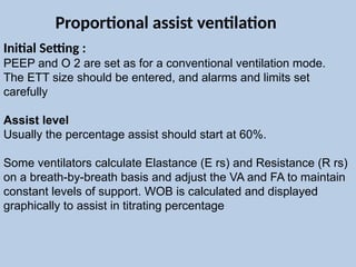
Initial Setting :
PEEPand O 2 are set as for a conventional ventilation mode.
The ETT size should be entered, and alarms and limits set
carefully
Assist level
Usually the percentage assist should start at 60%.
Some ventilators calculate Elastance (E rs) and Resistance (R rs)
on a breath-by-breath basis and adjust the VA and FA to maintain
constant levels of support. WOB is calculated and displayed
graphically to assist in titrating percentage
Proportional assist ventilation
54.
Support levels :
Inventilators where E rs and R rs are calculated manually, there
must be frequent remeasurement of parameters as E rs and R rs
change with the clinical condition
Weaning :
If the patient is comfortable and respiratory rate and blood gases
satisfactory, the percentage assist may be reduced in 10–20%
increments.
Proportional assist ventilation
PSV is patienttriggered, pressure targeted, and flow cycled
synchronized mode of ventilator support.
No mandatory breaths.
One of the most commonly used weaning modes
Pressure support ventilation
57.
Pressure support ventilation
•Inspirationis triggered by the patient ( by changes in pressure or
flow )
•When inspiration has been triggered, the ventilator raises airway
pressure to the set PS level.
•Flow pattern can vary between a decelerating flow pattern in
mainly passive patients and a sine wave flow pattern in patients
making effort throughout inspiration.
•Inspiration ends when inspiratory flow falls below a certain
percentage of the peak flow (usually 20 - 25%).
58.
Advantage :
•Reduce sedationrequirements
•Decrease respiratory muscle disuse atrophy
•Compensate for the additional WOB imposed by the underlying
disease process, the ETT and the breathing circuit.
Pressure support ventilation
59.
Disadvantage :
•Precise controlof tidal volume, MV, mean Paw and I:E ratios is
not possible.
•Unidentified ventilator patient dysynchrony
•Excessive support
•Poor sleep
Pressure support ventilation
•Synchronized mode ofventilator support.
•Unloads inspiratory muscles while upholding spontaneous breathing
•As the respiratory muscles and the ventilator receive the same signal,
synchronization is improved compared with other spontaneous modes
of ventilatory support.
•The patient–ventilator synchrony is equally efficient during both
invasive and non-invasive application of NAVA.
Neurally adjusted ventilatory assist
63.
Diaphragm electrical activity:
• The electrical activity of the diaphragm (EAdi, measured in μV) is
measured trans-oesophageally with microelectrodes situated
near the tip of the NAVA catheter.
• The electrodes are positioned at the level of the oesophageal
hiatus, using esophageal ECG to assist placement.
• The NAVA catheter also functions as a standard feeding tube.
• The EAdi comprises the temporo-spatial summation of the
neural output to the diaphragm transmitted via the phrenic
nerves, and hence is a representation of the neural drive to the
diaphragm.
Neurally adjusted ventilatory assist
65.
Triggering :
• Breathsare triggered by the EAdi.
• Breaths can also be triggered by a conventional pneumatic
signal ( If the EAdi signal late or inadequate).
• The trigger settings in NAVA are adjustable.
Cycling :
• The assist is cycled off when the EAdi decreases to a percentage
of the peak EAdi (40–70% of the peak EAdi, depending on the
amplitude of the signal).
• The cycling-off criterion during NAVA are non-adjustable.
Neurally adjusted ventilatory assist
66.
Pressure delivery :
•Pressure delivery is controlled by the EAdi signal.
• Setting the NAVA level (cm H2O/μV) determines the scale of
support.
• The pressure delivered (cm H2O above PEEP) is proportional to
the EAdi (μV).
• The proportionality can be adjusted by changing the NAVA level
(e.g.increasing the NAVA level: for a given EAdi, the pressure
delivered increases).
• This allows ventilatory demand and neural afferents to regulate
the assist, but within limits set by the caregiver.
Neurally adjusted ventilatory assist
Arguments Against NewModes
Lack high-level evidence for better patient outcomes
Potential for harm
Improved gas exchange does not necessarily improve outcomes: high
tidal volume, iNO, prone
New is not necessarily better
70.
The Evidence forNew Ventilator Modes …
It’s not the ventilator mode that makes a
difference …
It’s the skills of the clinician that makes the
difference.
Any ventilator mode has the potential to do harm!
High level evidence is lacking that any new ventilator
mode improves patient outcomes compared to existing
lung-protective ventilation strategies.
- Dean Hess