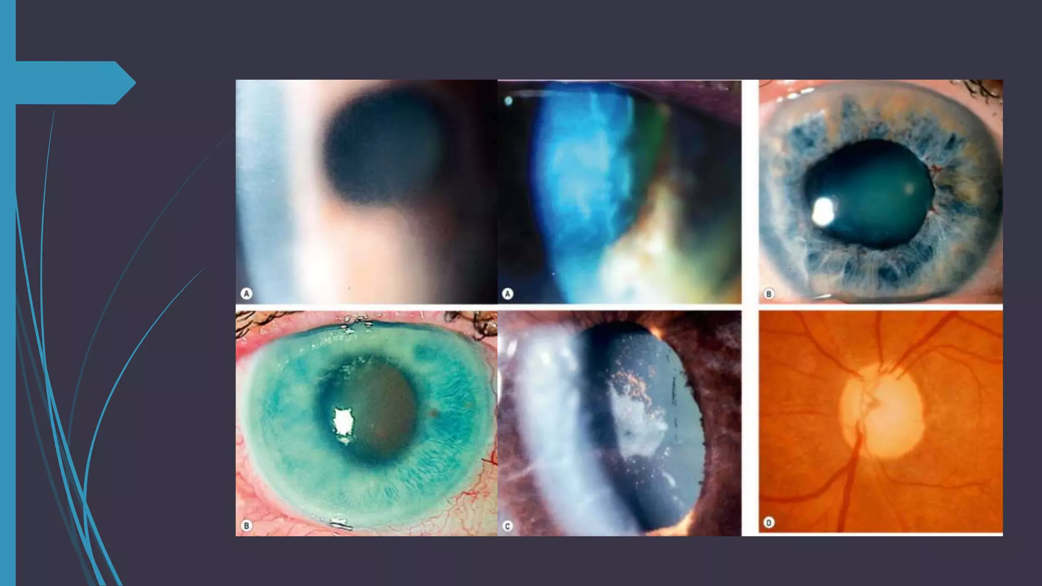
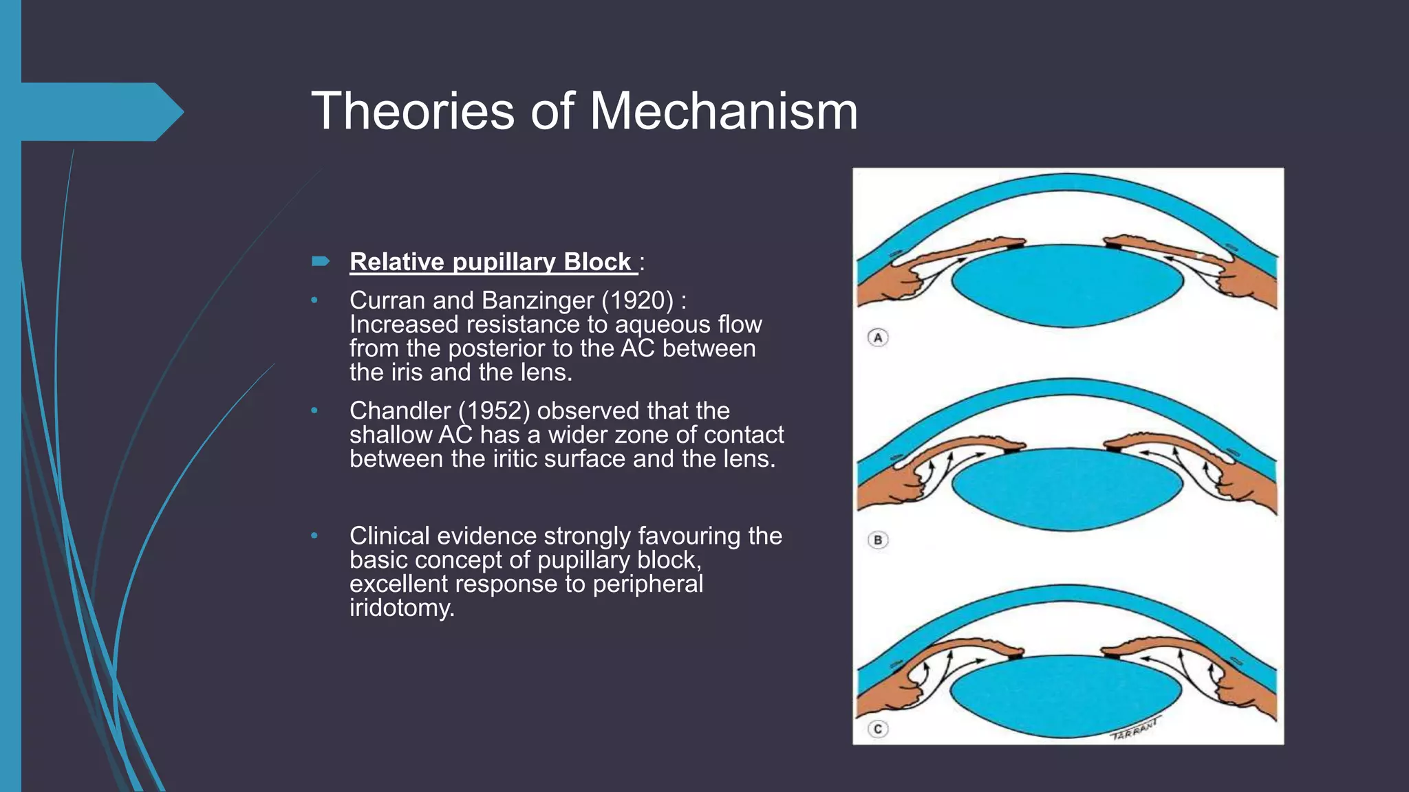
APPROACH TO THE CASE
OF PACG
Bipin Bista
Resident
Ophthalmology
National medical College
& Teaching Hospital
Introductory
 Characterised by apposition of the peripheral iris against the TM, resulting in
obstruction of aqueous outflow.
 Glaucoma is used when there’s evidence of GOA.
Pupillary Block Glaucoma
common form of ACG.
 Initiative event is from increased resistance to flow of aqueous humour between
pupillary portion of iris and anterior lens surface.
Forms of PBG
1. Acute angle-closure glaucoma : Sudden, severe with marked pain, blurred vision &
red eye. Associated with nausea and vomiting.
2. Sub-acute angle closure glaucoma : Mild or absent; repeated subclinical or sub
acute attacks, before finally developing PAS with chronic IOP elevation.
3. Chronic angle closure glaucoma : portions of AC angle are permanently closed by
PAS , Synechial closure may result from a prolonged acute attack or repeated sub-
acute attacks
4. Combined mechanism glaucoma :appears to have open-angle and angle-closure
mechanism
Epidemiology
 Less common than POAG
 > 40 years
 Female predilection is higher
 Asian has higher incidence than Africans
 Smaller corneal diameter
 Hypermetropic
Risk Factors
 Age : depth and volume diminish with age d/t thickening and forward displacement
of the lens.
 Race
 Sex
 Refractive Error : Occurring more with hypermetropia and also possible in Myopia.
 Family history
 Systemic Disorders : inverse correlation between DM2 and PBG.
Findings on Routine Examination
 IOP : Reveals normal IOP before and between attacks, unless peripheral attacks
synechia are presents.
 Evaluation of PAS : revealed AC depths, volumes and diameters smaller than
normal. Most important step in diagnosis : evaluate the depth and configurate AC
angle.
• Penlight examination : kept obliquely , with illuminated light from temporal side, a
relatively flat iris is seen, whereas in bowed iris , there will be shadow on the nasal
side.
• Slitlamp Examination : estimation of central AC depth, van Herick method, depth
less than one fourth of corneal thickness, potential to be occluded.
Gonioscopy
 Peripheral anterior chamber depth is thought to be shallow.
 Most important use is to know whether is appositional or synechial.
van Herick technique
 Grade 1 : PAC< ¼ CT
 Grade 2 : PAC = ¼ CT
 Grade 3 : PAC = ¼ to ½ CT
 Grade 4 : PAC > 1 CT
Schie gonioscopic classification
Classification Gonioscopic
Appearance
Wide open All structures visible
Grade I narrow Hard to see over iris
root into recess
Grade II narrow Ciliary body band
obscured
Grade III narrow Posterior trabeculae
obscured
Grade IV narrow Only schwalbe line
visible
Schaffer Gonioscopic classification
A. Wide open : 20-45 degrees , Closure
improbable
B. Moderately narrow : 10-20 degrees,
Closure possible.
C. Extremely narrow : Closure possible
D. Partially or totally closed : Closure
present
Spaeth Gonioscopic Classification
A. Angular width
B. Configuration of the peripheral iris
C. Apparent insertion of the iris root
Newer Techniques
 High frequency USG : relationship of the iris, posterior chamber, lens, zonules and
ciliary body.
 AC depth measurement : OCT
 Scheimpflug video imaging : qualitative assessment and observed longitudinally
Precipitating factors
Factors that produce
Mydriasis:
 Dim Illumination
 Emotional Stress
 Drugs : anti-cholinergics,
antihistaminics, antiparkinsonian,
antipsychotic, and GI spasmolytic
drugs, adrenergic drugs
Factors that produce
Miosis:
 Echothiophate iodide, di-isopropyl
fluorophosphates
 Increase in the relative pupillary block
d/t a wider zone of contact between
iris and lens and relaxation of the
lens zonules
Symptoms of Angle Closure Attacks
 Acute angle closure : pain, redness and blurred vision . Pain is typically a severe ,
deep ache that follows the trigeminal distribution and may be associated with
nausea, vomiting, bradycardia and profuse sweating. Blurred vision : stretching of
corneal lamellae initially and later on as corneal edema progresses.
 Subacute angle-closure Glaucoma : no recognizable symptoms, dull retrobulbar
aching, colored halos d/t corneal epithelial edema act as a diffraction grating : blue-
green central and yellow-red peripheral halo.
 Chronic angle closure Glaucoma : typically asymptomatic , until advances to visual
field loss
Clinical findings : External
 Hyperaemic conjunctiva
 Cloudy cornea
 Irregular, mid-dilated pupil : as a result of paralysis of the sphincter caused by
reduction in circulation induced by elevated IOP and possibly by degeneration of
the ciliary ganglion.
Clinical findings : Slitlamp Examination
 Confirms presence of corneal edema, cleared off by glycerine.
 Shallow AC, formed typically centrally with anterior bowing of peripheral iris, often
making contact with peripheral cornea.
 Pigment dispersion
 Sectoral atrophy of iris
 Posterior synechiae
 Glaukomflecken
Clinical findings : Gonioscopy
 A closed anterior chamber angle
 PAS
 Presence and extent of the synechiae
 Compressive Gonioscopy : degree of synechial closure , forces aqueous into
peripheral portion of the AC, which deepens it and facilitates visualisation of the
angle.
Clinical findings : Fundoscopy
 ONH : Hyperemic and oedematous
 Pallor without cupping in acute cases
 Pallor with cupping in chronic cases
 Associated secondarily with CRVO : Neovascular Glaucoma
Clinical findings : Visual Fields
 Constricted upper field.
 Rest of the changes are similar to COAG
Theories of Mechanism
 Relative pupillary Block :
• Curran and Banzinger (1920) :
Increased resistance to aqueous flow
from the posterior to the AC between
the iris and the lens.
• Chandler (1952) observed that the
shallow AC has a wider zone of contact
between the iritic surface and the lens.
• Clinical evidence strongly favouring the
basic concept of pupillary block,
excellent response to peripheral
iridotomy.
Anatomic factors predisposing to pupillary
block
 Thicker, anteriorly placed lens
 Smaller diameter and shorter posterior curvature of the cornea
 Shorter axial length of the globe
 Abnormal anterior lens position without an increase in lens thickness
 More anterior insertion of the iris into the ciliary body
 Narrower approach to the recess of the AC angle
 More anterior peripheral convexity of the iris.
Management : Medical therapy
 Although Closure cases gets desirable control surgically but first we need to opt for
Medical cure.
 Reduction of IOP : Oral or IV CAI Inhibitors, beta-blockers, alpha-2 agonists,
prostaglandin analogues. In difficult cases, hyperosmotic agents, IV Mannitol or
Urea, oral glycerol or isosorbide. Avoid Topical CAI Inhibitors as they exaggerate
corneal oedema.
Surgical Management
 Peripheral iridotomy
 Laser iridotomy
 Filtration surgery
 Lensectomy and cataract extraction surgery
o In cases of partial synechial closure even after compressive gonioscopy, best to
proceed with laser iridotomy.
o Caution in filtering surgery : Malignant glaucoma
o Prophylactic peripheral iridotomy for a fellow eye is recommended.
o Combined cataract extraction with filtration surgery is recommended.
Reference :
1.Glaucoma – Shield Textbook of Glaucoma 6th edition
2.Recent advances – Curbside Consultation in Glaucoma : Steven J. Gedde
3.Myron yanoff and jay s duker 4th edition
Thank You

Approach to the case of pacg

  • 1.
    APPROACH TO THECASE OF PACG Bipin Bista Resident Ophthalmology National medical College & Teaching Hospital
  • 2.
    Introductory  Characterised byapposition of the peripheral iris against the TM, resulting in obstruction of aqueous outflow.  Glaucoma is used when there’s evidence of GOA.
  • 3.
    Pupillary Block Glaucoma commonform of ACG.  Initiative event is from increased resistance to flow of aqueous humour between pupillary portion of iris and anterior lens surface.
  • 4.
    Forms of PBG 1.Acute angle-closure glaucoma : Sudden, severe with marked pain, blurred vision & red eye. Associated with nausea and vomiting. 2. Sub-acute angle closure glaucoma : Mild or absent; repeated subclinical or sub acute attacks, before finally developing PAS with chronic IOP elevation. 3. Chronic angle closure glaucoma : portions of AC angle are permanently closed by PAS , Synechial closure may result from a prolonged acute attack or repeated sub- acute attacks 4. Combined mechanism glaucoma :appears to have open-angle and angle-closure mechanism
  • 5.
    Epidemiology  Less commonthan POAG  > 40 years  Female predilection is higher  Asian has higher incidence than Africans  Smaller corneal diameter  Hypermetropic
  • 6.
    Risk Factors  Age: depth and volume diminish with age d/t thickening and forward displacement of the lens.  Race  Sex  Refractive Error : Occurring more with hypermetropia and also possible in Myopia.  Family history  Systemic Disorders : inverse correlation between DM2 and PBG.
  • 7.
    Findings on RoutineExamination  IOP : Reveals normal IOP before and between attacks, unless peripheral attacks synechia are presents.  Evaluation of PAS : revealed AC depths, volumes and diameters smaller than normal. Most important step in diagnosis : evaluate the depth and configurate AC angle. • Penlight examination : kept obliquely , with illuminated light from temporal side, a relatively flat iris is seen, whereas in bowed iris , there will be shadow on the nasal side. • Slitlamp Examination : estimation of central AC depth, van Herick method, depth less than one fourth of corneal thickness, potential to be occluded.
  • 8.
    Gonioscopy  Peripheral anteriorchamber depth is thought to be shallow.  Most important use is to know whether is appositional or synechial.
  • 9.
    van Herick technique Grade 1 : PAC< ¼ CT  Grade 2 : PAC = ¼ CT  Grade 3 : PAC = ¼ to ½ CT  Grade 4 : PAC > 1 CT
  • 10.
    Schie gonioscopic classification ClassificationGonioscopic Appearance Wide open All structures visible Grade I narrow Hard to see over iris root into recess Grade II narrow Ciliary body band obscured Grade III narrow Posterior trabeculae obscured Grade IV narrow Only schwalbe line visible
  • 11.
    Schaffer Gonioscopic classification A.Wide open : 20-45 degrees , Closure improbable B. Moderately narrow : 10-20 degrees, Closure possible. C. Extremely narrow : Closure possible D. Partially or totally closed : Closure present
  • 12.
    Spaeth Gonioscopic Classification A.Angular width B. Configuration of the peripheral iris C. Apparent insertion of the iris root
  • 13.
    Newer Techniques  Highfrequency USG : relationship of the iris, posterior chamber, lens, zonules and ciliary body.  AC depth measurement : OCT  Scheimpflug video imaging : qualitative assessment and observed longitudinally
  • 14.
    Precipitating factors Factors thatproduce Mydriasis:  Dim Illumination  Emotional Stress  Drugs : anti-cholinergics, antihistaminics, antiparkinsonian, antipsychotic, and GI spasmolytic drugs, adrenergic drugs Factors that produce Miosis:  Echothiophate iodide, di-isopropyl fluorophosphates  Increase in the relative pupillary block d/t a wider zone of contact between iris and lens and relaxation of the lens zonules
  • 15.
    Symptoms of AngleClosure Attacks  Acute angle closure : pain, redness and blurred vision . Pain is typically a severe , deep ache that follows the trigeminal distribution and may be associated with nausea, vomiting, bradycardia and profuse sweating. Blurred vision : stretching of corneal lamellae initially and later on as corneal edema progresses.  Subacute angle-closure Glaucoma : no recognizable symptoms, dull retrobulbar aching, colored halos d/t corneal epithelial edema act as a diffraction grating : blue- green central and yellow-red peripheral halo.  Chronic angle closure Glaucoma : typically asymptomatic , until advances to visual field loss
  • 16.
    Clinical findings :External  Hyperaemic conjunctiva  Cloudy cornea  Irregular, mid-dilated pupil : as a result of paralysis of the sphincter caused by reduction in circulation induced by elevated IOP and possibly by degeneration of the ciliary ganglion.
  • 17.
    Clinical findings :Slitlamp Examination  Confirms presence of corneal edema, cleared off by glycerine.  Shallow AC, formed typically centrally with anterior bowing of peripheral iris, often making contact with peripheral cornea.  Pigment dispersion  Sectoral atrophy of iris  Posterior synechiae  Glaukomflecken
  • 19.
    Clinical findings :Gonioscopy  A closed anterior chamber angle  PAS  Presence and extent of the synechiae  Compressive Gonioscopy : degree of synechial closure , forces aqueous into peripheral portion of the AC, which deepens it and facilitates visualisation of the angle.
  • 20.
    Clinical findings :Fundoscopy  ONH : Hyperemic and oedematous  Pallor without cupping in acute cases  Pallor with cupping in chronic cases  Associated secondarily with CRVO : Neovascular Glaucoma
  • 21.
    Clinical findings :Visual Fields  Constricted upper field.  Rest of the changes are similar to COAG
  • 22.
    Theories of Mechanism Relative pupillary Block : • Curran and Banzinger (1920) : Increased resistance to aqueous flow from the posterior to the AC between the iris and the lens. • Chandler (1952) observed that the shallow AC has a wider zone of contact between the iritic surface and the lens. • Clinical evidence strongly favouring the basic concept of pupillary block, excellent response to peripheral iridotomy.
  • 23.
    Anatomic factors predisposingto pupillary block  Thicker, anteriorly placed lens  Smaller diameter and shorter posterior curvature of the cornea  Shorter axial length of the globe  Abnormal anterior lens position without an increase in lens thickness  More anterior insertion of the iris into the ciliary body  Narrower approach to the recess of the AC angle  More anterior peripheral convexity of the iris.
  • 24.
    Management : Medicaltherapy  Although Closure cases gets desirable control surgically but first we need to opt for Medical cure.  Reduction of IOP : Oral or IV CAI Inhibitors, beta-blockers, alpha-2 agonists, prostaglandin analogues. In difficult cases, hyperosmotic agents, IV Mannitol or Urea, oral glycerol or isosorbide. Avoid Topical CAI Inhibitors as they exaggerate corneal oedema.
  • 25.
    Surgical Management  Peripheraliridotomy  Laser iridotomy  Filtration surgery  Lensectomy and cataract extraction surgery o In cases of partial synechial closure even after compressive gonioscopy, best to proceed with laser iridotomy. o Caution in filtering surgery : Malignant glaucoma o Prophylactic peripheral iridotomy for a fellow eye is recommended. o Combined cataract extraction with filtration surgery is recommended.
  • 27.
    Reference : 1.Glaucoma –Shield Textbook of Glaucoma 6th edition 2.Recent advances – Curbside Consultation in Glaucoma : Steven J. Gedde 3.Myron yanoff and jay s duker 4th edition Thank You