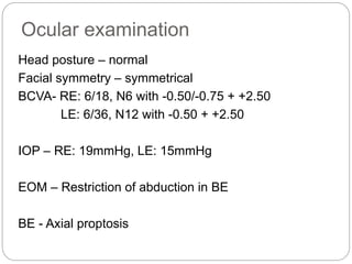
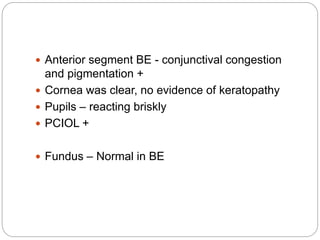
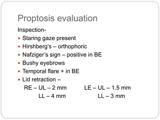
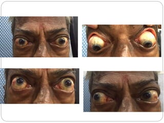
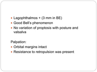
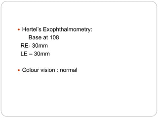
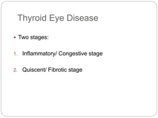
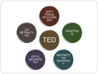
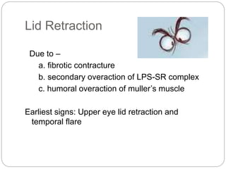
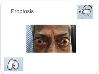
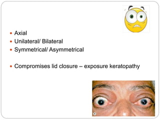
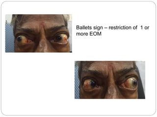
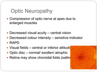
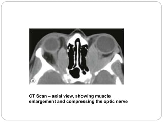
Dr.Maithri
DNB
Ophthalmology
THYROID EYE DISEASE
- Case and Discussion
History
 Chief complaints:
67 year old male
Decreased vision for distance and near in BE
since many years
Prominence of BE since 2 years
 Decreased vision in BE is gradually progressive and
painless.
 Vision improved to an extent after cataract surgery in BE
which was done 3 years ago but has again decreased
over the past few months
 H/O prominence of BE which was insidious and
simultaneous in onset, gradually progressive over 2
years with increased progression in the past 2 months.
 Associated with redness and mild discomfort
 No H/o pain, diplopia
 H/o of loss of appetite and loss of weight since 1 year
 No h/o neck swelling, tremors, skin pigmentation,
palpitations.
 No h/o trauma, fever
Past history:
 H/o Cataract surgery in BE 3 years ago
 H/o thyroid disease since 2 years on oral
medications
 Not a k/c/o DM/HTN/Asthma/ cardiac disease
Family history: Nothing significant
Personal History:
 Known smoker since 40 years, 4-5 cigarettes per
day
 Not alcoholic
General examination
 67 year old male conscious, alert, well oriented to
time, place and person.
 PR: 92 bpm
 Temp: Afebrile to touch
 BP: 130/80 mmHg
 CVS: S1, S2 heard, no murmurs
 RS/ PA/ CNS: Normal
 No pretibial myxedema
 No clubbing
Ocular examination
Head posture – normal
Facial symmetry – symmetrical
BCVA- RE: 6/18, N6 with -0.50/-0.75 + +2.50
LE: 6/36, N12 with -0.50 + +2.50
IOP – RE: 19mmHg, LE: 15mmHg
EOM – Restriction of abduction in BE
BE - Axial proptosis
 Anterior segment BE - conjunctival congestion
and pigmentation +
 Cornea was clear, no evidence of keratopathy
 Pupils – reacting briskly
 PCIOL +
 Fundus – Normal in BE
Proptosis evaluation
Inspection-
 Staring gaze present
 Hirshberg’s – orthophoric
 Nafziger’s sign – positive in BE
 Bushy eyebrows
 Temporal flare + in BE
 Lid retraction –
RE – UL – 2 mm LE – UL – 1.5 mm
LL – 4 mm LL – 3 mm
 Lagophthalmos + (3 mm in BE)
 Good Bell’s phenomenon
 No variation of proptosis with posture and
valsalva
Palpation:
 Orbital margins intact
 Resistance to retropulsion was present
 Hertel’s Exophthalmometry:
Base at 108
RE- 30mm
LE – 30mm
 Colour vision : normal
 Visual fields 30-2: Not reliable due to fixation loss
 MRI orbit – Showed compression of optic nerve
 VISA score:
 V- yes
 I – 2/10
 S – 1/3
 A – severe
Bilateral Thyroid Eye Disease – severe but no active
inflammation
Thyroid Eye Disease
 Two stages:
1. Inflammatory/ Congestive stage
2. Quiscent/ Fibrotic stage
TED
SOFT
TISSUE
INVOLVEM
ENT
PROPTOS
IS
RESTRICT
IVE
MYOPATH
Y
OPTIC
NEUROPA
THY
LID
RETRACTI
ON
Soft tissue Involvement
 Symptoms – irritation, grittiness, photophobia, dull
ache
 Signs – Epibulbar hyperemia
Periorbital edema – more in morning
Superior limbic keratoconjunctivitis
 Goldzeihers sign – conjunctival congestion
Lid Retraction
Due to –
a. fibrotic contracture
b. secondary overaction of LPS-SR complex
c. humoral overaction of muller’s muscle
Earliest signs: Upper eye lid retraction and
temporal flare
LID SIGNS
Eponym Sign
KOCHERS Staring appearance
VIGOUROUX Eyelid fullness or puffiness
VON GRAEFES Upper lid lag on downgaze
DALRYMPLES Upper eyelid retraction
STELLWAG Infrequent blinking
GIFFORDS Difficulty in everting upper lid
ENROTH Edema of lower lid
GRIFFITH Lower lid lag on upgaze
Proptosis
 Axial
 Unilateral/ Bilateral
 Symmetrical/ Asymmetrical
 Compromises lid closure – exposure keratopathy
Restrictive Myopathy
 30 – 50%
 Initially due to inflammation, later fibrosis
 1st muscle – Inferior rectus – contracture leads
to elevation defect
 Abduction defect – fibrosis of MR
 Depression defect – fibrosis of SR
 Adduction defect – fibrosis of LR
 Moebius sign – unable to converge eyes
Ballets sign – restriction of 1 or
more EOM
Optic Neuropathy
 Compression of optic nerve at apex due to
enlarged muscles
 Decreased visual acuity – central vision
 Decreased colour intensity – sensitive indicator
 RAPD
 Visual fields – central or inferior altitudinal defects
 Optic disc – normal/ swollen/ atrophic
 Retina may show choroidal folds (sallmanns)
CT Scan – axial view, showing muscle
enlargement and compressing the optic nerve
Conclusion
 Thyroid eye disease is a consortium of signs each of
which have a diagnostic and prognostic value
 EOM restriction shouldn’t be confused with nerve
palsies, optic neuropathy should be differentiated
from glaucoma
 To make the patient aware of the disease and
potential complications is important
 Each of the 5 manifestations need to be dealt with
individually and as a whole keeping in mind the
Thank you

Thyroid Eye Disease - Case and Discussion

  • 1.
  • 2.
    History  Chief complaints: 67year old male Decreased vision for distance and near in BE since many years Prominence of BE since 2 years
  • 3.
     Decreased visionin BE is gradually progressive and painless.  Vision improved to an extent after cataract surgery in BE which was done 3 years ago but has again decreased over the past few months  H/O prominence of BE which was insidious and simultaneous in onset, gradually progressive over 2 years with increased progression in the past 2 months.  Associated with redness and mild discomfort  No H/o pain, diplopia  H/o of loss of appetite and loss of weight since 1 year  No h/o neck swelling, tremors, skin pigmentation, palpitations.  No h/o trauma, fever
  • 4.
    Past history:  H/oCataract surgery in BE 3 years ago  H/o thyroid disease since 2 years on oral medications  Not a k/c/o DM/HTN/Asthma/ cardiac disease Family history: Nothing significant Personal History:  Known smoker since 40 years, 4-5 cigarettes per day  Not alcoholic
  • 5.
    General examination  67year old male conscious, alert, well oriented to time, place and person.  PR: 92 bpm  Temp: Afebrile to touch  BP: 130/80 mmHg  CVS: S1, S2 heard, no murmurs  RS/ PA/ CNS: Normal  No pretibial myxedema  No clubbing
  • 6.
    Ocular examination Head posture– normal Facial symmetry – symmetrical BCVA- RE: 6/18, N6 with -0.50/-0.75 + +2.50 LE: 6/36, N12 with -0.50 + +2.50 IOP – RE: 19mmHg, LE: 15mmHg EOM – Restriction of abduction in BE BE - Axial proptosis
  • 7.
     Anterior segmentBE - conjunctival congestion and pigmentation +  Cornea was clear, no evidence of keratopathy  Pupils – reacting briskly  PCIOL +  Fundus – Normal in BE
  • 8.
    Proptosis evaluation Inspection-  Staringgaze present  Hirshberg’s – orthophoric  Nafziger’s sign – positive in BE  Bushy eyebrows  Temporal flare + in BE  Lid retraction – RE – UL – 2 mm LE – UL – 1.5 mm LL – 4 mm LL – 3 mm
  • 10.
     Lagophthalmos +(3 mm in BE)  Good Bell’s phenomenon  No variation of proptosis with posture and valsalva Palpation:  Orbital margins intact  Resistance to retropulsion was present
  • 11.
     Hertel’s Exophthalmometry: Baseat 108 RE- 30mm LE – 30mm  Colour vision : normal
  • 12.
     Visual fields30-2: Not reliable due to fixation loss  MRI orbit – Showed compression of optic nerve
  • 13.
     VISA score: V- yes  I – 2/10  S – 1/3  A – severe Bilateral Thyroid Eye Disease – severe but no active inflammation
  • 16.
    Thyroid Eye Disease Two stages: 1. Inflammatory/ Congestive stage 2. Quiscent/ Fibrotic stage
  • 17.
  • 18.
    Soft tissue Involvement Symptoms – irritation, grittiness, photophobia, dull ache  Signs – Epibulbar hyperemia Periorbital edema – more in morning Superior limbic keratoconjunctivitis  Goldzeihers sign – conjunctival congestion
  • 19.
    Lid Retraction Due to– a. fibrotic contracture b. secondary overaction of LPS-SR complex c. humoral overaction of muller’s muscle Earliest signs: Upper eye lid retraction and temporal flare
  • 20.
    LID SIGNS Eponym Sign KOCHERSStaring appearance VIGOUROUX Eyelid fullness or puffiness VON GRAEFES Upper lid lag on downgaze DALRYMPLES Upper eyelid retraction STELLWAG Infrequent blinking GIFFORDS Difficulty in everting upper lid ENROTH Edema of lower lid GRIFFITH Lower lid lag on upgaze
  • 21.
  • 22.
     Axial  Unilateral/Bilateral  Symmetrical/ Asymmetrical  Compromises lid closure – exposure keratopathy
  • 23.
    Restrictive Myopathy  30– 50%  Initially due to inflammation, later fibrosis  1st muscle – Inferior rectus – contracture leads to elevation defect  Abduction defect – fibrosis of MR  Depression defect – fibrosis of SR  Adduction defect – fibrosis of LR  Moebius sign – unable to converge eyes
  • 24.
    Ballets sign –restriction of 1 or more EOM
  • 25.
    Optic Neuropathy  Compressionof optic nerve at apex due to enlarged muscles  Decreased visual acuity – central vision  Decreased colour intensity – sensitive indicator  RAPD  Visual fields – central or inferior altitudinal defects  Optic disc – normal/ swollen/ atrophic  Retina may show choroidal folds (sallmanns)
  • 26.
    CT Scan –axial view, showing muscle enlargement and compressing the optic nerve
  • 27.
    Conclusion  Thyroid eyedisease is a consortium of signs each of which have a diagnostic and prognostic value  EOM restriction shouldn’t be confused with nerve palsies, optic neuropathy should be differentiated from glaucoma  To make the patient aware of the disease and potential complications is important  Each of the 5 manifestations need to be dealt with individually and as a whole keeping in mind the
  • 28.